Blog

Simple Breast Cyst

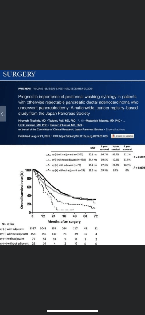
  • The patient has a BIRADS 2 simple cyst
  • To be regarded as a simple cyst, a breast nodule must meet three criteria:
    • The margins must be circumscribed:
      • i.e., a margin “that is well defined or sharp, with an abrupt transition between the lesion and surrounding tissue”
    • It must be anechoic:
      • i.e., “without internal echoes”
    • It must show posterior acoustical enhancement:
      • i.e., “a column that is more echogenic deep to the mass
  • Simple cysts:
    • Are almost never associated with cancer in the absence of other abnormalities seen on mammogram or ultrasound
  • There are numerous reflectors in breast tissue, and all of the sound waves that are reflected do not make it back to the transducer:
    • Many of them bounce back and forth (reverberate) between reflectors in the tissue, and with each reverberation, part of the echoes return to the transducer, are recorded, and part undergo another excursion between the reflectors
    • Most of these echoes are obscured by all of the other echoes in the tissue, but when an otherwise anechoic window (a cyst) is present:
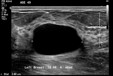
      • The reverberation echoes can be seen in the anterior part of the cyst:
        • In the image shown, a hyperechoic band can be seen within or just above the anterior wall of the cyst, especially on the left side of the image:
          • The acoustic mismatches between this band and the less echogenic tissue superficial to it and the anechoic fluid deep to it, cause echoes that reverberate, creating the artifact in the near field of the cyst
  • References
    • D’Orsi CJ, Sickles EA, Mendelson EB, Morris EA. ACR BI-RADS® Atlas: Breast Imaging Reporting and Data System, 5th ed. Reston, VA: American College of Radiology; 2013.
    • Kremkau FW. Diagnostic Ultrasound: Principles and Instruments, 7th ed. Elsevier; 2006:274-292. 1Image/Figure 2: Click on space below to upload image. Use highest available resolution and file size .png format: recommended .jpg format: acceptable .gif format: only recommended for text-based images and basic black and white charts Image/Figure 2 Reprint Permission: Obtain required reprint permissions when using copyrighted material Courtesy of the American Society of Breast Surgeons Breast Ultrasound Certification Reviewers.☐ Reprint permission obtained/attached Image/Figure 2 Caption: Include brief, informative caption for image/figure Ultrasound imaging of the palpable lesion.

#Arrangoiz #BreastSurgeon #CancerSurgeon #BreastCancer #CASO #CenterforAdvancedSurgicalOncology

Criteria for the Selection of a Lesion Suitable for a Supraglottic Partial Laryngectomy.

  1. At least 5 mm margin at anterior commissure.
  2. True vocal cords must be mobile.
  3. Only one arytenoid may be removed.
  4. No cartilage invasion by tumor.
  5. Tongue mobility should be normal.
  6. No extension to interarytenoid or post-cricoid area.
  7. Apex of pyriform sinus should be free.
  8. Generally, lesions < 3 cm.

#Arrangoiz #CancerSurgeon #HeadandNeckSurgeon #SurgicalOncologist #CASO #CenterforAdvancedSurgicalOncology #Miami

The 7 steps to prevent cancer

1. Don’t use tobacco

2. Eat a healthy diet

3. Maintain a healthy weight and be physically active

4. Protect yourself from the sun

5. Get vaccinated

6. Avoid risky

6. Avoid risky behaviors

7. Get regular medical care

#Arrangoiz #CancerSurgeon #Surgeon #Prevention #Endcancer #CenterforAdvsncedSurgicalOncology #CASO

Clinical Manifestations of Pregnancy Associated Breast Cancer (PABC) Part II

Any palpable abnormality in a pregnant or postpartum woman persisting for more than a few weeks needs a careful investigative workup to rule out PABC.

Although up to 90% of PABC presents as a palpable mass, there can be atypical presentations, including milk rejection from the affected breast or bloody nipple discharge.

Bloody nipple discharge can be especially difficult to differentiate from a benign or malignant origin as it may happen due to pregnancy-induced changes leading to delicate intraductal epithelial spurs that are easily traumatized and shed and thus not considered pathologic in all cases.

In the absence of clinically detectable masses or abnormality on cytology, these patients may be followed closely.

However, if bloody nipple discharge persists after several months postpartum, diagnostic studies should be performed to rule out pathology.

#Arrangoiz #BreastSurgeon #CancerSurgeon #SurgicalOncology #PregnancyAssociatedBreastCancer #BreastCancer #CASO #CenterforAdvancedSurgicalOncology

Clinical Features of Pregnancy Associated Breast Cancer (PABC) Part 1

Patients with PABC are often found to have advanced disease at diagnosis, with the median size of tumor ranging from 3.4 to 4 cm and the reported incidence of axillary lymph node metastasis between 56% and 73%.

Delay in diagnosis is often due to the physiologic changes that occur in breasts during pregnancy that can pose challenges for differentiating a new mass from engorged and nodular breast tissue on a physical examination.

In the first and second trimesters, there is active proliferation and differentiation of the lobules, alveoli, and lactiferous ducts, leading to enlargement and increased overall density of breast tissue.

The prolactin level rises during the third trimester, which stimulates milk-producing cells to differentiate, and alveoli and milk ducts are filled with colostrum.

Around parturition, the breasts undergo lactogenesis, which leads to the secretion of colostrum and then milk.

#Arrangoiz #BreastSurgeon #PregnancyAssociatedBreastCancer #CancerSurgeon #SurgicalOncology #CASO #PABC #CenterforAdvancedSurgicalOncology

Molecular Basis of Pregnancy Associated Breast Cancer

PABC shares histology and some prognostic features with breast cancer occurring in other young women.

Invasive ductal carcinoma is the most common histologic subtype, followed by invasive lobular carcinoma.

The majority of tumors are grade II or III, and lymphovascular invasion is common.

However, the molecular subtypes of PABC seem to differ from those of non-PABC.

Several case-control studies have demonstrated that the hormone receptor status in PABC is more likely to be negative when compared with that in breast cancer in age-matched cohorts.

In contrast, there are conflicting reports of the HER-2 receptor status for PABC.

In a case-control study published in 1993 by Elledge and colleagues, 58% of the tumors from pregnant patients were positive for HER-2 on immunohistochemistry, whereas only 16% of age-matched non-pregnant patients had HER-2-positive tumors.

However, in a more contemporary report from the MD Anderson Cancer Center series of pregnant women by Middleton and colleagues, 28% of PABC cases had positive membrane staining for HER-2, and there was no significant difference when compared with breast carcinoma in young nonpregnant women.

#Arrangoiz #BreastSurgeon #CancerSurgeon #SurgicalOncology #PregnancyAssociatedBreastCancer #BreastCancer #PABC #CASO #CenterforAdvancedSurgicalOncology

Pregnancy Associated Breast Cancer

Pregnancy-associated breast cancer (PABC) is defined as breast cancer diagnosed during pregnancy or in the first postpartum year.

Breast cancer is the second most common malignancy affecting pregnant women.

It is estimated that one in 3,000 pregnant women is diagnosed with breast cancer and that up to 3% of all breast cancers are associated with pregnancy.

In general, breast cancer in young women is rare; the estimated incidence of breast cancer in women younger than 40 years of age was less than 4% in 2015 in the United States.

However, by definition, PABC occurs in women of reproductive age, and the average age of women with PABC is between 32 and 38 years of age.

The birth rate for women age 30 to 44 years has been steadily increasing in the past few decades, and as more women are delaying childbearing for various reasons, the incidence of PABC has risen and is expected to continue to rise.

As in non-PABC, the risk of PABC seems to be age related.

A large Canadian population cohort study using birth data from a national registry reported that PABC appeared to be more common among women with a first-term pregnancy occurring after the age of 35.

#Arrangoiz #BreastSurgeon #CancerSurgeon #PABC #PregnancyAssociatedBreastCancer #SurgicalOncology #Miami #SurgicalOncologist #CASO #CenterforAdvancedSurgicalOncology

Peritoneal Washing in Pancreatic Cancer

Peritoneal washing cytology (CY)+ in Pancreatic ductal adenocarcinoma (PDAC) is classified as M1 disease by the American Joint Committee on Cancer staging system and NCCN guidelines, whereas the Japanese General Rules for Pancreatic Cancer have not yet included the CY status for staging.

Hiroyoshi Tsuchida et al analysed well-maintained, nationwide database of 1970 patients with PDAC to evaluate the relationship among CY status, clinical factors, and overall survival and to clarify the clinical implications for operative resection in patients who are CY+ status.

It demonstrated that OS in patients who underwent resection was markedly worse in patients who were CY+ than in patients who were CY– who underwent resection and that peritoneal recurrence appeared earlier in patients who were CY+.

Read full text article at
https://www.surgjournal.com/article/S0039-6060(19)30411-8/fulltext

#Arrangoiz #CancerSurgeon #surgjournal #surgery #surgeryjournal #pancreas #peritonealWashingCytology

Classification of Glossectomies

  • TYPE I GLOSSECTOMY (MUCOSECTOMY):Definition:
      • Incision of the mucosa in healthy tissue with appropriate safety margins (0.5 cm to 1.0 cm depending on whether or not the lesion is well defined):
        • The mucosa and submucosa are included up to the intrinsic muscle fibers of the tongue
        • The deep resection margin should include:
          • A thin layer of the intrinsic muscles:Because of a possible invasion of the submucosa
    • Generally:
      • The wound is left to heal by secondary intention
      • Although the defect may be:Partially closed primarilyOr covered with a skin graft
    • Indication:Precancerous, superficial suspicious lesions, limited to the epithelium of the tongue without previous biopsy:
        • The aim of surgery:
          • Is to remove all the lesion with adequate margins up to the healthy tissue with both diagnostic and curative intent

Presentation1

  • TYPE II GLOSSECTOMY (PARTIAI GLOSSECTOMY):Definition: It includes the lesion and adjacent normal mucosa, submucosa, and the intrinsic muscles up to the surface of the extrinsic muscles (when the directions of the muscle fibers change)
      • With appropriate safety margins (approximately 1.5 cm):The resection usually is diamond shaped on the surface, while more deeply, it is shaped like a truncated cone with the intrinsic muscles as the apex. The terminal branches of the lingual artery should be ligated
        • The lingual nerve is usually preserved
    • Closure may be partial or total with the objective of avoiding:Bleeding, postoperative edema, and retracted scars
  • Indication: Lesions infiltrating submucosa and superficially into intrinsic muscles, but not extrinsic muscles, or infiltration less than 10 mm deep

Presentation1

  • TYPE III GLOSSECTOMY:Type IIIa glossectomy (hemiglossectomy):Definition: The specimen includes the mucosa, submucosa, and intrinsic and extrinsic muscles ipsilateral to the lesion:The mucosa is resected up to healthy tissue with appropriate safety margins (at least 1.5 cm)
          • The lingual artery must be ligated and removed en bloc with the lingual and hypoglossal nerves, in the specimen of the primary tumor and neck nodes
          • The base of the ipsilateral tongue is preserved
          • The tip of the tongue can be preserved or not
      • Indication: Lesions infiltrating the intrinsic and minimally extrinsic muscles or infiltration greater than 10 mm but confined within the ipsilateral tonguePresentation1
    • Type IIIb glossectomy (compartmental hemiglossectomy):Definition: The specimen includes the mucosa, submucosa, intrinsic and extrinsic muscles ipsilateral to the lesion, genioglossus, hyoglossus and styloglossus muscles, and the inferior portion of the palatoglossus muscle.
        • Medially:The midline raphe is included in the resection
        • The lingual nerve is resected as far cranially as possible
        • The hypoglossal nerve is removed after the ansa
        • The lingual artery and vein is ligated in proximity to the horn of the hyoid bone, and removed en bloc with specimen and neck nodes
      • Indication: Lesions massively infiltrating the intrinsic and extrinsic muscles but confined to the ipsilateral tongue

Presentation1

  • TYPE IV GLOSSECTOMY:Type IVa glossectomy (subtotal glossectomy):Definition: This is an anterior subtotal glossectomy
        • With preservation of both sides of the base of the tongue, posterior hyoglossus muscle, and hypoglossal and lingual nerves, from the less involved side.
      • Indication:Lesions that arise in the anterior portion of the mobile tongue and exceed the hemilingual area of origin involving the contralateral genioglossus muscle but limited to mobile tongue

Presentation1

    • Type IVb (near-total glossectomy):Definition: Type IVa glossectomy with extension to the ipsilateral base of the tongue:The following contralateral structures are preserved:Hyoglossus and styloglossus muscles, hypoglossal and lingual nerves, and lingual artery (functional unit of the base of the tongue)Indication: Massive lesions that exceed the border of the hemilingual area of origin infiltrating the ipsilateral base of the tongue and the contralateral genioglossus muscle

Presentation1

  • TYPE V GLOSSECTOMY (TOTAL GLOSSECTOMY):Definition: The specimen includes all of the mobile tongue and the base of the tongue transected at the level of the lingual arteries, hypoglossal, lingual nerves, and the floor of the mouth.Indication:Massive infiltrating lesions, for instance, those of the anterior ventral surface of the tongue, dorsum of the tongue, or the tongue base, which bilaterally involve the extrinsic genioglossus, hyoglossus, and styloglossus with impairment of the mobility of the tongue

Presentation1

cropped-18403652_10206829497335208_5004404657991480104_n1.jpg

  • What is Head and Neck Surgery?:
    • It is a surgical sub-specialty that deals mainly with benign and malignant tumors of the head and neck region, including:
      • The scalp, facial region, eyes, ears, nose, nasal fossae, paranasal sinuses, oral cavity, pharynx (nasopharynx, oropharynx, hypopharynx), larynx (supraglotic larynx, glottis larynx, subglotic larynx), thyroid gland, parathyroid gland, salivary glands (parotid glands, submandibular glands, sublingual glands, minor salivary glands), soft tissues of the neck, skin of the head and neck region.
        • The head and neck surgeon’s work area:Does not cover tumors or diseases of the brain and other areas of the central nervous system or those of the cervical spine:This is the neurosurgeon field.
    • Among the diagnostic procedures performed by the head and neck surgeon,  are the following:
      • Nasopharyngolaryngoscopy:
        • Performed to examine, evaluate and, possibly perform a biopsy, of oral cavity, pharyngeal and laryngeal lesions.
    • The surgeries most commonly performed by the head and neck surgeon are:
      • Total or near total thyroidectomies
      • Hemithryoidectomies (lobectomies)
      • Comprehensive neck dissections
      • Selective neck dissections
      • Maxillectomies:
        • Total maxillectomy
        • Subtotal maxillectomy
        • Infrastructure maxillectomy
        • Suprastructure maxillectomy
        • Medial maxillectomy
      • Mandibulectomy:
        • Segmental
        • Marginal
      • Tracheostomy
      • Salivary gland surgeries:
        • Parotid gland operations:
          • Limited superficial parotidectomy with identification and preservation of the facial nerve
          • Superficial parotidectomy with identification and preservation of the facial nerve
          • Near total parotidectomy with identification and preservation of the facial nerve
          • Total parotidectomy
        • Submandibular gland resection
        • Sublingual gland resection
      • Resection of tumors of the oral cavity:
        • Glossectomy
        • Resection of the floor of the mouth tumors
      • Resection of tumors of the pharynx
      • Resection of tumors of the larynx
      • Split-thickness skin grafts
      • Full-thickness skin grafts
      • Sentinel lymph node mapping and sentinel lymph node biopsy
      • Resection of malignant skin tumors (BCC, SCC, melanoma) of the head and neck region
  • The formation of the head and neck surgeon includes mastering the following subjects:
    • Surgical Anatomy
    • History and Basic Principles of Head and Neck Surgery
    • Epidemiology, Etiology, and Pathology of Head and Neck Diseases
    • Diagnostic Radiology of the Head and Neck Region
    • Tumors of the Scalp, Skin and Melanoma
    • Eyelids and Orbit
    • Nasal Cavity and Paranasal Sinuses
    • Skull Base and Temporal Bone
    • Lips and Oral Cavity
    • Pharynx and Esophagus
    • Larynx and Trachea
    • Cervical Lymph Nodes
    • Thyroid and Parathyroid Glands
    • Salivary Glands
    • Neurogenic Tumors and Paragangliomas
    • Soft Tissue Tumors
    • Bone Tumors and Odontogenic Lesions
    • Reconstructive Surgery
    • Oncologic Dentistry and Maxillofacial Prosthetics
    • Principles of Radiation Oncology
    • Principles of Chemotherapy
    • Molecular Oncology, Genomics and Immunology
    • Nutrition
    • Biostatistic

Rodrigo Arrangoiz MS, MD, FACS a head and neck surgeon / endocrine surgeon / surgical oncologist and is a member of Center for Advanced Surgical Oncology at Palmetto General Hospital:

prof_739_20190417135234

Training:

• General surgery:

• Michigan State University:

• 2004 al 2010

• Surgical Oncology / Head and Neck Surgery / Endocrine Surgery:

• Fox Chase Cancer Center (Filadelfia):

• 2010 al 2012

• Masters in Science (Clinical research for health professionals):

• Drexel University (Filadelfia):

• 2010 al 2012

• Surgical Oncology / Head and Neck Surgery / Endocrine Surgery:

• IFHNOS / Memorial Sloan Kettering Cancer Center:

• 2014 al 2016

#Arrangoiz

#Teacher

#Surgeon

#Cirujano

#ThyroidExpert

#ThyroidSurgeon

#CirujanodeTiroides

#ExpertoenTiroides

#ExpertoenParatiroides

#Paratiroides

#Hiperparatiroidismo

#CancerdeTiroides

#ThyroidCancer

#PapillaryThyroidCancer

#SurgicalOncologist

#CirujanoOncologo

#CancerSurgeon

#CirujanodeCancer

#HeadandNeckSurgeon

#CirugiaEndocrina

#CirujanodeTumoresdeCabezayCuello

#OralCavityCancer

#CASO

#CenterforAdvancedSurgicalOncology

#PalmettoGeneralHospitalwww.centerforadvancedsurgicaloncology.com

CovidSurg Collaboration Head and Neck Surgery

👉The most recent publication of our CovidSurg collaboration showed head and neck surgery during the pandemic, represents a significant risk of contagion of # COVID19, which is significantly higher (an order of magnitude) than the risk of community transmission. 

👉The study can be seen in the Cancer journal of the American Cancer Society

👉bit.ly/3pcnZw2

#Arrangoiz #CancerSurgeon #HeadandNeckSurgery
#CovidSurg