Blog

Removal of Morphologically Abnormal Parathyroid Gland (s)

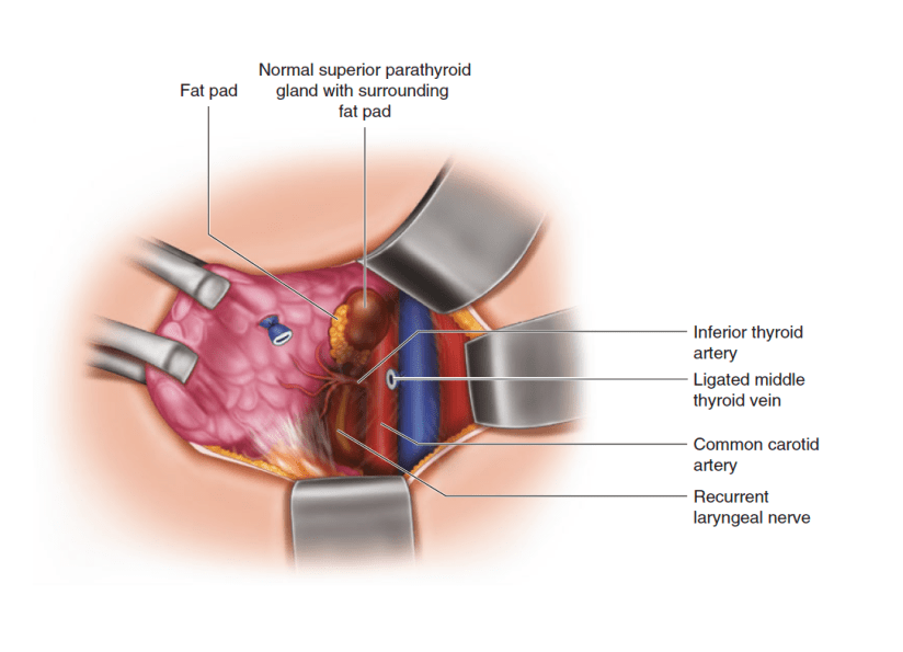
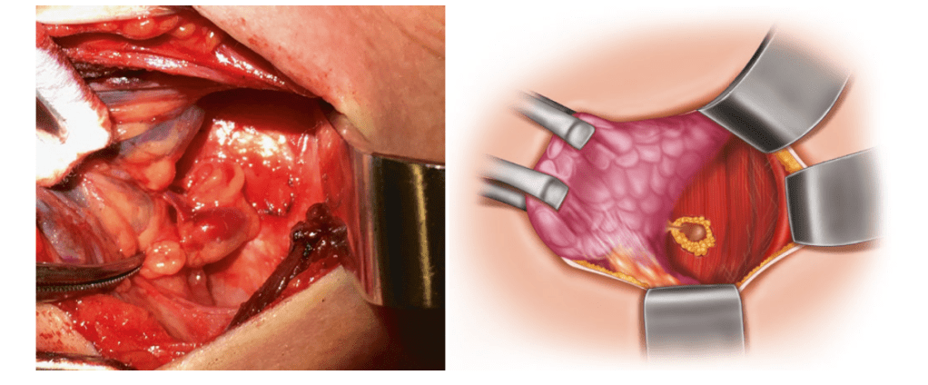
  • The gland is bluntly dissected from the surrounding tissue back toward its vascular pedicle, with particular care not to breach the capsule of the gland (Figure)
Exposure of the vascular pedicle. The gland has been dissected away from the surrounding tissue to expose its supplying vascular pedicle, which can then be ligated with a bipolar cautery or a small clip. The position of the recurrent laryngeal nerve, which can be seen in the bottom of the image, should be re-confirmed during this stage
  • The vascular pedicle is then ligated with bipolar cautery or a small clip:
    • After first re-confirming the position of RLN.
  • The excised gland(s) should then be sent to pathology
    • To confirm the weight and presence of parathyroid tissue, if available

#Arrangoiz #ParathyroidSurgeon #ParathyroidExpert #Parathyroidectomy #MSMC #MountSinaiMedicalCenter #HeadandNeckSurgeon #Surgeon #Teacher #EndocrineSurgery

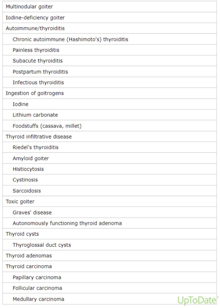
Etiology of Goiters

• Etiology:

• Iodine deficiency:

• Is the most common cause of goiter worldwide

• In mildly and moderately iodine-deficient regions in Denmark:

• Goiter (as determined by ultrasonography) is present in 15% and 22.6% of the population, respectively

• In the United States, where significant iodine deficiency does not exist:

• Multinodular goiter, chronic autoimmune (Hashimoto’s) thyroiditis, and Graves’ disease are more common causes of goiter:

• In older adults, multinodular goiter is most common

• Other less common causes of goiter include:

• Tumors

• Thyroiditis

• Infiltrative diseases

• The risk of thyroid cancer within a multinodular goiter:

• Is approximately 3% to 5%:

• Similar to the risk in a solitary thyroid nodule

• In a series of 718 patients operated on for goiter in Pakistan:

• 3% of patients were found to have a malignancy

• In a surgical series (with potential selection bias) of 3233 patients with multinodular goiter:

• In which all patients had preoperative ultrasounds and patients who had indeterminate, suspicious, or malignant (Bethesda 3 to 6) preoperative FNA were excluded:

• 31.7% had incidental thyroid cancers:

• Of which 56% were papillary microcarcinomas

• Pathophysiology:

• In patients with iodine deficiency or chronic autoimmune (Hashimoto’s) thyroiditis:

• An increase in thyroid-stimulating hormone (TSH) secretion:

• Is the predominant cause of goiter

• In contrast, most patients with sporadic nontoxic multinodular goiters:

• Have normal serum TSH concentrations:

• In these individuals, the thyroid enlargement is probably caused by several growth factors (including TSH):

• That act over time on thyroid follicular cells:

• That have different synthetic and growth potentials

• There is often a family history of goiter:

• Suggesting that genetic factors may also play a role

• The result is diffuse and later multinodular thyroid enlargement:

• Some nodules eventually become autonomous:

• Due to activating mutations in the TSH receptor or G proteins within the thyroid follicular cells

• The following observations support this sequence of events:

• Thyroid volume is larger in older patients

• The longer the patient has a goiter:

• The larger the size of the goiter

• The larger the size of the goiter:

• The lower the serum TSH concentration

• In patients with Graves’ disease:

• TSH receptor antibodies (TRAb):

• Stimulate the TSH receptor:

• To cause thyroid growth and excessive hormonal secretion

Clinical Presentation of Patients with Goiter

  • The clinical manifestations of goiter:
    • Depends upon the presence of thyroid dysfunction and upon the growth rate of the goiter
  • Some patients may have symptoms and biochemical evidence of hypothyroidism or hyperthyroidism:
    • However, the majority of patients with goiter:
      • Are asymptomatic and biochemically euthyroid
  • Patients with longstanding, large goiters:
    • May develop symptoms of obstruction:
      • Due to progressive compression of the trachea or sudden enlargement (usually accompanied by pain):
        • Secondary to hemorrhage into a nodule
  • Asymptomatic:
    • Most goiters grow very slowly over many decades:
      • Therefore, the majority of patients with goiter:
        • Are asymptomatic
  • The goiter may first be noted on:
    • Physical examination or
    • Found incidentally on cross-sectional imaging studies performed for unrelated reasons
  • Thyroid dysfunction:
    • If the goiter is due to Hashimoto’s thyroiditis or severe iodine deficiency:
      • Patients may have symptoms of hypothyroidism:
        • For example:
          • Fatigue, constipation, cold intolerance
    • If due to multinodular goiter (with autonomy) or Graves’ disease:
      • Patients may have symptoms of hyperthyroidism:
        • For example:
          • Palpitations, dyspnea on exertion, unexplained weight loss
  • Obstructive symptoms:
    • Patients with longstanding goiters (cervical or substernal) may develop symptoms of obstruction:
      • Due to progressive compression of the trachea or sudden enlargement (usually accompanied by pain):
        • Secondary to hemorrhage into a nodule
    • The majority of patients with obstructive cervical goiters:
      • Have had a visible goiter for many years
  • Most patients with substernal goiter (77% to 90% in two series) also have visible goiters:
    • Although some are found incidentally on imaging studies performed for unrelated reasons
  • In those without visible goiter:
    • Substernal goiters may also be found:
      • Because of obstructive symptoms
  • Since goiters tend to grow slowly:
    • Substernal goiters are most commonly discovered:
      • During the fifth and sixth decades of life:
        • And are found more often in women than men
  • The most common symptom in patients with obstructive cervical or substernal goiter:
    • Is exertional dyspnea:
      • Which is present in 30% to 60% of patients:
        • This symptom usually occurs when:
          • The tracheal diameter is less than 8 mm
  • In some patients with substernal goiter:
    • Dyspnea is primarily positional or nocturnal:
      • And it occurs primarily during maneuvers that force the thyroid into the thoracic inlet:
        • Such as reaching and bending
  • When tracheal compression becomes severe (luminal diameter less than 5 mm):
    • Stridor or wheezing occurs at rest:
      • This upper airway wheezing must be distinguished from asthma
  • An upper respiratory illness:
    • May exacerbate upper airway obstruction
  • Cough:
    • Is present in 10% to 30% of patients:
      • And it may be positional
  • Pain is unusual
  • Choking sensation is common
  • Goiter may contribute to obstructive sleep apnea:
    • And thyroidectomy may improve symptoms:
      • In a study of 45 patients with snoring symptoms who were undergoing thyroidectomy for any reason (42% for goiter or compressive symptoms):
        • 29% had improved snoring frequency, apnea, and frequency of daytime somnolence after surgery
  • A variety of other symptoms can be induced by obstructive goiter:
    • Dysphagia:
      • Is a less common complaint because of the posterior position of the esophagus
  • Compression of a recurrent laryngeal nerve:
    • May cause transient or permanent vocal cord palsy:
      • Resulting in hoarseness
  • Phrenic nerve paralysis
  • Horner’s syndrome:
    • Due to compression of the cervical sympathetic chain
  • Rarely:
    • Jugular vein compression or thrombosis
    • Cerebrovascular steal syndromes
    • Even the superior vena cava syndrome

#Arrangoiz #ThyroidExpert #ThyroidSurgeon #HeadandNeckSurgeon #CancerSurgeon #SurgicalOncologist #Miami #Florida #MSMC #MountSinaiMedicalCenter #Mexico

Goiter Introduction

• The term goiter refers to:

• An abnormal growth of the thyroid gland

• Goiters can be:

• Diffuse or nodular:

• Depending on the etiology

• May be associated with:

• Normal, decreased, or increased thyroid hormone production

• The clinical manifestations vary with:

• Thyroid function and with the size and location of the goiter

• Anatomical Relationships:

• In healthy adults without iodine deficiency:

• A normal thyroid gland is approximately:

• 4.0 cm to 4.8 cm x 1.0 cm to 1.8cm x 0.8 cm to 1.6 cm in size

• Mean sonographic volume of:

• 7 mL to 10 mL:

• Thyroid volume measured by ultrasonography:

• Is slightly greater in men than women

• Increases with age and body weight

• Decreases with increasing iodine intake

• Weight of:

• 10 grams to 20 grams

• The normal thyroid gland:

• Is immediately caudal to the larynx and encircles the anterolateral portion of the trachea

• The thyroid gland is bordered by:

• The trachea and esophagus posteriorly

• The carotid sheath laterally

• Enlarging thyroid lobes:

• Usually grow outward:

• Because of their location in the anterior neck in front of the trachea:

• Covered only by thin strap muscles, subcutaneous tissue, and skin

• As a result of this outward growth:

• Even very large goiters may not compress the trachea or impinge on the great vessels lateral to the lobes:”

• However, in patients with substantial enlargement of one lobe or asymmetric enlargement of both lobes:

• The trachea, esophagus, or blood vessels:

• May be displaced or, less often, compressed

• Bilateral lobar enlargement:

• Especially if the goiter extends posterior to the trachea:

• May cause either:

• Compression or concentric narrowing of the trachea

• Compression of the esophagus

• Compression of the jugular veins

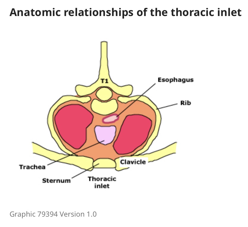
• The thoracic inlet:

• Is an ovoid area that measures approximately 5 cm x 10 cm:

• Boundaries:

• The sternum anteriorly

• The first thoracic vertebral body posteriorly

• The first ribs laterally

• The inlet is traversed by the:

• Trachea

• Esophagus

• Blood vessels

• Nerves

• The inferior pole of each thyroid lobe:

• Normally lies above the thoracic inlet:

• However, with some goiters, there is growth of one or both lobes through the inlet into the thoracic cavity:

• Which can result in obstruction of any of the structures in the inlet:

• Such goiters are called substernal:

• Although retrosternal is probably a more precise term

• Most substernal goiters are in the:

• Anterolateral mediastinum

• But approximately 10%:

• Are located primarily in the posterior mediastinum

• The prevalence of substernal goiter as a percentage of thyroidectomies:

• Ranges from 2% to 19%

#Arrangoiz #ThyroidSurgeon

Identification of the Inferior Parathyroid Gland

  • Visual:
    • Attention is then turned to identifying the structures related to the inferior gland:
      • As with the superior gland, careful exposure and mobilization of the thyroid gland may be all that is required to identify the inferior parathyroid glands
    • A “tongue” of thymic tissue can often be seen extending with the inferior pole vessels and “pointing” toward the inferior pole of the thyroid:
      • The inferior gland is usually located along this path
    • The inferior glands arise from the third branchial pouch:
      • In association with the thymus, and run a much longer course on descent
      • As such, they have a more variable location, including within the thymus
    • Begin by looking for a fat pad where a tongue of cervical thymus “points” to the inferior pole of the thyroid:
      • The inferior gland is often found on or within the posterior surface of this fat pad (Figure 1)
    • Continue inspecting from the lower pole of the thyroid along to the tongue of the cervical thymus
  • Additional Maneuvers:
    • If careful blunt dissection of the inferior pole vessels fails to identify the parathyroid gland:
      • The thymus should be isolated and mobilized:
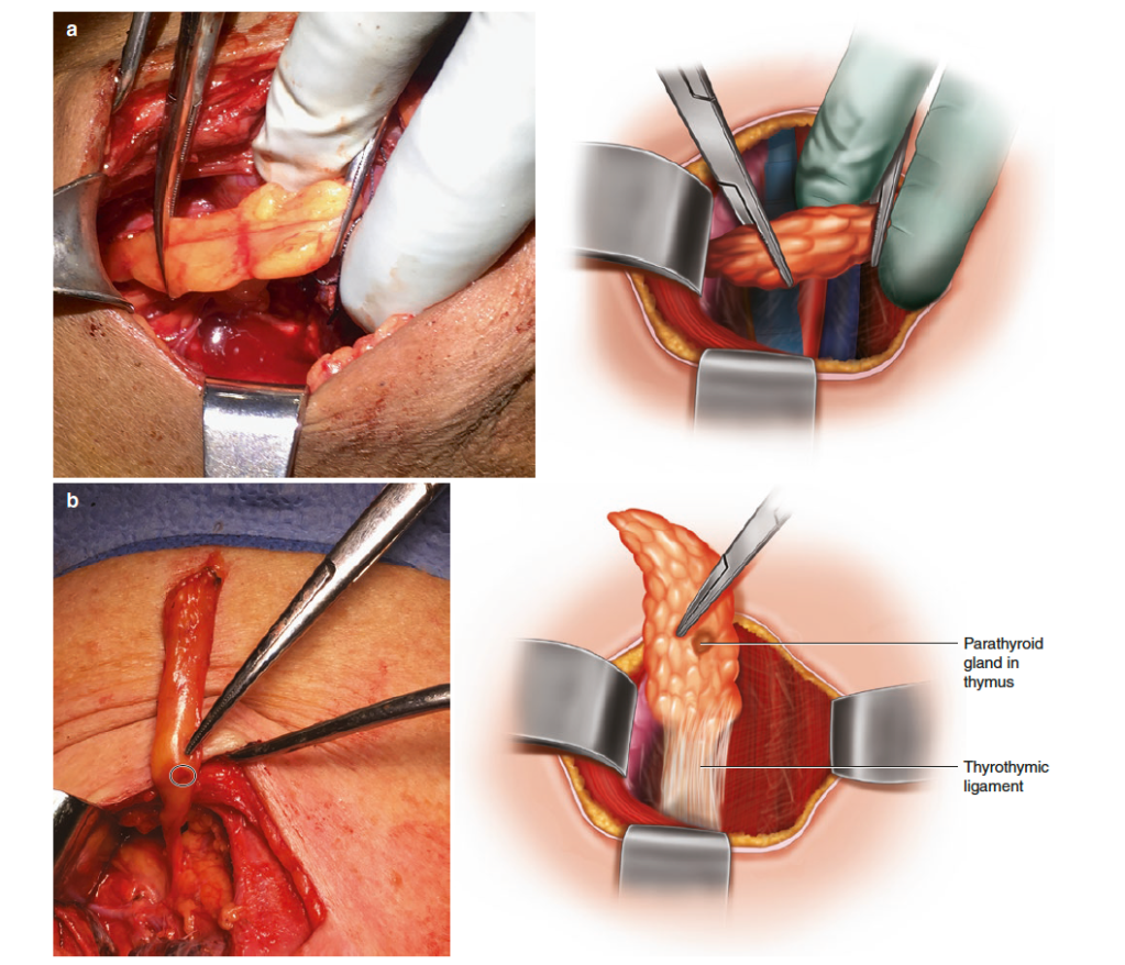
        • To do this step, the thyrothymic ligament is divided and the thymus is delivered into the wound with gentle retraction using hemostats, while pushing away any loose fibroareolar tissue
      • Care must be taken not tear the capsule of the thymus (Figure 2)
    • If the gland is still missing, the inferior parathyroid glands may also be found in the superior and anterior mediastinum, and along the carotid sheath as high as the carotid bifurcation:
      • The accessible portion of the superior mediastinum should be palpated and explored, and the carotid sheath opened and inspected from the root of the neck to the base of the skull
    • Lack of thymic tissue caudal to the thyroid gland on inspection is suggestive of a non-descended third branchial pouch, and should invite closer examination along its course of descent
    • Finally, if the missing abnormal gland has still not been found, consideration should be given to performing a thyroid lobectomy, as intra-thyroidal parathyroid glands have been reported in about 3% of patients:
      • These may have been previously reported as a thyroid nodule on preoperative ultrasound (hence this is a good time to re-check any preoperative imaging), or they can sometimes also be seen on intra-operative ultrasound
Mobilization of the thymus for a missing inferior parathyroid gland. If the inferior gland is not identified after dissection of the inferior thyroid pole and cervical thymus does not demonstrate it, the thymus should be mobilized from its position in the chest. In image (a), after division of the thyrothymic ligament, the thymus is being gently retracted into the wound with hemostats, taking care to keep the capsule intact. In image (b) from the same patient, an inferior parathyroid gland demonstrating Cope’s sign is identified in the distal portion of the thymus

#Arrangoiz #ParathyroidSurgeon #ParathyroidExpert #Hyperparathyroidism #HeadandNeckSurgeon #EndocrineSurgery #MSMC #MountSinaiMedicalCenter #Miami #Teacher #Surgeon Mexico

Omission of Adjuvant Radiation Therapy after Breast Conserving Surgery for DCIS

• Omission of RT for low-risk DCIS

• Radiation therapy (RT) reduces the odds of in-breast recurrence:

• But likely does not change the odds of distant recurrence or decrease mortality

• For patients with low-risk disease that has been fully resected with widely negative margins:

• The absolute reduction of in-breast recurrence:

• May not be large enough to justify the risks associated with RT:

• In such patients, it is reasonable to omit RT:

• Especially in the setting of:

• Comorbidity

• Advanced age

• Patient preference

• Some authors have a higher threshold to omit radiation:

• For young women given some data suggesting RT is more likely to benefit these patients

• While the results from the NSABP B-17 trial:

• Have been used to argue for RT in all women who undergo lumpectomy for pure DCIS:

• Methodological issues such as suboptimal pathologic evaluation and uncertainty about the completeness of excision:

• May have led to an overestimation of the benefit of RT in this study

• Moreover, RT is expensive, time consuming, and may be accompanied by significant side effects:

• So omission for patients likely to derive the least benefit is reasonable

• Since RT reduces the risk of ipsilateral recurrence without changing the risk of developing contralateral disease:

• Omission of RT would be a reasonable approach:

• For patients with an ipsilateral recurrence risk:

• Approximately equal to the risk of developing contralateral disease

• Among women diagnosed with DCIS:

• The long-term risk of developing a contralateral breast cancer or DCIS:

• Is approximately 3% to 10%

• Studies have tried to identify such a low-risk population using:

• Histopathologic and gene expression analysis

• While it is difficult to identify a clear-cut low-risk population:

• The benefit of RT becomes less clear:

• As the risk of ipsilateral recurrence approaches that of contralateral recurrence

• Histopathologic criteria:

• While there are no strict criteria for “low risk,” many authors have defined low risk as DCIS that is:

• Low- or intermediate-grade

• Small (less than 2.5 cm in size)

• Resected with widely negative margins (≥ 1 cm)

• Omission of RT in such patients is reasonable:

• Although associated with a small risk of ipsilateral recurrence

• Several studies suggest that clinical pathologic criteria:

• May define a low-risk cohort of patients with DCIS:

• For whom RT may be reasonably omitted given a low risk of recurrence

• These studies are summarized below:

• The Eastern Cooperative Oncology Group (E5194):

• Was an observational study:

• That investigated excision without RT in women with low- to intermediate-grade versus high-grade DCIS

• Eligible patients had:

• Less than 2.5 cm of low- to intermediate-grade DCIS or

• Less than 1.0 cm for high-grade DCIS

• Margins ≥ 3 mm were required and a negative post-excision mammogram was obtained for all participants

• Tamoxifen following excision:

• Was allowed but not mandated

• With a median follow-up of 6.7 years, the following local recurrent rates (LRR) were reported:

• Five-year LRR for low- or intermediate-grade DCIS (n = 565) was:

• 6.1% (95% CI 4.1-8.2)

• Five-year LRR for high-grade DCIS (n = 105) was:

• 15.3% (95% CI 8.2-22.5)

• The 12-year LRRs for the low- or intermediate-grade group was:

• 14.4%

• The 12-year LRRs for the high-grade group was:

• 24.6%

• These results suggest that patients with low- to intermediate-grade DCIS:

• May be better candidates for local excision alone than those with high-grade lesions who have a higher risk of recurrence

• However, a 10-year LRR approaching 15% in patients with low- or intermediate-grade DCIS:

• May not be low enough to justify the routine omission of post-excision RT even in this patient population:

• This 10-year rate of LRR occurred despite a median tumor size of only 6 mm

• The Radiation Therapy Oncology Group 9804 trial:

• Which was closed early due to low accrual:

• Investigated outcomes of RT omission in the setting of low-risk DCIS

• Randomizing 636 patients with low-risk disease to either RT or observation after surgery

• In this study, low risk consisted of:

• Low- or intermediate-grade DCIS measuring less than 2.5 cm with resection to negative margins of ≥ 3 mm

• Median tumor size was 5 mm

• While recurrence rates were decreased with RT:

• The recurrence rate was also low in the control group

• With a median follow-up of seven years, RT resulted in:

• A reduced risk of a local recurrence compared with observation:

• 0.9% versus 6.7% (HR 0.11, 95% CI 0.03-0.47)

• A higher rate of mild to moderate (grade 1 or 2) toxicities (76% versus 30%):

• Although the rate of serious toxicities was similar in both arms (4%)

• Of patients treated with RT, grade 1, 2, or 3 late toxicities were seen in 30%, 5%, and 0.7%, respectively

• No difference in either:

• Disease-free survival or

• Overall survival

• Results at longer follow-up:

• Also showed lower local recurrence rates with RT:

• 15-year ipsilateral breast recurrence rates of:

• 7.1% versus 15.1% without versus with RT, respectively (HR 0.36, 95% CI 0.20-0.66)

• Identifying patients who can safely be managed with surgical excision alone using clinicopathologic data:

• Remains a challenge, and some patients may value the reduction in recurrence (DCIS and invasive) enough to warrant pursuing post-excision RT regardless of their risk factors

• Gene expression analysis:

• Such as the Oncotype DX DCIS recurrence score has been studied as a tool for identification of patients for whom post-lumpectomy RT may reasonably be omitted:

• But data regarding its utility are still limited

• Although gene expression analyses in DCIS patients are not routine:

• If a DCIS recurrence score has already been obtained:

• It should be considered within the context of:

• Known prognostic factors (ie, tumor size, grade, and margin width) as well as radiation-related factors (ie, cost, convenience, and possible side effects) in consideration for omission of RT after lumpectomy

• The DCIS recurrence score:

• Utilizes a multigene assay that was prospectively evaluated in 327 patients with DCIS who participated in the E5194 trial

• In one analysis, patients were stratified by recurrence score into three groups that were associated with the following risks of an ipsilateral breast event (DCIS or invasive breast cancer):

• Low (< 39) – 12%

• Intermediate (39 to 54) – 25%

• High (≥ 55) – 27%

• Similar results were found in a large study of over 1200 patients with DCIS who were treated with breast-conserving surgery, with or without radiation:

• At a median follow-up of 9.4 years, the 10-year rate of developing a local recurrence, stratified by recurrence score, in patients treated with surgery and radiation was as follows:

• Low-risk – 7.5%

• Intermediate-risk – 13.6%

• High-risk – 20.5%

• Patients with a high-risk DCIS recurrence score:

• Had higher rates of local recurrence and experienced a greater absolute benefit from the addition of RT to surgery relative to those with a low-risk score

• Adjusting for propensity score and year of diagnosis in the high-risk group:

• The 10-year risk of local recurrence was 33% versus 20%, without or with radiation, respectively

• Adjusting for propensity score and year of diagnosis in the low-risk group:

• The 10-year risk of local recurrence was 16% versus 9%, without or with radiation, respectively

• These results demonstrate that patients with a low DCIS recurrence score have a lower risk of in-breast recurrence than those with intermediate or high DCIS scores and may derive a lesser benefit from radiation:

• However, the risk of ipsilateral recurrence in those in the low-risk group who did not undergo radiation therapy was still higher than the expected rate of contralateral breast disease

• Further validation of these results is required before the multigene assay can become a standard part of clinical practice

• Pathologic examination:

• For patients with DCIS, complete tissue processing is important to exclude small foci of invasive carcinoma, determine the size and / or extent of DCIS, ascertain the presence of contiguous or multifocal distribution, and evaluate the distance to the resection margins (margin width)

• However, for large specimens this may not be practical, and in such cases we focus on complete examination of the fibrous parenchyma (omitting the fatty tissue)

• Key pathologic components:

• The pathology report should include the following:

• Nuclear grade and necrosis:

• Low, intermediate, or high

• Presence or absence of comedo necrosis

• The size or extent of the lesion

• The distance to the closest margin, including:

• Whether the margins were only focally or extensively involved

• Specimen orientation by the surgeon:

• To identify specific margins and allow for targeted re-excision if necessary

• Estrogen receptor expression

• This result guides systemic therapy decisions

• The role of human epidermal growth factor receptor 2 (HER2) expression in DCIS is evolving:

• However, at present, consensus guidelines do not recommend routine testing of pure DCIS:

• For HER2 overexpression

• Margin width:

• The margin width (distance between the edge of the DCIS and the inked margins):

• Reflects the completeness of excision and is an important determinant of local recurrence in DCIS:

• Particularly for patients considering foregoing radiotherapy after breast-conserving surgery

References

• Sagara Y, Freedman RA, Vaz-Luis I, et al. Patient Prognostic Score and Associations With Survival Improvement Offered by Radiotherapy After Breast-Conserving Surgery for Ductal Carcinoma In Situ: A Population-Based Longitudinal Cohort Study. J Clin Oncol 2016; 34:1190.

• Shurell E, Olcese C, Patil S, et al. Delay in radiotherapy is associated with an increased risk of disease recurrence in women with ductal carcinoma in situ. Cancer 2018; 124:46.

• Shah C, McGee M, Wilkinson JB, et al. Clinical outcomes using accelerated partial breast irradiation in patients with ductal carcinoma in situ. Clin Breast Cancer 2012; 12:259.

• Ciervide R, Dhage S, Guth A, et al. Five year outcome of 145 patients with ductal carcinoma in situ (DCIS) after accelerated breast radiotherapy. Int J Radiat Oncol Biol Phys 2012; 83:e159.

• Shah C, Vicini F, Wazer DE, et al. The American Brachytherapy Society consensus statement for accelerated partial breast irradiation. Brachytherapy 2013; 12:267.

• Offersen BV, Alsner J, Nielsen HM, et al. Hypofractionated Versus Standard Fractionated Radiotherapy in Patients With Early Breast Cancer or Ductal Carcinoma In Situ in a Randomized Phase III Trial: The DBCG HYPO Trial. J Clin Oncol 2020; 38:3615.

• Fisher B, Costantino J, Redmond C, et al. Lumpectomy compared with lumpectomy and radiation therapy for the treatment of intraductal breast cancer. N Engl J Med 1993; 328:1581.

• Cuzick J, Sestak I, Pinder SE, et al. Effect of tamoxifen and radiotherapy in women with locally excised ductal carcinoma in situ: long-term results from the UK/ANZ DCIS trial. Lancet Oncol 2011; 12:21.

• Allred DC, Anderson SJ, Paik S, et al. Adjuvant tamoxifen reduces subsequent breast cancer in women with estrogen receptor-positive ductal carcinoma in situ: a study based on NSABP protocol B-24. J Clin Oncol 2012; 30:1268.

• Donker M, Litière S, Werutsky G, et al. Breast-conserving treatment with or without radiotherapy in ductal carcinoma In Situ: 15-year recurrence rates and outcome after a recurrence, from the EORTC 10853 randomized phase III trial. J Clin Oncol 2013; 31:4054.

• Hughes LL, Wang M, Page DL, et al. Local excision alone without irradiation for ductal carcinoma in situ of the breast: a trial of the Eastern Cooperative Oncology Group. J Clin Oncol 2009; 27:5319.

• Solin LJ, Gray R, Hughes LL, et al. Surgical Excision Without Radiation for Ductal Carcinoma in Situ of the Breast: 12-Year Results From the ECOG-ACRIN E5194 Study. J Clin Oncol 2015; 33:3938.

• McCormick B, Winter K, Hudis C, et al. RTOG 9804: a prospective randomized trial for good-risk ductal carcinoma in situ comparing radiotherapy with observation. J Clin Oncol 2015; 33:709.

• McCormick B, Winter KA, Woodward W, et al. Randomized Phase III Trial Evaluating Radiation Following Surgical Excision for Good-Risk Ductal Carcinoma In Situ: Long-Term Report From NRG Oncology/RTOG 9804. J Clin Oncol 2021; 39:3574.

• Solin LJ, Gray R, Baehner FL, et al. A multigene expression assay to predict local recurrence risk for ductal carcinoma in situ of the breast. J Natl Cancer Inst 2013; 105:701.

• Rakovitch E, Nofech-Mozes S, Hanna W, et al. A large prospectively designed study of the DCIS score: Recurrence risk after local excision for ductal carcinoma in situ patients with and without irradiation. Cancer Res 2015; 75S: SABCS #S5-04.

• Rakovitch E, Sutradhar R, Nofech-Mozes S, et al. 21-Gene Assay and Breast Cancer Mortality in Ductal Carcinoma In Situ. J Natl Cancer Inst 2021; 113:572.

• Rakovitch E, Nofech-Mozes S, Hanna W, et al. Multigene Expression Assay and Benefit of Radiotherapy After Breast Conservation in Ductal Carcinoma in Situ. J Natl Cancer Inst 2017; 109.

The Radiation Therapy Oncology Group 9804 Trial

• The Radiation Therapy Oncology Group 9804 trial:

•Which was closed early due to low accrual:

• Investigated outcomes of RT omission in the setting of low-risk DCIS

• Randomizing 636 patients with low-risk disease to either RT or observation after surgery

• In this study, low risk consisted of:

• Low- or intermediate-grade DCIS measuring less than 2.5 cm with resection to negative margins of ≥ 3 mm

• Median tumor size was 5 mm

• While recurrence rates were decreased with RT:

• The recurrence rate was also low in the control group

• With a median follow-up of seven years, RT resulted in:

• A reduced risk of a local recurrence compared with observation:

• 0.9% versus 6.7% (HR 0.11, 95% CI 0.03-0.47)

• A higher rate of mild to moderate (grade 1 or 2) toxicities (76% versus 30%):

• Although the rate of serious toxicities was similar in both arms (4%)

• Of patients treated with RT, grade 1, 2, or 3 late toxicities were seen in 30%, 5%, and 0.7%, respectively

• No difference in either:

• Disease-free survival or

• Overall survival

• Results at longer follow-up:

• Also showed lower local recurrence rates with RT:

• 15-year ipsilateral breast recurrence rates of:

• 7.1% versus 15.1% without versus with RT, respectively (HR 0.36, 95% CI 0.20-0.66)

#Arrangoiz #BreastSurgeon #CancerSurgeon #RTOG9804 #DCIS #OmissionRadiation

Thyroid Function Recovery After Lobectomy for Low-Risk Papillary Thyroid Cancer

Dou Y, Chen Y, Hu D, Su X 2021 The recovery of thyroid function in low-risk papillary thyroid cancer after lobectomy: A 3-year follow-up study. Front Endocrinol (Lausanne) 11:619841. PMID: 33633689.

Background

The incidence of low-risk papillary thyroid cancer has been increasing, due in part to the early diagnosis of thyroid nodules and thyroid cancer. For these low-risk tumors, the outcomes for lobectomy are similar to those for total thyroidectomy; lobectomy also has the advantage of a lower risk of complications such as vocal-fold palsy and hypoparathyroidism and likely a lower need for thyroid hormone replacement.

However, many studies have begun to show that a significant proportion of patients who undergo lobectomy will need hormone supplementation owing to high levels of serum thyrotropin (TSH) (>2 mIU/L), in line with recommendations for postthyroidectomy thyroid cancer treatment. This study evaluated the factors related to the maintenance of thyroid function after lobectomy for patients with low-risk papillary thyroid cancer and the proportion of these patients who recovered thyroid function in the first year after surgery.

Methods

This is a retrospective study of 190 patients who underwent lobectomy for low-risk papillary thyroid cancer. The follow-up period ranged from 20 to 36 months. The clinicopathological variables analyzed were age, sex, tumor size, tumor location, body-mass index, thyroid lobe resected (left or right), Hashimoto’s thyroiditis, preoperative TSH, and postoperative TSH.

None of the patients received any thyroid hormone therapy in the immediate postoperative period; thyroid function was monitored. The authors evaluated thyroid function and related symptoms every 3 months in the first postoperative year and every 6 months in the second and third years. Patients with TSH levels >10 mlU/L or TSH levels > 5.9 mIU/L with marked signs of hypothyroidism were started on levothyroxine. Patients with subclinical hypothyroidism without symptoms were observed without hormone replacement until the subsequent follow-up visit. None of the patients showed any neck metastasis or disease recurrence in the remaining thyroid lobe during the follow-up period.

Results

Of all 190 subjects, 113 (47%) continued to have normal thyroid function, while 77 (53%) developed temporary or permanent hypothyroidism. The univariate analysis showed no difference in in the two groups regarding age, sex, tumor size or location, body-mass index, and serum thyroperoxidase (TPO) autoantibody positivity. Interestingly, the laterality of the resected lobe was significant in the development of postoperative hypothyroidism (70.1% of patients who underwent right lobectomy vs. 49.6% of patients who underwent left lobectomy).

High preoperative serum TSH levels (>2.62 mIU/L) was the most important independent risk factor for the development of postoperative hypothyroidism, followed by Hashimoto’s thyroiditis and right lobectomy.

High levels of serum thyroglobulin autoantibody were also associated with postoperative hypothyroidism, but not as an independent factor. Twenty-eight (36.4%) of the patients in the hypothyroidism group recovered normal thyroid function, and 49 (63.6%) remained hypothyroid. Only a high TSH preoperative level was associated with permanent hypothyroidism in this latter group. In the follow-up period, both the euthyroid and recovery groups maintained a relatively stable TSH level, in a range similar to that of the preoperative TSH level. An acute fluctuation in the TSH level was seen owing to initiation of or changes in levothyroxine therapy. The time to recover thyroid function varied from 3 to 9 months, and all patients who recovered normal thyroid function during this immediate period remained euthyroid during long-term follow-up.

Conclusions

In this cohort of patients who underwent thyroid lobectomy for low-risk thyroid cancer, higher levels of preoperative serum TSH (>2.62 mIU/L), Hashimoto’s thyroiditis, and right lobectomy were independent factors associated with postoperative hypothyroidism.

This study addresses a topic that is of great interest when caring for patients with low-risk papillary carcinoma who undergo partial thyroidectomies: anticipating the need for thyroid hormone replacement in the postoperative period.

The acceptance of thyroid lobectomy as an optimal surgical option for appropriate patients is expanding. This approach appears to have oncologic outcomes similar to those for total thyroidectomy, yet with a low level of complications such as recurrent laryngeal-nerve damage, hypoparathyroidism, and hypothyroidism. However, many lobectomy patients do develop transient or permanent hypothyroidism that requires hormone replacement. The factors that influence thyroid function after lobectomy remain unclear, as is the goal for serum thyroid function in this group of patients. Although the American Thyroid Association guidelines recommend the maintenance of TSH levels at <2.0 mlU/L, many studies suggest that in this group of patients, thyroid hormone replacement may not be a strong driver of preventing disease recurrence. Also, in some patients who develop hypothyroidism after lobectomy, thyroid function could still recover up to 1 year after surgery.

This study’s strength is its analysis of all the factors related to the increased incidence of postoperative hypothyroidism and those associated with the recovery of thyroid function. The study corroborates the findings by several other authors, who have reported that TSH levels slightly higher than those currently recommended did not increase the chance of recurrence of low-risk papillary cancers treated by lobectomy (1,2,7). Another important conclusion is that the most significant predictor of postoperative hypothyroidism is an elevated preoperative TSH, besides the presence of Hashimoto’s thyroiditis, which is similar to observations from our clinical practice. However, in contrast to the present study’s findings, we do not perceive a significative difference in serum thyroid function among patients who undergo a right or left lobectomy.

The overall message of the study is to show that in patients with low-risk papillary thyroid cancer who undergo lobectomy, waiting up to 1 year before starting hormone replacement, especially in patients with low preoperative TSH levels and no evidence of Hashimoto’s thyroiditis, may be reasonable. We have followed these principles in our daily practice as well, and have noted appropriate oncologic outcomes and good quality of life of our patients.

#Arrangoiz #ThyroidSurgeon

Vitamin D Synthesis

  • Vitamin D synthesis:
    • Begins in the:
      • Keratinocytes of the skin
    • Subsequently, hydroxylation occurs:
      • In the liver to yield 25-hydroxyvitamin D
    • The final step in the conversion of vitamin D to its active form occurs in the kidney:
      • Where a second hydroxylation reaction takes place:
        • To yield 1,25-dihydroxyvitamin D
  • Sun- light plays a key role:
    • In the initial synthesis step in the skin:
      • Persons who are not exposed to sunlight:
        • Require supplemental vitamin D through dietary intake
Synthesis and metabolism of vitamin D:
7-dehydrocholesterol (provitamin D3) in the skin absorbs ultraviolet B (UVB) radiation with wavelengths of 290 to 315 nm and is converted to pre-vitamin D3.
Pre-vitamin D3 undergoes thermal isomerization to vitamin D3
Continued exposure to UVB radiation can result in the breakdown of pre-vitamin D3 and vitamin D3 to inactive photoproducts
Dietary vitamin D2 (ergocalciferol) and vitamin D3 (cholecalciferol) are absorbed in the gastrointestinal tract, incorporated into chylomicrons, and transported via the lymphatic system into systemic circulation.
Vitamin D (vitamin D2 and vitamin D3) from the diet and skin enters the circulation bound to the vitamin D-binding protein.
As a fat-soluble molecule, it can be taken up by adipose tissue and stored for later use.
Circulating vitamin D is metabolized in the liver to 25-hydroxyvitamin D [25(OH)D] by the enzyme vitamin D-25-hydroxylase.
Vitamin D-25-hydroxylase activity is inhibited by 25(OH)D (negative feedback).
25(OH)D re-enters the circulation and is metabolized in the kidney and other tissues to the active metabolite 1,25-dihydroxyvitamin D [1,25(OH)2D] by 25(OH)D-1α-hydroxylase.
Renal production of 1,25(OH)2D is inhibited by elevated serum levels of phosphorus, calcium and fibroblast growth factor 23 (FGF-23).
Parathyroid hormone enhances renal production of 1,25(OH)2D.
Catabolism of 25(OH)D and 1,25(OH)2D into biologically-inactive molecules is primarily mediated by the cytochrome P-450 enzymes CYP24 and CYP3A4.

#Arrangoiz #ParathyroidSurgeon #ParathyroidExpert #CancerSurgeon #HeadandNeckSurgeon #EndocrineSurgery #Miami #VitaminDSynthesis #CalciumHemostasis #Hyperparathyroidism #MountSinaiMedicalCenter