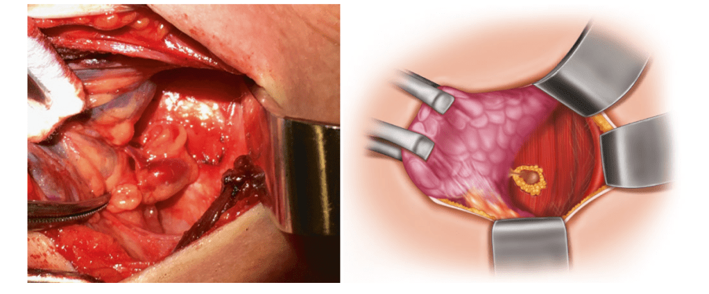
The gland is bluntly dissected from the surrounding tissue back toward its vascular pedicle, with particular care not to breach the capsule of the gland (Figure)
Exposure of the vascular pedicle. The gland has been dissected away from the surrounding tissue to expose its supplying vascular pedicle, which can then be ligated with a bipolar cautery or a small clip. The position of the recurrent laryngeal nerve, which can be seen in the bottom of the image, should be re-confirmed during this stage
The vascular pedicle is then ligated with bipolar cautery or a small clip:
After first re-confirming the position of RLN.
The excised gland(s) should then be sent to pathology
To confirm the weight and presence of parathyroid tissue, if available
My name is Rodrigo Arrangoiz I am a breast surgeon/ thyroid surgeon / parathyroid surgeon / head and neck surgeon / surgical oncologist that works at Center for Advanced Surgical Oncology in Miami, Florida.
I was trained as a surgeon at Michigan State University from (2005 to 2010) where I was a chief resident in 2010. My surgical oncology and head and neck training was performed at the Fox Chase Cancer Center in Philadelphia from 2010 to 2012. At the same time I underwent a masters in science (Clinical research for health professionals) at the University of Drexel. Through the International Federation of Head and Neck Societies / Memorial Sloan Kettering Cancer Center I performed a two year head and neck surgery and oncology / endocrine fellowship that ended in 2016.
Mi nombre es Rodrigo Arrangoiz, soy cirujano oncólogo / cirujano de tumores de cabeza y cuello / cirujano endocrino que trabaja Center for Advanced Surgical Oncology en Miami, Florida.
Fui entrenado como cirujano en Michigan State University (2005 a 2010 ) donde fui jefe de residentes en 2010. Mi formación en oncología quirúrgica y e n tumores de cabeza y cuello se realizó en el Fox Chase Cancer Center en Filadelfia de 2010 a 2012. Al mismo tiempo, me sometí a una maestría en ciencias (investigación clínica para profesionales de la salud) en la Universidad de Drexel. A través de la Federación Internacional de Sociedades de Cabeza y Cuello / Memorial Sloan Kettering Cancer Center realicé una sub especialidad en cirugía de cabeza y cuello / cirugia endocrina de dos años que terminó en 2016.
View all posts by Rodrigo Arrangoiz MS, MD, FACS, FSSO