Blog

Kidney Disease and Primary Hyperparathyroidism (PHPT)

  • Approximately 80% of patients with PHPT:
    • Have some degree of renal dysfunction or symptoms / signs
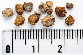
  • Kidney stones were previously reported in up to 80% of patients with PHPT:
    • But now occur in roughly 15% to 20% of the cases PHPT:
      • The calculi are typically composed of:
        • Calcium oxalate or
        • Calcium phosphate
      • In contrast, PHPT is found to be the underlying disorder:
        • In only 3% to less than  5% of patients presenting with nephrolithiasis
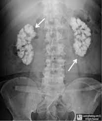
  • Nephrocalcinosis:
    • Which refers to renal parenchymal calcification:
      • Is found in less than 5% of patients with PHPT and is more likely to lead to renal dysfunction
  • Chronic hypercalcemia also can impair the concentrating ability of the kidney:
    • Thereby resulting in:
      • Polyuria
      • Polydipsia
      • Nocturia
  • The incidence of hypertension is variable but has been reported to occur:
    • In up to 50% of patients with PHPT:
      • Hypertension appears to be more common in:
        • Older patients and correlates with the magnitude of renal dysfunction:
          • In contrast to other symptoms:
            • It is least likely to improve after parathyroidectomy
  • Among normocalcemic patients with nephrolithiasis:
    • PHPT should be suspected if:
      • The serum calcium concentration is in the:
        • High-normal range:
          • Because the hypercalcemia may be intermittent and detected only by multiple measurements:
            • In one series of 48 patients with nephrolithiasis and PHPT:
              • 30 patients (63%) had:
                • Serum calcium concentrations between 10.2 and 11 mg/dL (2.55 and 2.75 mmol/L)
  • Most stones in patients with PHPT are composed of:
    • Calcium oxalate:
      • Although a slightly alkaline urine:
        • May favor the precipitation of calcium phosphate leading to calcium phosphate stones
    • A possible contributing factor for stone formation in PHPT:
      • Is hypercalciuria:
        • Although PTH directly stimulates the distal tubular reabsorption of calcium:
          • This effect is overshadowed by the increase in filtered calcium due to the hypercalcemia:
            • Leading to increased urinary calcium excretion:
              • In 35% to 40% of patients with PHPT
        • However, urinary calcium excretion per gram of creatinine:
          • Does not necessarily differentiate patients with or without stones:
            • This is likely due to the limited precision of a 24-hour urine calcium collection and to the complexity of factors that determine stone formation
        • Urinary calcium concentration:
          • Is only one of at least six urinary risk factors that determine the urine saturation of the calcium salts that lead to calcium stone formation:
            • It is for this reason that elevated levels of urinary calcium excretion are no longer, in and of itself, considered an indication for surgery in PHPT
    • A high serum calcitriol concentration:
      • Caused by PTH stimulation of renal hydroxylation of 25-hydroxyvitamin D:
        • May contribute to both hypercalciuria and stone formation:
          • This was illustrated in a report of 50 patients with PHPT:
            • In which 19 of the 30 patients who had:
              • High serum calcitriol concentrations, hypercalciuria, and increased dietary calcium absorption:
                • Had kidney stones
            • As compared with only 3 of 20 patients who had:
              • Normal to high-normal serum calcitriol concentrations and normal urinary calcium excretion and dietary calcium absorption
          • However, this distinction was not apparent in another series of 70 patients with mild PHPT, 18% of whom had kidney stones;
            • There was no difference in serum PTH, calcium, or calcitriol concentrations between patients with and without kidney stones, but more of the kidney stone group had hypercalciuria
    • In several studies, clinically silent kidney stones were reported in:
      • 7% to 21% of patients with PHPT:
        • Patients with undiagnosed (subclinical) nephrocalcinosis or calcium kidney stones are regarded as having symptomatic disease:
          • Regardless of the absence of symptoms:
            • Thus, these patients meet criteria for surgical intervention
  • For patients who do not have other overt indications for surgery:
    • Some UpToDate authors and editors obtain renal imaging (ultrasound, computed tomography [CT], or abdominal radiograph) to look for nephrocalcinosis or asymptomatic nephrolithiasis at the time of the original evaluation for PHPT:
      • Ultrasound is typically the imaging modality used

#Arrangoiz #ParathyroidSurgeon #ParathyroidExpert #Hyperparathyroidism #PrimaryHyperparathyroidism #CancerSurgeon #EndocrineSurgery #Teacher #Surgeon #HeadandNeckSurgeon #SurgicalOncologist #ParathyroidAdenoma #Hypercalcemia #ElevatedCalciumLevels #Miami #MountSinaiMedicalCenter #MSMC #Mexico #Hialeah
AMAROS Trial

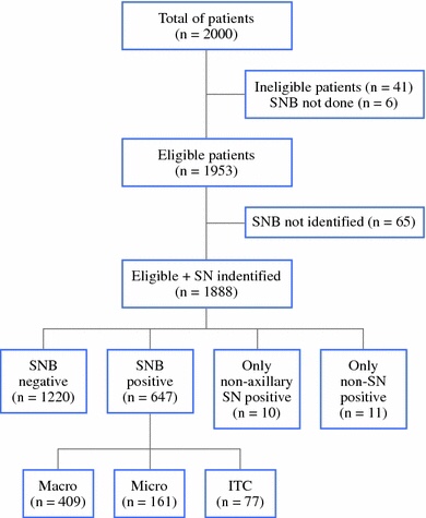
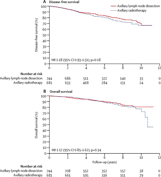
👉The AMAROS trial is a phase III non-inferiority study comparing ALND with axillary radiation therapy in patients with T1 to T2 clinically node-negative breast cancer patients with a positive sentinel node.

👉The primary endpoint was axillary recurrence.

👉The trial showed low 5-year rates of regional recurrence in the ALND and axillary radiation therapy groups (0.43% vs 1.19%, respectively), but the risk of patient perceived (subjective) or measured (objective) lymphedema was twice as high in the ALND arm compared to the radiation arm (subjective, 23% vs 11%; objective, 13% vs 5%) after 5 years of follow-up.

REFERENCES

  1. Rutgers EJ, Donker M, Straver ME. Radiotherapy or surgery of the axilla after a positive sentinel node in breast cancer patients: final analysis of the EORTC AMAROS trial (10981/22023). J Clin Oncol. 2013;31 (suppl; abstr LBA1001). Available at: http://meetinglibrary.asco.org/content/109779-132. Accessed November 7, 2013.
  2. Straver ME, Meijnen P, van Tienhoven G, et al. Sentinel node identification rate and nodal involvement in the EORTC 10981-22023 AMAROS trial. Ann Surg Oncol. 2010;17:1854-1861.

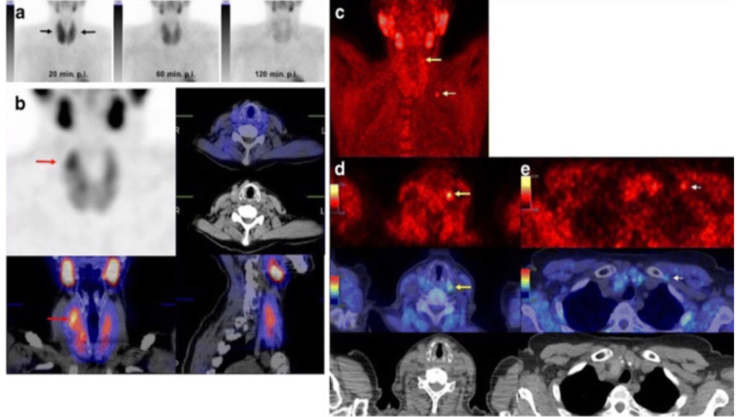
PETC/CT with 18F-Choline Localizes Hyperfunctioning Parathyroid Adenomas Equally well in Normocalcemic Hyperparathyroidism as in overt Hyperparathyroidism.

 PET

Abstract

PURPOSE:

  • Identification of pathologic parathyroid glands in primary hyperparathyroidism, traditionally based on neck ultrasound (US) and / or 99mTc-Sestamibi scintigraphy:
    • Can be challenging
  • PET / CT with 18F-Fluorocholine (18F-FCH):
    • Might improve the detection of pathologic parathyroid glands
  • The aim of this study was to compare the diagnostic performance of 18F-FCH-PET / CT with that of dual-phase dual-isotope parathyroid scintigraphy and neck US

METHODS:

  • Thirty-four consecutive patients with primary hyperparathyroidism were prospectively enrolled, seven had normocalcemic hyperparathyroidism, and 27 had classic hypercalcemic hyperparathyroidism.
  • All patients underwent:
    • High-resolution neck US
    • Dual-phase dual-isotope 99mTc-Pertechnetate / 99mTc-Sestamibi scintigraphy
    • 18F-FCH-PET / CT

RESULTS:

  • In the whole patients’ group, the detection rates of the abnormal parathyroid gland were:
    • 68% for neck US
    • 71% for 18F-FCH-PET / CT
    • 15% for 99mTc-Sestamibi scintigraphy
  • The corresponding figures in normocalcemic and hypercalcemic hyperparathyroidism were:
    • 57% and 70% for neck US
    • 70% and 71% for 18F-FCH-PET / CT
    • 0% and 18% for 99mTc-Sestamibi scintigraphy
  • In the 17 patients in whom the abnormal parathyroid gland was identified, either at surgery or at fine needle aspiration cytology / biochemistry:
    • The correct detection rate was:
      • 82% for neck US
      • 89% for 18F-FCH-PET / CT
      • 17% for 99mTc-Sestamibi scintigraphy

CONCLUSIONS:

  • 18F-FCH-PET / CT can be considered a first-line imaging technique for the identification of pathologic parathyroid glands in patients with normocalcemic and hypercalcemic hyperparathyroidism, even when the parathyroid volume is small
SUMMARY:

👉The diagnostic performance of 18F-FCH-PET / CT may be better than that of dual-phase dual-isotope parathyroid scintigraphy or neck US in normocalcemic or hypercalcemic primary hyperparathyroidism.

👉https://www.ncbi.nlm.nih.gov/pubmed/30094743/

#Arrangoiz #ParathyroidSurgeon #ParathyroidExpert #Hyperparathyroidism #PrimaryHyperparathyroidism #CancerSurgeon #EndocrineSurgery #Teacher #Surgeon #HeadandNeckSurgeon #SurgicalOncologist #ParathyroidAdenoma #Hypercalcemia #ElevatedCalciumLevels #Miami #MountSinaiMedicalCenter #MSMC #Mexico #Hialeah
Surgical Most Commons

  • What is the most common:
    • Type of melanoma?
      • Superficial spreading
    • Type of breast cancer?
      • Infiltrating ductal
    • Site of breast cancer?
      • Upper outer quadrant
    • Vessel involved with a bleeding duodenal ulcer?
      • Gastroduodenal artery
    • Cause of common bile duct obstruction?
      • Secondary Choledocholithiasis
    • Cause of small bowel obstruction (SBO) in adults in the United States?
      • Postoperative peritoneal adhesions
    • Cause of SBO in children?
      • Hernias
    • Cause of emergency abdominal surgery in the United States?
      • Acute appendicitis
    • Electrolyte deficiency causing ileus?
      • Hypokalemia
    • Cause of blood transfusion resulting in death?
      • Clerical error (wrong blood types)
    • Site of distant metastasis of sarcoma?
      • Lungs (hematogenous spread)
    • Position of anal fissure?
      • Posterior
    • Acute pancreatitis?
      • Biliary / Gallstones
    • Chronic pancreatitis?
      • Alcohol
    • Cause of large bowel obstruction?
      • Colon cancer
    • Cause of fever less than 48 postoperative hours?
      • Atelectasis
    • Bacterial cause of urinary tract infection (UTI)?
      • Escherichia coli
    • Abdominal organ injured in blunt abdominal trauma?
      • Liver
    • Benign tumor of the liver?
      • Hemangioma
    • Malignancy of the liver?
      • Metastasis
    • Pneumonia in the ICU?
      • Gram-negative bacteria
    • Cause of epidural hematoma?
      • Middle meningeal artery injury
    • Cause of lower GI bleeding?
      • Upper GI bleeding
    • Cancer in females?
      • Breast cancer
    • Cancer in males?
      • Prostate cancer
    • Type of cancer causing DEATH in males and females?
      • Lung cancer
    • Cause of free peritoneal air?
      • Perforated peptic ulcer disease
    • Cause of death ages 1 to 44?
      • Trauma

#Arrangoiz #Surgeon #Teacher #SurgicalOncologist #CancerSurgeon #ThyroidSurgeon #ParathyroidSurgeon #HeadandNeckSurgeon #MSMC #MountSinaiMedicalCenter #Miami #Mexico

Pearls for Identification of Parathyroid Glands

  • Aside from the classic description of a normal parathyroid gland being “London tan” in color and about the size of a grain of rice, there are a number of other subtle and less frequently described morphological features that aid in their identification:
    • Bloodless field:
      • A bloodless surgical field is of high importance when attempting to identify parathyroid glands:
        • As any blood staining impairs the ability to identify and assess the morphological features of the gland
    • Parathyroid glands are not palpable unless abnormal:
      • A normal parathyroid gland is soft and quite compressible on digital palpation:
        • This important feature can help to differentiate a normal gland from a small lymph node, which is normally rubbery and palpable
    • Fat pads (Figure 1):
      • The parathyroid glands are often encased in a “fat pad”:
        • Located in a region where the thymus “points” to the inferior pole of the thyroid gland (inferior gland)
        • Cranial to the ITA and generally posterior to the RLN (superior gland)
    • Vascular pedicle:
      • A small vascular pedicle can often be seen entering the parathyroid gland
    • Cope’s sign (Figure 2):
      • Bruising of the parathyroid gland that can occur with mobilization or dissection
    • “Kissing glands” (Figure 3):
      • This term is used to describe two parathyroid glands that are so close in position that they appear to a single, bilobed gland
      • Although uncommon, this possible configuration should be borne in mind when searching for a missing gland
      • Kissing glands can be differentiated from a true bilobar gland by identifying a cleavage plane between the capsules of the two glands
    • Slides under fascia:
      • The parathyroid glands are usually found within the thyroids pretracheal fascial capsule, but not adherent to it
      • This feature allows the gland to be gently slid or rolled under the fascia by a fine instrument such as a Crile
Fat pads. Visual inspection can yield many clues to aid in the identification of the parathyroid glands. Inspection should begin by looking for a fat pad located where the thymus points to the inferior pole of the thyroid gland for the inferior gland, and 1 to 2 cm cranial to the inferior thyroid artery on the posterior surface of the thyroid gland and anterior to the recurrent laryngeal nerve (as shown)
Cope’s sign. Mobilization in the area of the parathyroid gland can cause bruising and discoloration of the parathyroid gland. This sign is often subtle, but it can be an important visual cue to direct further dissection. This image shows a red-purple discoloration at the top of the thymus in the region of the inferior parathyroid gland. Further dissection allowed morphologic identification of a normal inferior gland
“Kissing” left upper and lower parathyroid glands. Parathyroid tissue was identified in a fat pad at the level of the inferior thyroid artery, which initially appeared to be a single, bilobed gland. Closer inspection revealed a cleavage plane between the lobes, which was carefully dissected, and separate upper and lower glands were able to be morphologically identified

#Arrangoiz #ParathyroidSurgeon #ParathyroidExpert #Hyperparathyroidism #Surgeon #HeadandNeckSurgeon #Teacher #MSMC #MountSinaiMedicalCenter #Miami #Mexico #EndocrineSurgery

Surgical Treatment of Ductal Carcinoma In Situ

  • The diagnosis of DCIS is followed by the surgical treatment with:
    • Breast-conserving surgery:
      • Also referred to as:
        • Segmental mastectomy
        • Partial mastectomy
        • Lumpectomy
        • Quadrantectomy
        • Wide local excision
    • Mastectomy:
      • Total or simple mastectomy
      • Skin sparing mastectomy
      • Nipple sparing mastectomy
  • Most patients who undergo breast-conserving surgery:
    • Receive postoperative radiation therapy:
      • To improve local control
  • Postoperative endocrine therapy:
    • With tamoxifen or an aromatase inhibitor:
      • Should also be considered for those patients whose tumors are:
        • Hormone (estrogen and / or progesterone) receptor positive
  • Mastectomy Versus Breast-conserving Therapy:
    • Historically:
      • DCIS was treated with mastectomy
    • The rationale for performing total mastectomy in patients with DCIS was:
      • Based on the high incidence of:
        • Multifocality
        • Multicentricity:
          • The reported incidence of multicentricity may depend on the extent of the pathologic review and therefore varies from 18% to 60%
        • As well as on the risk of occult invasion associated with the disease:
          • The incidence of microinvasion in DCIS varies according to the size and extent of the index lesion:
            • Lagios et al. (1989):
              • Reported a 2% incidence of microinvasion in patients with DCIS measuring less than 25 mm in diameter, compared with a 29% incidence of microinvasion in those with lesions larger than 26 mm
              • The incidence of microinvasion is also higher in patients with high-grade or comedo-type DCIS with necrosis and in patients with DCIS who present with a palpable mass or nipple discharge
    • Thus, mastectomy remains the standard with which other proposed therapeutic modalities are compared:
      • However, in patients with DCIS, there are no prospective trials comparing outcomes after mastectomy with those after breast-conserving surgery
    • A retrospective review by Balch et al. (1993):
      • Documented a:
        • Local relapse rate of 3.1% and a mortality rate of 2.3% after mastectomy for DCIS
    • The cancer-related mortality rate following mastectomy for DCIS was:
      • 1.7% in a series reported by Fowble (1989)
      • Ranged from 0% to 8% in a review by Vezeridis and Bland (1994)
  • In one of the largest studies comparing breast-conserving therapy with mastectomy, Silverstein et al. (1992):
    • Examined 227 cases of DCIS without microinvasion
    • In this nonrandomized study:
      • Patients with tumors smaller than 4 cm with microscopically clear margins:
        • Underwent breast-conserving surgery and radiation therapy
      • Whereas patients with tumors larger than 4 cm or with positive margins:
        • Underwent mastectomy
    • The rate of disease-free survival at 7 years was:
      • 98% in the mastectomy group compared with 84% in the breast-conserving surgery group (P = 0.038)
    • With no difference in overall survival rates
  • In a meta-analysis, Boyages et al. (1999):
    • Reported a recurrence rate of:
      • 22.5% – following breast-conserving surgery alone
      • 8.9% – following breast-conserving surgery with radiation therapy
      • 1.4% – mastectomy
    • In patients who underwent breast-conserving surgery alone:
      • Approximately 50% of the recurrences were invasive cancers
    • Although recurrence rates are higher in patients who undergo breast-conserving surgery than in patients who undergo mastectomy:
      • No survival advantage:
        • Has been shown for patients treated with mastectomy’s
  • Technique of Breast-conserving Surgery:
    • The goal of breast-conserving surgery is to:
      • Remove all suspicious calcifications and obtain negative surgical margins:
        • SSO / ASCO consensus guidelines recommend a 2 mm margin
    • Because DCIS is usually nonpalpable:
      • Breast-conserving surgery can be performed with mammographically or sonographically guided placement of a localizing wire or radioactive seed:
        • Seed localization of nonpalpable breast lesions is increasingly used in US and through multidisciplinary collaboration with surgeons, radiologists, and pathologists:
          • Has the advantage of allowing a less obtrusive marker to be placed at a time that is uncoupled from the time of surgery
        • At most centers where seed localization is performed:
          • An I-125 radioactive seed is placed by a radiologist within the area of disease:
            • In much the same way wire localization has historically been performed
        • The seed is localized intraoperatively using a gamma probe placed on the appropriate setting to detect radioactive decay for the seed’s isotope label, and the surrounding tissue is excised
        • Resection of the seed, lesion, and previously placed clip:
          • Are confirmed intraoperatively by pathologic and radiologic review
        • Nonradioactive methods of seed localization including ultrasound and magnetic seeds are also available
    • Intraoperative orientation of the specimen:
      • With two or more marking sutures is critical for margin analysis
      • In addition, specimen radiography is essential:
        • To confirm the removal of all microcalcifications
      • In patients with extensive calcifications:
        • Bracketing of the calcifications with two or more seeds or wires may assist in the excision of all suspicious calcifications
    • After whole-specimen radiography:
      • The specimen should be inked and then serially sectioned for pathologic examination to evaluate the margin status and extent of disease
    • Chagpar et al. (2003):
      • Demonstrated that intraoperative margin assessment with the use of sectioned-specimen radiography:
        • Enabled reexcisions to be performed at the same surgery if the microcalcifications extended to the cut edge of the specimen:
          • Minimizing the need for second procedures for margin control
      • After margins are deemed adequate intraoperatively:
        • The boundary of the resection cavity is marked with radiopaque clips to aid in the planning of postoperative radiation therapy and to facilitate mammographic follow-up
    • The goal of breast-conserving surgery is to:
      • Obtain tumor-free margins
    • A detailed pathologic study of DCIS, reported by Holland et al. (1990):
      • Demonstrated that up to 44% of lesions extended more than 2 cm further on histologic examination than that estimated by mammography:
        • However, in most women, a 1- to 2-cm margin around the lesion is not feasible since the cosmetic result would be poor
    • A new guideline put out by three national cancer organizations says that:
      • 2 mm (about one-eighth of an inch) clean margins should be the standard for women diagnosed with ductal carcinoma in situ (DCIS) treated with lumpectomy and whole-breast radiation
      • The guideline was published online on Aug. 15, 2016 by the:
        • Journal of Clinical Oncology. Read “Society of Surgical Oncology–American Society for Radiation Oncology–American Society of Clinical Oncology Consensus Guideline on Margins in Breast Conserving Surgery with Whole Breast Irradiation in Patients with DCIS
      • In 2015 a multidisciplinary panel was convened by the American Society of Clinical Oncology, the American Society for Radiation Oncology, and the Society of Surgical Oncology to review a meta-analysis and other literature concerning margin width and ipsilateral breast tumor recurrence (IBTR) in patients with DCIS undergoing breast-conserving therapy (Morrow, 2016):
        • The meta-analysis included 20 studies with 7,883 patients and 865 IBTRs with a median follow-up of 78.3 months
        • The panel reported that positive margins:
          • Were associated with a twofold increased risk of IBTR compared with patients who had negative margins
        • They also reported that margins of at least 2 mm were associated with a lower risk of IBTR compared with narrower margins
        • Patients treated with wide local excision alone without radiation therapy:
          • Had substantially higher rates of IBTR compared with patients undergoing wide local excision and whole breast irradiation regardless of the margin width
        • Rates of IBTR and contralateral breast cancer:
          • Were reduced in patients who received tamoxifen, however:
            • There was no significant impact on IBTR in patients with negative margins when compared with patients receiving placebo
        • The panel concluded that a 2-mm margin was adequate in patients undergoing breast-conserving surgery and whole breast irradiation for DCIS:
          • Clinical judgment should be utilized in deciding on the need for reexcision in patients with margins less than 2 mm.
    • The 2016 guidelines of the National Comprehensive Cancer Network (NCCN) dictate that close (less than 1 mm) margins are inadequate for DCIS and should be reexcised:
      • But for patients in whom the border of disease is the fibroglandular boundary (i.e., chest wall) or skin:
        • Radiation with a boost to the surgical scar is an acceptable alternative to reexcision

#Arrangoiz #CancerSurgeon #BreastSurgeon #SurgicalOncologist #BreastCancer #LCIS #DCIS #DuctalCarcinomaInsitu #LobularNeoplasia #LobularCarcinomaInsitu #Surgeon #Teacher #Miami #Mexico #MSMC #MountSinaiMedicalCenter

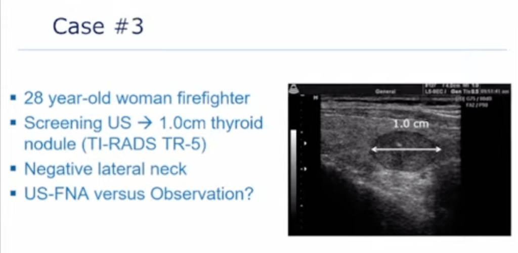
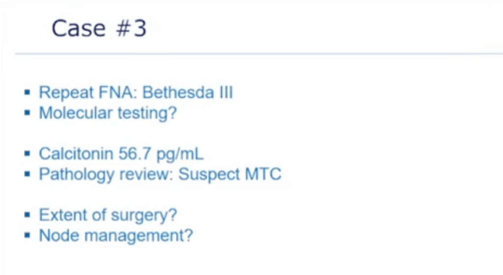
Thyroid Nodule Case

Repeat biopsy vs molecular testing?
Family history, pathologist stain for calcitonin, CEA, rule out MEN Type II, genetic testing and counseling (before or after surgery?), plan surgery (total thyroidectomy central neck compartment dissection)

#Arrangoiz #ThyroidSurgeon #CancerSurgeon #SurgicalOncologist #HeadandNeckSurgeon #EndocrineSurgery #ThyroidCancer #Miami #Mexico #MountSinaiMedicalCenter #Surgeon #Teacher #ThyroidNodules #RadioactiveIodine #RAI #PTC #MSMC

Diagnostic Biopsy for DCIS

  • Stereotactic core-needle or vacuum-assisted biopsy:
    • Is the preferred method for diagnosing DCIS
  • Calcifications that appear faint on mammogram or that are deep in the breast and close to the chest wall:
    • May be difficult to target with stereotactic biopsy
  • In addition, use of stereotactic biopsy in patients above the weight limit of the stereotactic system (about 150 kg) and in patients with very small breasts:
    • May be impossible
  • Patients who cannot remain prone or who cannot cooperate for the duration of the procedure:
    • Are also not good candidates for stereotactic biopsy
  • Bleeding disorders and the concomitant use of anticoagulants:
    • Are relative contraindications
  • Biopsy specimens:
    • Should be radiographed to document the sampling of suspicious microcalcifications
  • Care should be taken to mark the biopsy site with a metallic clip:
    • In the event that all microcalcifications are removed with the biopsy procedure
  • In the final biopsy procedure report:
    • It is important to report the:
      • Needle gauge used
      • How many cores were obtained
      • An estimate as to what percentage of the calcifications was removed
  • Because stereotactic core-needle and vacuum-assisted biopsy specimens represent only a sample of an abnormality observed on mammography:
    • The results are subject to sampling error
  • Invasive carcinoma:
    • Is found on excisional biopsy in 20% of patients:
      • In whom DCIS was diagnosed by a stereotactic core-needle biopsy
  • Thus, if the core-needle biopsy results are discordant with the findings of imaging studies:
    • A wire- or seed-localized excisional biopsy can be performed to establish the diagnosis
  • After diagnosis using stereotactic core-needle biopsy:
    • Approximately 20% to 30% of patients with ADH, up to 20% of patients with radial scar, approximately 5% to 10% of patients with flat epithelial atypia (FEA):
      • Are found to have a coexistent carcinoma near the site of the biopsy:
        • When complete excision is performed
    • Therefore, when the final pathologic studies from core-needle biopsy procedures indicate either of these diagnoses (ADH, radial scar, FEA):
      • Consideration should be made for excisional biopsy:
        • Though in the case of pure FEA:
          • There is some evidence to suggest that surgical excision is not necessary if all calcifications are removed at the time of biopsy (Calhoun et al., 2015)
  • Patients who are not candidates for stereotactic biopsy or who have stereotactic biopsy results that are inconclusive or discordant with the mammographic findings:
    • Should undergo excisional biopsy:
      • This technique is performed with the assistance of preoperative wire or seed localization of the mammographic abnormality in conjunction with the previously placed metallic clip marking the biopsy site
      • Postexcision specimen radiograph:
        • Is essential to confirm the removal of microcalcifications or targeted lesion
      • The excisional biopsy should be performed with the aim of obtaining a margin-negative resection:
        • That can serve as the definitive surgery

#Arrangoiz #CancerSurgeon #BreastSurgeon #SurgicalOncologist #BreastCancer #LCIS #DCIS #DuctalCarcinomaInsitu #LobularNeoplasia #LobularCarcinomaInsitu #Surgeon #Teacher #Miami #Mexico #MSMC #MountSinaiMedicalCenter

An International Survey of Thyroid Nodule Risk Stratification System Use and Preferences: Are We Moving Towards a Universal System?

Clin Thyroidol 2022;34:246–249.

  • Background:
    • Multiple ultrasound-based risk stratification systems (RSSs) have been developed to assist clinicians in the evaluation and management of thyroid nodules
    • Currently available systems differ in their approach to nodule categorization and suggest different fine-needle aspiration (FNA) size thresholds
    • Head-to-head comparisons of the performance of widely available RSS have yielded inconsistent results, making it difficult for individual practitioners to choose and implement an appropriate system
    • An International Thyroid Nodule Ultrasound Working Group (ITNUWG):
      • Is currently working on an international RSS to harmonize current systems
    • The present study is an international survey conducted by members of the ITNUWG to assess RSS use patterns and practitioner preferences
  • Methods:
    • A 22-question online international survey was developed by four members of the ITNUWG steering committee and administered via Survey-Monkey.com
    • The focus of the survey was on choice and usage of RSS, practitioner and practice type and demographics, geographic region, specialty, level of training, experience, and volume of thyroid nodule ultrasound and fine-needle aspiration
    • Invitations to complete the survey were disseminated by email by professional organizations including the American Thyroid Association (ATA), Associazione Medici Endocrinologi (AME), European Thyroid Association, Korean Society of Thyroid Radiology, and the Society of Radiologists in Ultrasound
    • The survey focused on five widely available RSSs including the:
      • American Association for Clinical Endocrinology (AACE), American College of Endocrinology (ACE) and AME unified guidelines, ACR TI-RADS, the ATA guidelines, EU-TIRADS, and K-TIRADS
    • It was designed to be completed in less than 15 minutes and participation was voluntary without reward; respondents did not have to answer all questions
  • Results:
    • A total of 875 respondents from 52 countries participated in the survey
    • 724 respondents answered questions about RSS awareness, value, and use
    • The response rate could not be calculated due to overlapping membership across the 5 disseminating societies
    • The majority of respondents were attending physicians in academic practice
    • There were 54% of respondents from Europe and 28.3% from North America; 61.5% were endocrinologists, 20.6% were radiologists, and 11.4% were surgeons
    • A reported 94.6% of respondents were at least somewhat familiar with an RSS, and a 91% found value in RSS usage
    • There were 95.6% of respondents who stated that an RSS was used in their practice, and 30.8% used more than one
    • Alone or in combination, RSS usage was as follows:
      • ATA guidelines (34%)
      • ACR TI-RADS (33.7%)
      • EU-TIRADS (29.6%)
      • AACE/ACE/AME guidelines 20.5%
      • K-TIRADS (14.6%)
      • Other (4.6%)
      • None (5%)
    • Geography and clinician specialty were major determinates of which RSS was used:
      • Surgeons and “others” were most likely to use multiple RSSs (40%)
    • A total of 271 respondents stated they did not personally use an RSS, most who favored narrative descriptions, and cited lack of institutional requirement (19.6%), multiplicity of options (18.1%), preference for using suspicious features (14.8%), contention that expertise is as or more effective (12.9%)
    • There were 62% of the 724 respondents who felt a universal lexicon with illustrative images would improve inter-observer variability
    • 54% supported a comprehensive online atlas
    • 44.9% supported a universal lexicon endorsed by societies
    • 95.2% preferred no more than 5 risk categories
  • Conclusions:
    • An international survey of RSS use patterns and practitioner characteristics and preferences was performed to inform development of a unifying international RSS
    • The survey demonstrated wide variability in practice patterns across geography and specialty, with majority support for a comprehensive atlas and unified lexicon with no more than 5 risk categories
  • Summary:
    • Ultrasound-based RSSs have attempted to codify and standardize the evaluation of thyroid nodules and have largely succeeded in articulating a shared lexicon of sonographic features that have predictive value
    • While RSSs differ in their specific format (e.g. pattern recognition versus point systems), definitions and weight of each sonographic criterion, risk categories, FNA size thresholds, and the presence and specifics of recommended surveillance intervals, most systems have more similarities than differences, and perform well with robust negative predictive value
    • The present study is one of the largest and most inclusive efforts to date to understand who uses which systems, and to seek to understand determinants of use, as well as clinician preferences
    • While the study’s methods traded some statistical rigor (no calculable response rate, high risk of sampling and non-response biases, etc.) for ease of execution (SurveyMonkey.com) and wide breadth and exposure (use of professional organizations to elicit international respondents, high respondent heterogeneity), it confirms the intuitive observation that clinicians choose systems informed by their geography and specialty, both of which select for involvement with particular professional societies, many of which have their own validated systems
    • the multiplicity of systems is particularly bewildering for primary care providers and patients (especially in the era of “open” notes), especially when radiologists and/or clinicians are using multiple RSSs with differing management recommendations
    • This confusion had led to widespread efforts to compare the performance of RSSs
    • While many comparative analyses have attempted to identify the “best” system, they employ heterogenous methods and outcomes, often looking at biopsy rates, and diagnostic accuracy
    • To date, there has not been a clear winner, and most of the discrepancies across systems are in lower risk strata
    • However, recommendations for lower risk categories are important, especially given that most thyroid cancers are small indolent papillary thyroid cancers and up to 11% to 14% of adults, perhaps more, have incidental papillary thyroid cancer found on biopsy
    • Overdiagnosis, and attendant overtreatment, are a source of cost and morbidity, although headway is being made
    • An ideal RSS would catch all clinically significant thyroid cancers through an up-front FNA biopsy or follow up sonographic surveillance, and minimize the cost and inconvenience of long-term follow up for innocent nodules and cancers
    • It is interesting that the survey did not query why some practitioners use more than one RSS
    • While most of physicians have settled on one particular system for day-to-day use, most of us would admit to selective application of one of several RSSs to justify a specific recommendation or express evidence-based support for patient preferences voiced in shared decision-making:
      • For instance, in older adults with multiple comorbidities and a conservative approach to healthcare, the 2015 ATA guidelines option to survey rather than biopsy spongiform nodules larger than 2 cm, and / or use ACR TI-RADS for more conservative FNA size thresholds, at least in part because of mindfulness around overdiagnosis and increasing data on the safety of an active surveillance strategy for the right patient and low-risk tumor characteristics
    • In the end, most clinically meaningful feature of this survey study is the effort that bore it
    • The results are clear that a majority of clinicians would welcome a universal lexicon and risk stratification system, and it is exciting to see an international effort underway to elicit clinician practice patterns and preferences as part of a larger collaboration to reconcile, improve, and unify current RSSs in the context of the unique biology and epidemiology of thyroid cancer
  • References:
    • Hoang JK, Asadollahi S, Durante C, Hegedus L, Papini E, Tessler FN 2022 An International Survey on Utilization of Five Thyroid Nodule Risk Stratification Systems: A Needs Assessment with Future Implications. Thyroid. ePub 2022 Mar 1.
    • Grani G, Sponziello M, Pecce V, Ramundo V, Durante C 2020 Contemporary Thyroid Nodule Evaluation and Management. J Clin Endocrinol Metab 105.

#Arrangoiz #ThyroidSurgeon #ThyroidExpert #HeadandNeckSurgeon #CancerSurgeon #SurgicalOncologist #EndocrineSurgery #ThyroidNodules #UltrasoundThyroid #MSMC #MountSinaiMedicalCenter #Miami #Mexico

Prognosis of Anaplastic Thyroid Cancer

  • ATC has a rapidly progressive course and early dissemination
  • The most common sites of distant spread include, in descending order:
    • The lung, bone, and brain
  • Metastases, particularly in the lung:
    • Are likely to be present at diagnosis in more than 50% of cases
  • The overall 5-year survival rate:
    • Is reportedly less than 10%, and most patients do not live longer than a few months after diagnosis
  • One study has shown that patients younger than 60 years who have ATC confined to the thyroid:
    • Have a better prognosis than patients who are older and have distant metastases
  • A retrospective study from Korea found that:
    • Age less than 60 years, tumor size less than 7 cm, and lesser extent of disease:
      • Were independent predictors of lower disease-specific mortality 
  • While some studies have suggested that postoperative radiotherapy:
    • May be of benefit in terms of survival, definitive prospective trials are lacking
  • Akaishi et al conducted a review of 100 patients with ATC in a single hospital (Ito Hospital) from 1993-2009:
    • The 1-year survival rates were as follows:
      • Stage IVA – 72.7%
      • Stable IVB- 24.8%
      • Stable IVC – 8.2%
  • Multivariate analysis demonstrated worse prognosis with:
    • Age older than 70 years, white blood cell count of 10,000/μL or more, extrathyroidal invasion, and distant metastases at the time of diagnosis
  • Survival was significantly better if the patient received complete resection, external radiation at doses of 40 Gy or more, or both
  • Orita et al developed a prognostic index that can predict prognosis and assist in the early treatment of ATC

#Arrangoiz #ThyroidSurgeon #CancerSurgeon #SurgicalOncologist #HeadandNeckSurgeon #EndocrineSurgery #ThyroidCancer #Miami #Mexico #MountSinaiMedicalCenter #Surgeon #Teacher #ThyroidNodules #RadioactiveIodine #RAI #PTC #MSMC