Blog

Sentinel Lymph Node Biopsy in Ductal Carcinoma In Situ (DCIS)

  • DCIS is noninvasive:
    • By definition, is unable to metastasize:
      • However, studies have shown that up to 15% of patients with pure DCIS:
        • Have isolated tumor cells (ITCs) or micrometastasis on nodal evaluation
      • However, these small tumor deposits:
        • Likely have little prognostic significance:
          • They may represent only cell clusters displaced by biopsy
  • In patients with DCIS detected by core biopsy:
    • There is a 15% to 20% associated risk:
      • Of an invasive component when excised
  • Patients undergoing mastectomy for DCIS:
    • Should be offered SLNB:
      • Since it would not be feasible to perform following mastectomy:
        • If invasive carcinoma is subsequently identified
  • ASCO consensus guidelines recommend that:
    • Patients with DCIS who undergo breast-conserving operation should not routinely have SLNB:
      • However, SLNB could be discussed with patients undergoing breast conservation:
        • Who have a core biopsy diagnosis of DCIS and:
          • A large area of DCIS on imaging (2 cm to 5 cm)
          • High-grade DCIS
          • Comedonecrosis
          • When a physical examination or imaging:
            • Shows a discrete mass
      • These findings have been associated with an increased risk of invasive cancer:
        • And SLNB at the time of the initial operation could avoid a second operation

#Arrangoiz #BreastSurgeon #CancerSurgeon #SurgicalOncologist #Surgeon #Teacher #BreastCancer #BreastDiseases #AxillaryDissection #BreastSurgery #MountSinaiMedicalCenter #MSMC #Miami #Mexico #SLNM #SLNB #SentinelLymphNodeBiopsy

SENTINEL LYMPH NODE BIOPSY – Technique and Indications

  • The lymph node that is the most likely to harbor metastatic disease:
    • Is the sentinel lymph node
  • In a landmark study published in 1994:
    • A vital blue dye:
      • Was used to identify the first node in the lymphatic chain to harbor metastatic tumor
    • An axillary dissection was then completed:
      • The pathologists evaluated the sentinel lymph node separately from the remaining axillary contents:
        • The sentinel node was identified with a specificity of 96% in 174 consecutive patients with various stages of breast cancer
  • As the sentinel node technique was further explored:
    • Additional studies demonstrated that SLNB could be reliably preformed using:
      • Radiolabeled colloid
      • Vital blue dye:
        • Methylene blue
        • Isosulfan blue
        • Blue patent
      • Both techniques:
        • Radiolabeled colloid and vital blue dye
  • The American Society of Clinical Oncology (ASCO):
    • 2014 Sentinel Lymph Node Biopsy Early-Stage Breast Cancer Update:
      • Evaluated the performance of SLNB and reported that:
        • False negative rates of the procedure ranged from:
          • 4.6% to 16.7%
        • An overall accuracy of:
          • 93% to 97.6%
      • Factors that increase the false negative rate of SLNB are:
        • Obesity
        • Tumor location
        • Failure to localize a lymph node on lymphoscintigraphy
        • Patient age
        • Surgeon experience
  • When radiolabeled sulfur colloid is used:
    • 0.4 mCi to 2 mCi of technetium-99m (99mTc)-labeled particles:
      • Measuring 50 to 200 nm in diameter are injected into the breast:
        • From 30 minutes to 24 hours before SLNB
      • Higher doses:
        • Are often used if the patient is injected the day prior to the operation
    • After injection:
      • The area is massaged gently for approximately 5 minutes:
        • To improve the lymphatic drainage
    • Patients may undergo static scintigraphy images:
      • To aid localization
  • In the operating room:
    • A gamma probe is used to identify the radioactive node
      • “Hot” nodes:
        • Nodes that qualify as sentinel lymph nodes are those that have:
          • Counts greater than 10% of the ex vivo counts of the most radioactive lymph node
  • Using a threshold of more than 10% of the hottest node:
    • Has been validated and correctly identifies:
      • 98% of sentinel lymph nodes
  • 99mTc-labeled sulfur colloid and lymphoscintigraphy:
    • Can identify alternate lymphatic drainage:
      • And are particularly useful in patients with previous axillary operations
  • Most surgeons find the use of both radioisotope and blue dye to be more successful than one method alone:
    • However, either may be used alone in experienced hands
  • If vital blue dye is used:
    • 3 ml to 5 mL of solution is injected:
      • Followed by at least 5 minutes of massage is used:
        • To improve lymphatic drainage
  • With either technique:
    • A transverse incision is then made:
      • Along the inferior margin of the hair-bearing region of the axilla
    • Blunt and sharp dissection:
      • Is used to identify a lymphatic track or blue-stained or “hot” node
    • Following removal of the sentinel lymph node:
      • The surgeon should palpate the axilla for any additional suspicious nodes and remove these as well
    • Although most patients have one sentinel lymph node:
      • Multiple nodes may be identified and should be removed
    • Care should be taken to remove only the targeted lymph node or nodes:
      • Separating them carefully from investing axillary fat:
        • As excessive dissection and tissue removal:
          • May increase rates of postprocedure seroma or lymphedema remove these as well
  • Complications following SLNB are less frequent than complications following ALND:
    • Especially with respect to:
      • Lymphedema, sensory loss, and mobility
  • Patients following SLNB:
    • Report improved quality of life and a more rapid return to work:
      • However, the International Cooperative Group Trial:
        • Reported lymphedema and seroma in:
          • 7% of patients treated with SLNB
        • Wound infection and hematoma rates:
          • Were both 1%
        • Shoulder or arm pain and decreased abduction are:
          • Also occasionally seen with SLNB.
  • Both methylene blue and 1% isosulfan blue dye:
    • Are commonly used to identify sentinel lymph nodes:
      • Isosulfan blue has a documented risk of allergic reaction, resulting in:
        • Rash, urticaria, and hypotension:
          • In as many as 3% of patients
        • Anaphylaxis:
          • In less than 1%
    • Although methylene blue is less costly and has a lower risk of anaphylaxis:
      • It is associated with skin necrosis in up to 10% of patients:
        • When injected intradermally or subcutaneously
      • For this reason, it should be diluted 1:2 in saline:
        • And an intradermal injection should be avoided
    • Multiple studies have tried to prove the superiority of one or other of these dyes:
      • They appear equivalent
  • Debate remains about the location of injection:
    • Peritumoral, subareolar, subdermal, and intradermal injection sites have all been used
    • Studies have shown that there is little difference between areolar injections and peritumoral injections:
      • But a randomized controlled trial:
        • Suggested that areolar intradermal injections:
          • Have the highest rates of localization and decreased harvest time of the first sentinel lymph node
    • In tumors that are large and in the upper outer quadrant of the breast:
      • Peritumoral injection is probably superior
    • In patients who undergo nipple-sparing mastectomy:
      • Subareolar injection should be avoided
    • Skin tattooing:
      • Can occur with intradermal injection
  • Patients with early-stage invasive breast cancer (clinically T1 and T2):
    • Who do not have clinically positive lymph nodes (cN0):
      • Should undergo SLNB
  • The ASCO reviewed the literature and created guidelines for the use of SLNB:
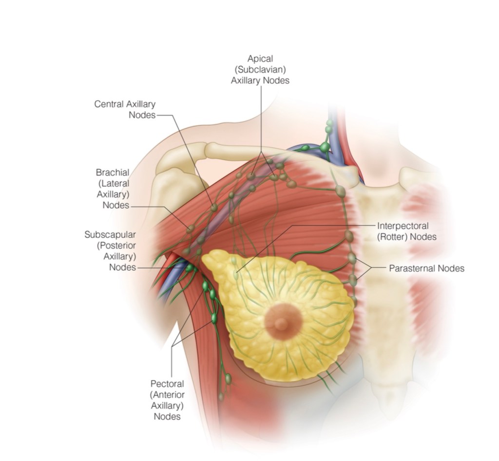
    • The current ASCO guidelines for SLNB are displayed in the second table
  • SLNB is widely applicable:
    • But there are some circumstances:
      • In which it is contraindicated
      • And others in which its utility is questionable
    • Given the dermal and pervasive lymphatic involvement in inflammatory carcinoma:
      • An ALND should be performed in patients with inflammatory cancer:
        • These patients have lymphatic vessels largely obstructed by tumor emboli:
          • Making sentinel node localization unreliable with high false negative rates:
            • Even after neoadjuvant chemotherapy (NAC)
    • Patients with large or locally advanced breast cancers (clinically T3 / T4):
      • Have historically:
        • Not been offered SLNB
      • Small case series have shown a large variability in:
        • The accuracy (85% to 98%) and a false negative rate (3% to 18%) of SLNB for patients with large tumors:
          • And these patients made up a small group in large, randomized sentinel lymph node clinical trials
      • Clinically node-negative patients with large tumors:
        • Also have high rates of positive SLNB as well as additional involved nonsentinel lymph nodes:
          • Ranging from 40% to 80%
        • Although many practitioners perform SLNB for patients with clinically T3 tumors;
          • Current ASCO guidelines argue that there is still insufficient evidence to support the practice

#Arrangoiz #BreastSurgeon #CancerSurgeon #SurgicalOncologist #Surgeon #Teacher #BreastCancer #BreastDiseases #AxillaryDissection #BreastSurgery #MountSinaiMedicalCenter #MSMC #Miami #Mexico

Surgical Evaluation of the Axilla in Breast Cancer

  • A physical examination alone:
    • Cannot accurately predict the presence of axillary disease
  • The accuracy of a physical examination to detect axillary metastasis:
    • Ranges from 61% to 68%:
      • When compared with resection
  • Ultrasonography, magnetic resonance imaging, and positron emission tomography–computed tomography:
    • Have all been used to evaluate the axilla:
      • And although these imaging modalities may improve on physical examination:
        • They are not as accurate as lymphadenectomy for small deposits:
          • And have a higher rate of false positives
  • A level I and II axillary lymph node dissection (ALND):
    • Has been the gold standard:
      • For evaluating the extent of axillary disease
    • Unfortunately, the incidences of:
      • Lymphedema, chronic pain, seroma development, future cellulitis, numbness, and limits to mobility:
        • Are all significant sequelae following ALND
  • Approximately 70% of patients who are clinically node negative:
    • Will have no evidence of disease detected with ALND:
      • Putting these patients needlessly at risk for complications
  • In 1991, the technique of sentinel lymph node biopsy (SLNB) was proposed as an alternative to ALND in breast cancer patients:
    • The development of SLNB has now replaced ALND:
      • As a highly accurate and less morbid axillary staging procedure for most patients

#Arrangoiz #BreastSurgeon #CancerSurgeon #SurgicalOncologist #Surgeon #Teacher #BreastCancer #BreastDiseases #AxillaryDissection #BreastSurgery #MountSinaiMedicalCenter #MSMC #Miami #Mexico

Surgical Evaluation of the Axilla in Breast Cancer

  • A physical examination alone:
    • Cannot accurately predict the presence of axillary disease
  • The accuracy of a physical examination to detect axillary metastasis:
    • Ranges from 61% to 68%:
      • When compared with resection
  • Ultrasonography, magnetic resonance imaging, and positron emission tomography–computed tomography:
    • Have all been used to evaluate the axilla:
      • And although these imaging modalities may improve on physical examination:
        • They are not as accurate as lymphadenectomy for small deposits:
          • And have a higher rate of false positives
  • A level I and II axillary lymph node dissection (ALND):
    • Has been the gold standard:
      • For evaluating the extent of axillary disease
    • Unfortunately, the incidences of:
      • Lymphedema, chronic pain, seroma development, future cellulitis, numbness, and limits to mobility:
        • Are all significant sequelae following ALND
  • Approximately 70% of patients who are clinically node negative:
    • Will have no evidence of disease detected with ALND:
      • Putting these patients needlessly at risk for complications
  • In 1991, the technique of sentinel lymph node biopsy (SLNB) was proposed as an alternative to ALND in breast cancer patients:
    • The development of SLNB has now replaced ALND:
      • As a highly accurate and less morbid axillary staging procedure for most patients

#Arrangoiz #BreastSurgeon #CancerSurgeon #SurgicalOncologist #Surgeon #Teacher #BreastCancer #BreastDiseases #AxillaryDissection #BreastSurgery #MountSinaiMedicalCenter #MSMC #Miami #Mexico

Clinical Evaluation of Axillary Disease

  • A thorough history and careful examination:
    • Help determine the extent of clinical disease in a patient with breast cancer
  • The examination is best done:
    • With the patient seated:
      • Arms removed from the gown and relaxed at the side
    • The examiner then palpates the axillary soft tissue beneath the relaxed arm:
      • Pressing it against the chest wall, where lymph nodes can be felt as they roll across the firm surface
    • Feeling beneath the lateral border of the pectoralis major muscle and high up along the thoracic outlet:
      • Can reveal important clinical findings
    • The axillary contents should also be assessed with the patient supine:
      • And the arm above the head
    • The presence or absence of palpable axillary metastasis determines further management

#Arrangoiz #BreastSurgeon #CancerSurgeon #SurgicalOncologist #Surgeon #Teacher #BreastCancer #BreastDiseases #AxillaryDissection #BreastSurgery #MountSinaiMedicalCenter #MSMC #Miami #Mexico

Mammary analog secretory carcinoma (MASC)

  • Mammary analog secretory carcinoma (MASC):
    • Is a recently described salivary gland tumor:
      • That was likely classified as acinic cell carcinoma in the past
  • The tumor has striking histologic and molecular similarities:
    • To secretory carcinoma of the breast
  • Secretory carcinoma of the breast (SC):
    • Has shown to have a recurrent balanced chromosomal translocation:
      • t(12;15) (p13;q25):
        • Which leads to an oncogenic fusion gene ETV6-NTRK3
    • This translocation is also present in MASC:
      • This fusion gene encodes a chimeric tyrosine kinase:
        • That is known to play an important role on its oncogenesis
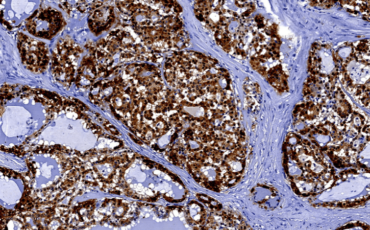
  • Immunohistochemical similarities between MASC and SC of the breast also include:
    • Being S100 protein, epithelial membrane antigen (EMA), and vimentin positive and “triple negative” (ER/PR/Her2 negative)
  • MASC predominantly affects:
    • Men and normally does not behave in an aggressive biology
  • The parotid gland is the most common affected gland by MASC
  • The official terminology of this entity:
    • Is now “secretory carcinoma”
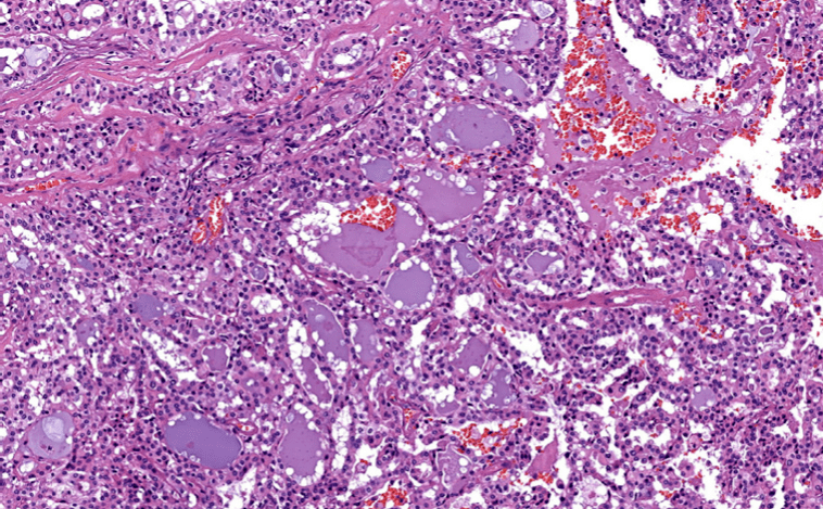
  • At the histologic level:
    • Tumor cells have eosinophilic or clear bubbly cytoplasm:
      • They may grow as tubules or microcysts, papillae, or macrocysts
    • Secretions are almost always present:
      • In the microcysts and / or macrocysts
  • MASC characteristically harbors:
    • A balanced chromosomal translocation (12, 15):
      • Resulting in the formation of the ETV6–NTRK3 fusion genes
  • Even though they have similar growth rate between MASC and ACC:
    • MASC is more likely to metastasize to the regional lymph nodes:
      • It should be considered as a more aggressive tumor compared with the regular low grade ACC
  • MASC usually presents as a painless:
    • It is a non-tender mass that increases in size overtime
  • The majority of MASC arise from the parotid gland;
    • Accounting for two thirds of the reported cases
  • The mean age for presentation of MASC is 47 years:
    • In contrast with SC of the breast that usually occurs in younger patients
  • MASC:
    • Is considered a low-grade carcinoma with a favorable prognosis:
      • According to Skálová et al:
        • It has moderate risk for local recurrence (15%)
        • Lymph node metastases (20%)
        • Low risk for distant metastases (5%)
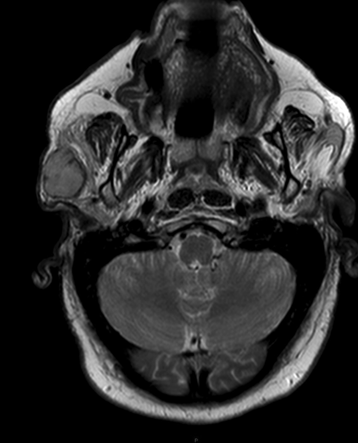
Neck magnetic resonance imaging of the head and neck showing the right parotid lesion.
Superficial parotidectomy with preservation of the facial nerve.
Hematoxylin and eosin staining.
Immunohistochemical study for S-100 protein.

#Arrangoiz #CancerSurgeon #HeadandNeckSurgeon #SurgicalOncologist #MASC #MammaryAnalogSecretoryCarcinoma #SalivaryGlandTumors #MSMC #MountSinaiMedicaCenter #Mexico #Miami

Epithelial-Myoepithelial Carcinoma of the Salivary Glands

  • Epithelial-myoepithelial carcinoma (EMC):
    • Is another typically indolent salivary gland tumor:
    • That is characterized by a multi-nodular pattern and biphasic or bilayered arrangement of inner ductal cells and outer myoepithelial cells, with classically clear cytoplasm
  • EMC:
  • Is a rare tumor with low malignant potential:
    • Accounting for less than 1 % of salivary neoplasms
  • Peak incidence occurs in seventh decade:
    • With a predilection for female
  • As early as 1956, EMC was reported as:
    • Adenomyoeipthelioma, clear cell adenoma, tubular solid adenoma and clear cell carcinoma:
      • Due to its varied histopathologic appearance
  • The term EMC was coined by Donath et al. in 1972:
    • This discrete entity was subsequently incorporated in WHO classification of salivary gland tumors in 1991
  • On histopathologic examination:
    • It shows dual cell population:
      • Composed of luminal ductal epithelial cells (inner layer) surrounded by large polygonal clear myoepithelial cells (outer layer):
        • Due to bidirectional differentiation of the stem cell
  • The most common location is the parotid gland:
    • Also the index case has parotid involvement:
      • But it is also known to occur in submandibular gland and minor salivary glands of palate, base of tongue and rarely in breast, lung, kidney, uterus
  • Rarely, it may show high grade transformation of epithelial or myoepithelial component:
    • Resulting in aggressive behavior
  • Because of rarity of EMC a standard treatment guideline is not yet known:
    • Surgical resection is the most widely used approach:
      • Although, some of the reported cases have utilized post-operative radiotherapy (PORT) extrapolated from other salivary gland histologies
  • Although it is a low grade tumor:
    • Local recurrence rates of 23% to 50 % have been reported:
      • With 25 % chance of distant metastasis
    • Local recurrence in as early as 6 months post operatively has been seen
  • Histopathologic markers such as:
    • Solid growth pattern, nuclear atypia, DNA aneuploidy, necrosis, positive surgical margins and high proliferative activity:
      • Have been identified by some authors to be associated with more aggressive behavior and high frequency of local recurrences and metastases
  • Surgery alone may not be sufficient in cases with histopathologic markers of aggressive behavior:
    • These patients may be candidates for PORT
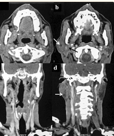
CT axial (a, b) and coronal (c, d) images of face and neck showing enlarged right superficial Parotid gland (awhite arrow) also involving deep lobe of right parotid gland (b black arrow) with small area of necrosis (c white arrow, d white arrow)
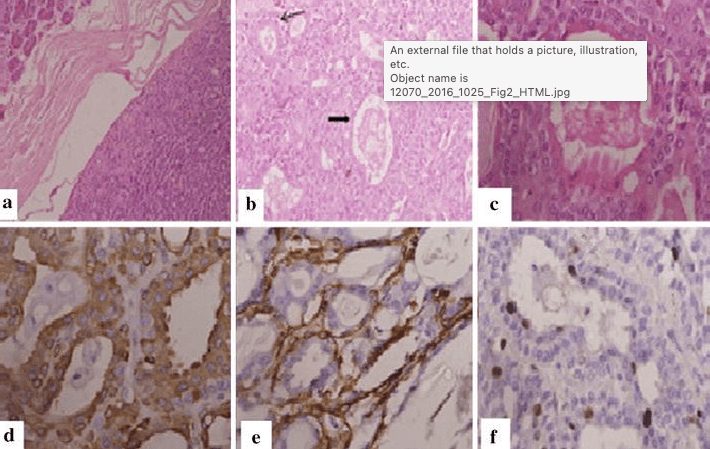
A well circumscribed tumor with thick fibrous capsule (a HE ×100). There is duct and tubule formation with ducts showing epithelial (broad arrow) and myoepithelial components (line arrow) (b HE ×200). High power view shows a duct composed of an outer rim of myoepithelial cells and inner dark epithelial cells having scant eosinophilic cytoplasm and bland nuclei (c HE ×400). Immunohistochemistry reveals epithelial component staining positively with cytokeratin (d IHC ×400) and SMA staining in the myoepithelial component (e IHC ×400) with MiB-1 labeled cells (f IHC ×400)

#Arrangoiz #CancerSurgeon #HeadandNeckSurgeon #SurgicalOncologist #SalivaryGlandTumors #EpithelialMyoepithelialCarcinoma #EMC #MSMC #MountSinaiMedicalCenter #Mexico #Miami

Acinic Cell Carcinoma (AcCC) of the Salivary Glands

• Acinic cell carcinomas (AcCC):

• Is the second most common malignant tumor:

• Involving the parotid gland

• They represent 15% of malignant parotid gland neoplasms

• AcCC accounts for approximately:

• 5% to 17% of all salivary gland tumors:

• 10% to 17% of all malignant salivary gland tumors

• The parotid gland is composed almost exclusively of serous type acini:

• And it is the most common site of AcCC:

• 80% to 90% of cases

• Other reported sites are:

• The palate (up to 15%)

• Submandibular gland (4%)

• Sublingual gland (1%)

• AcCC occurs most often in the fifth decade of life:

• Women are affected more often than in men (ratio 3:2)

• It is generally a low-grade carcinoma:

• With indolent behavior

• Recurrences are usually delayed:

• Sometimes after decades:

• In a series reported by Ellis et al:

• 12% of tumors recurred and 8% metastasized (bone, lung, and brain)

• The regional lymph nodes:

• Were the most common site of metastases:

• Spafford et al:

• Reported cervical lymphadenopathy to be present in 10% to 15% of cases of AcCC

• Spiro et al:

• In a series of 67 cases with AcCC treated before 1968, identified five cases (7.5%) with cervical metastases at the time of initial treatment

• It is interesting that this tumor is the most common bilateral malignant salivary gland neoplasm:

• Although its bilateral presentation is not nearly as common as the bilateral presentations of benign tumors:

• Warthin’s tumor and pleomorphic adenoma

• AcCC:

• Are typically encased in a fibrous capsule:

• Grossly resembling round circumscribed nodules with a tan surface

• The cut surface is solid but may show cystic degeneration and hemorrhage

• Histologically, there are five cell types:

• Serous acinar cells:

• Explaining the predilection for the parotid gland

• Cells with clear cytoplasm

• Intercalated ductal cell

• Nonspecific glandular cell

• Vacuolated cell

• The microscopic recognition of AcCC also requires a strong appreciation for its varied growth patterns:

• There are four histologic growth patterns:

• Solid

• Microcystic

• Papillary

• Follicular

• Caution must be taken not to misread the:

• Solid pattern as normal parotid parenchyma

• The papillary-cystic pattern as cystic mucoepidermoid carcinoma

• The follicular pattern as metastatic thyroid carcinoma

• Serous acinar differentiation:

• Is developed most fully in the acinic cell:

• These cells have dark round nuclei and granular purplish cytoplasm

• The diagnosis of AcCC may be difficult to establish:

• Especially when some other cell type dominates the histopathology picture

• Some examples of this are the predominance of clear cells might cause confusion with:

• Mucoepidermoid carcinoma, clear cell adenocarcinoma, and metastatic renal cell carcinoma:

• In these circumstances, the diagnostic acinic cells can be identified using a periodic acid-Schiff (PAS) reagent:

• Their cytoplasmic secretory granules are PAS positive and diastase resistant

• Overall survival has been crudely estimated to be about 84%:

• Survival at 5 years has been reported between 76% to 90%, but fell to 56% at 20 years:

• Emphasizing the need for long- term follow-up

Invasive Lobular Carcinoma of the Breast

Invasive Lobular Carcinoma of the Breast

  • Invasive lobular carcinoma:
    • Is the most common special histological subtype of invasive breast carcinoma
  • From an evolutionary point of view:
    • These tumors arise from a family of non-obligate precursor lesions called:
      • Atypical lobular hyperplasia (ALH) and lobular carcinoma in situ (LCIS):
        • Which may be collectively termed:
          • Lobular neoplasia (LN)
  • Even within this narrow spectrum of pre-invasive lesions and frank invasive carcinoma:
    • There is significant morphological and biological heterogeneity
  • The multistep model of breast cancer progression:
    • Contends that although lobular carcinomas:
      • Arise along the low-grade, ER-positive arm of the pathway (with low-grade, ER-positive ductal lesions):
        • De-differentiation to higher grade lesions: 
          • Can occur through acquisition of alterations in oncogenes such as:
            • ERBB2 and TP53:
              • Producing a spectrum of heterogenous proliferations
  • Lobular neoplasia (LN):
    • Are mostly an incidental finding:
      • Comprise neoplastic proliferation of characteristically discohesive cells:
        • Which fill and distend the terminal duct lobular units
    • LN encompasses both:
      • ALH and LCIS:
        • The boundary between the two is defined by:
          • An arbitrary cut-off using a quantitative measure:
            • Depending on the relative extent of involvement of the terminal duct lobular unit (TDLU):
              • If more than 50% of the TDLU is occupied:
                • The lesion is upgraded to LCIS
      • LN is considered to be a:
        • Non-obligate precursor of invasive cancer with:
          • ALH associated with a:
            • 4 to 5 times increased relative risk for subsequent cancer
        • LCIS associated with a:
          • 8 to 10 times increased relative risk for subsequent cancer:
            • The risk is bilateral:
              • But predominates for the ipsilateral breast
  • Classic invasive lobular carcinoma (ILC):
    • Typically demonstrates:
      • Single cell infiltration and a characteristic targetoid pattern of growth with minimal associated stromal response
    • This pattern of subtle invasion:
      • Is such that the size of the tumor often exceeds the imaging findings:
        • And obtaining clear surgical margins may be challenging
    • Although ILCs are generally palpable:
      • A high false-negative mammography rate is possible:
        • In 19% to 43%
    • In addition to the classic form of ILC:
      • Which is typically histological grade 2:
        • There are special morphological subtypes including:
          • Pleomorphic
          • Solid
          • Alveolar
          • Tubulo-lobular
      • These variants are rarely seen as pure form:
        • And are more likely to be present with the classical type
  • ILC and its subtypes are typified by:
    • A loss of cellular adhesion:
      • Frequently the result of biallelic inactivation (i.e. gene mutation combined with gene deletion) of the CDH1 gene:
        • Encoding E-cadherin:
          • Although other mechanisms of expression loss also feature
  • ILCs are normally estrogen (ER) and progesterone (PR) receptor positive:
    • And as such patients are indicated for hormone therapy
  • Whilst the biological characteristics of ILC afford patients a good prognosis in the short term:
    • It has become clear that the longer-term prognosis of ILC is frequently worse than for patients with the more commonly diagnosed invasive breast carcinoma of no special type (IBC-NST; invasive ductal carcinoma, IDC)
  • The metastatic presentation of ILC:
    • Has long been considered unique:
      • With a predilection for common sites (liver, lung, bone), but also gastrointestinal and gynecological sites of colonization:
        • Recent studies further support this:
          • Inoue et al. showed that lung metastases were less prevalent:
            • But peritoneal metastases are significantly higher in ILC (assumed predominantly classic ILC) compared to ER-positive IBC-NST
          • A recent study of metastatic spread to gynecological site:
            • Demonstrated an association with ILC and young age at diagnosis and confirmed earlier reports of the wide metastatic colonization of ILC
      • Immunophenotyping:
        • Showed a heterogeneous interplay between:
          • Hormone receptors and their co-factors during progression, including:
            • Frequent downregulation of PR expression and variable changes between AR, GATA3, and FOXA1 seen in different metastases within the same patient
  • Rarer presentations are increasingly being published in the literature:
    • Further highlighting the peculiar natural history of ILC:
      • For example, numerous case reports of ILC seeding as orbital metastases appear to suggest these are more likely to arise from an ILC than other types, and in a sole example of a mixed ductal-lobular carcinoma, only the lobular component was found in the orbital metastasis
  • The WHO Classificationrecognizes three variants of LCIS:
    • Classic (CLCIS)
    • Pleomorphic (PLCIS)
    • Florid (FLCIS) 
  • The defining features of both PLCIS and FLCIS have recently been clarified: 
    • PLCIS:
      • Is characterized by cells with enlarged nuclei (4× size of lymphocytes) or similar cytological features to those seen in high-grade ductal carcinoma in situ (DCIS)
  • FLCIS:
    • Is characterized by confluent expansive growth, and there must be marked distention of involved acini with little intervening stroma or an expanded acinus or duct approximately 40 to 50 cells in diameter
  • PLCIS is therefore characterized by its degree of cytological atypia:
    • Whereas FLCIS describes an architectural pattern (confluent, mass-like growth) with proliferation that is of classic type (CLCIS)
  • Unlike CLCIS, PLCIS and FLCIS are more likely to have:
    • Comedo-necrosis and calcifications and hence clinical and radiological presentations
  • CLCIS:
    • Is invariably ER and PR positive, and HER2 negative
  • FLCIS exhibits a similar phenotype:
    • Although may occasionally be HER2 positive
  • PLCIS exhibits a more varied phenotype:
    • With less frequent hormone receptor positivity, and an increased likelihood for HER2 overexpression:
      • Particularly in the apocrine-type of PLCIS as well as a higher proliferative index
  • The natural history of PLCIS and FLCIS:
    • Is as yet not well understood, and as such, relative risk of progression to frank invasive disease remains unclear, and we await long-term outcome data
  • An accumulating volume of work has investigated the molecular characteristics of CLCIS and these special subtypes:
    • The application of genomic technologies (copy number profiling, whole exome and targeted panel sequencing):
      • To a large set of LCIS confirmed that these lesions were frequently:
        • Clonally related to other more malignant lesions found to co-occur in the same specimen (i.e. DCIS and ILC):
          • And that intralesion molecular heterogeneity was also identified within LCIS:
            • Particularly among those lesions clonally related to DCIS and / or ILC
    • The data further supports the idea that:
      • LCIS shares molecular characteristics with its invasive counterpart:
        • Suggesting they do indeed have a common clonal origin:
          • And that LCIS is a non-obligate precursor of ILC
      • Considerable genomic diversity may arise in LCIS whilst the lesion is still confined within the ductal architecture:
        • Accounting for some of the morphological and biological variability observed on the progression to invasive cancer
        • Some of this intralesional LCIS heterogeneity:
          • Accounts for the occurrence of PLCIS and FLCIS
      • There is a striking similarity in the genomic profiles of CLCIS, PLCIS, and FLCIS (and invasive tumors):
        • With recurrent gains of 1q and losses on 16q and CDH1 mutations: 
          • Suggesting they arise from a common etiology
      • FLCIS and PLCIS with apocrine differentiation:
        • Are more genomically complex than CLCIS and non-apocrine PLCIS:
          • With an increase in the number of amplifications, genomic losses, and breakpoints
      • An increased frequency of ERBB2 / HER2 mutations or amplifications:
        • Has been reported in PLCIS compared to classic LCIS
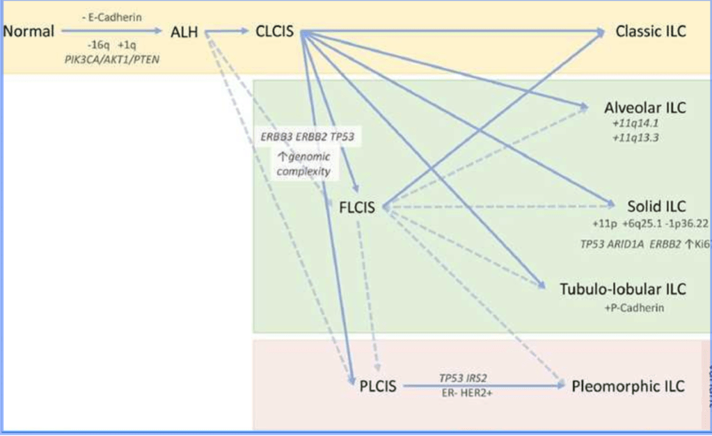
Multistep model of the evolution of classic ILC and its morphological variants.
A lineage of ‘lobular’ disease evolves from a normal epithelial cell on a background of a loss of E-cadherin expression and function, and key early somatic alterations involving gain of chromosome 1q, loss of 16q, and mutations in PIK3CA, AKT1, or PTEN.
The morphological and molecular diversity of in situ and invasive lobular lesions is likely to be a result of the subsequently arising pattern of molecular alterations that drive progression.
Atypical lobular hyperplasia (ALH) is distinguishable from lobular carcinoma in situ (LCIS) based on the extent of proliferation within the lobule.
Pleomorphic LCIS (PLCIS) and florid LCIS (FLCIS) can emerge either from ALH (presumably) or from classic LCIS (CLCIS), with an increasing level of genomic
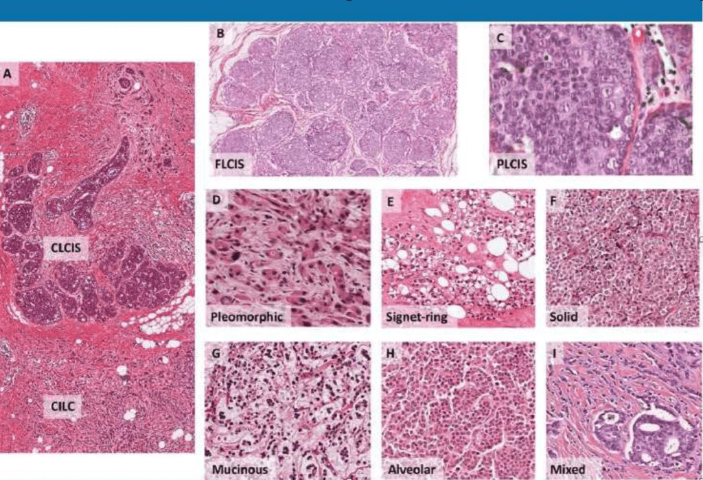
Histological examples of lobular variants.
a. CLCIS and CILC (as marked), × 40 magnification.
b. FLCIS shows the cytology of CLCIS with marked distention of lobular units to form a confluent mass-like lesion, × 40 magnification.
c. PLCIS with cytological atypia—nuclear pleomorphism with large vesicular nuclei and nucleoli—at least some × 4 the size of lymphocytes; × 600 magnification.
d. PILC, characteristic discohesion but with high-grade pleomorphic nuclei, with pink, foamy cytoplasm typical of an apocrine phenotype, × 400 magnification.
e. ILC with signet ring cell morphology, × 200 magnification.
f. Solid with sheets of classic type cells, × 200 magnification.
g. ILC showing mucinous / histiocytoid

#Arrangoiz #BreastSurgeon #CancerSurgeon #SurgicalOnologist #MSMC #MountSinaiMedicalCenter #BreastCancer #InvasiveLobularCarcinoma #ILC #LobularNeoplasia #Mexico #Miami

Auto-Transplantation of a Parathyroid Gland

  • The auto-transplantation site is directed by the pathological condition directing bilateral neck exploration (BNE):
    • As a general rule of thumb:
      • If the devascularized gland is morphologically normal:
        • It is transplanted into the sternocleidomastoid muscle
      • If the devascularized gland is morphologically abnormal:
        • The gland is transplanted into a site remote from the operative field:
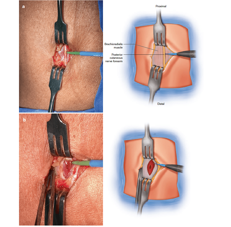
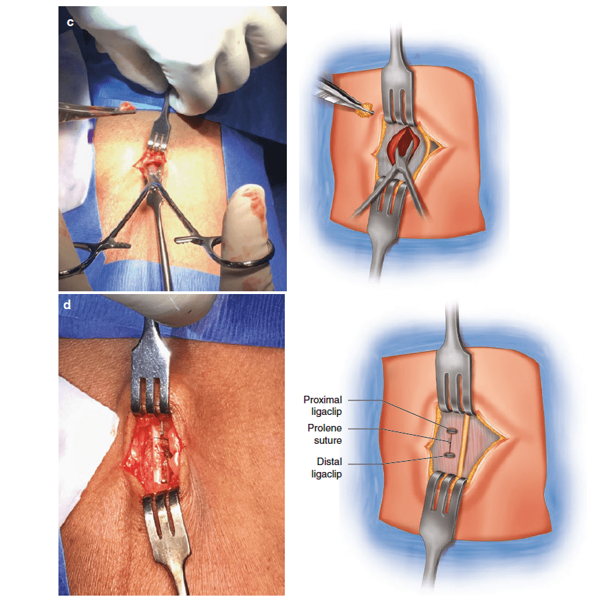
          • Such as the brachioradialis muscle
Procedure for auto-transplant into brachioradialis. These images demonstrate the steps taken to transplant an abnormal parathyroid gland into the right brachioradialis muscle of a patient with tertiary hyperparathyroidism. (a) A 2-cm incision is made over the brachioradialis muscle approximately 5 cm distal to the cubital crease. The dissection is continued down to the deep fascia, with care taken to identify and protect the posterior cutaneous nerve of the forearm (a). (b) The fascia is incised and a 1-cm pocket is created in the muscle. (c) The morcellated parathyroid tissue is placed into the pocket. (d) The fascia is closed, and the position of the auto-transplant is marked with a Hemoaclip at its proximal and distal extent, and a Prolene suture between these

#Arrangoiz #ParathyroidSurgeon #ParathyroidExpert #Parathyroidectomy #MSMC #MountSinaiMedicalCenter #HeadandNeckSurgeon #Surgeon #Teacher #EndocrineSurgery