- Visual:
- Attention is then turned to identifying the structures related to the inferior gland:
- As with the superior gland, careful exposure and mobilization of the thyroid gland may be all that is required to identify the inferior parathyroid glands
- A “tongue” of thymic tissue can often be seen extending with the inferior pole vessels and “pointing” toward the inferior pole of the thyroid:
- The inferior gland is usually located along this path
- The inferior glands arise from the third branchial pouch:
- In association with the thymus, and run a much longer course on descent
- As such, they have a more variable location, including within the thymus
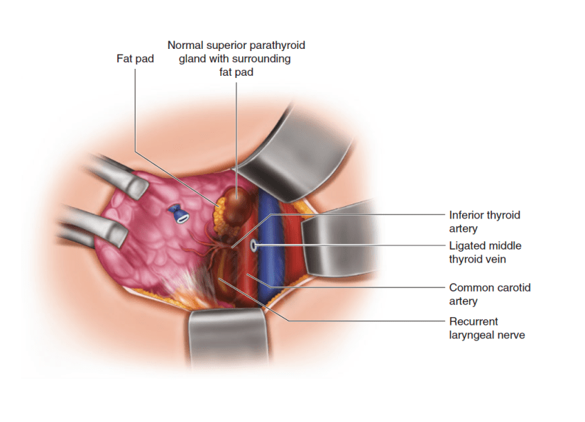
- Begin by looking for a fat pad where a tongue of cervical thymus “points” to the inferior pole of the thyroid:
- The inferior gland is often found on or within the posterior surface of this fat pad (Figure 1)
- Continue inspecting from the lower pole of the thyroid along to the tongue of the cervical thymus
- Attention is then turned to identifying the structures related to the inferior gland:
- Additional Maneuvers:
- If careful blunt dissection of the inferior pole vessels fails to identify the parathyroid gland:
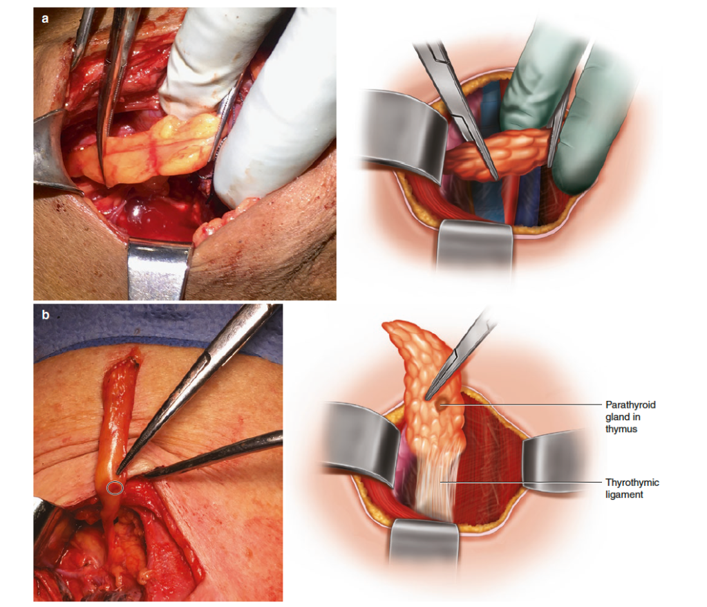
- The thymus should be isolated and mobilized:
- To do this step, the thyrothymic ligament is divided and the thymus is delivered into the wound with gentle retraction using hemostats, while pushing away any loose fibroareolar tissue
- Care must be taken not tear the capsule of the thymus (Figure 2)
- The thymus should be isolated and mobilized:
- If the gland is still missing, the inferior parathyroid glands may also be found in the superior and anterior mediastinum, and along the carotid sheath as high as the carotid bifurcation:
- The accessible portion of the superior mediastinum should be palpated and explored, and the carotid sheath opened and inspected from the root of the neck to the base of the skull
- Lack of thymic tissue caudal to the thyroid gland on inspection is suggestive of a non-descended third branchial pouch, and should invite closer examination along its course of descent
- Finally, if the missing abnormal gland has still not been found, consideration should be given to performing a thyroid lobectomy, as intra-thyroidal parathyroid glands have been reported in about 3% of patients:
- These may have been previously reported as a thyroid nodule on preoperative ultrasound (hence this is a good time to re-check any preoperative imaging), or they can sometimes also be seen on intra-operative ultrasound
- If careful blunt dissection of the inferior pole vessels fails to identify the parathyroid gland:
#Arrangoiz #ParathyroidSurgeon #ParathyroidExpert #Hyperparathyroidism #HeadandNeckSurgeon #EndocrineSurgery #MSMC #MountSinaiMedicalCenter #Miami #Teacher #Surgeon Mexico
