My name is Rodrigo Arrangoiz I am a breast surgeon/ thyroid surgeon / parathyroid surgeon / head and neck surgeon / surgical oncologist that works at Center for Advanced Surgical Oncology in Miami, Florida.
I was trained as a surgeon at Michigan State University from (2005 to 2010) where I was a chief resident in 2010. My surgical oncology and head and neck training was performed at the Fox Chase Cancer Center in Philadelphia from 2010 to 2012. At the same time I underwent a masters in science (Clinical research for health professionals) at the University of Drexel. Through the International Federation of Head and Neck Societies / Memorial Sloan Kettering Cancer Center I performed a two year head and neck surgery and oncology / endocrine fellowship that ended in 2016.
Mi nombre es Rodrigo Arrangoiz, soy cirujano oncólogo / cirujano de tumores de cabeza y cuello / cirujano endocrino que trabaja Center for Advanced Surgical Oncology en Miami, Florida.
Fui entrenado como cirujano en Michigan State University (2005 a 2010 ) donde fui jefe de residentes en 2010. Mi formación en oncología quirúrgica y e n tumores de cabeza y cuello se realizó en el Fox Chase Cancer Center en Filadelfia de 2010 a 2012. Al mismo tiempo, me sometí a una maestría en ciencias (investigación clínica para profesionales de la salud) en la Universidad de Drexel. A través de la Federación Internacional de Sociedades de Cabeza y Cuello / Memorial Sloan Kettering Cancer Center realicé una sub especialidad en cirugía de cabeza y cuello / cirugia endocrina de dos años que terminó en 2016.
In a meta-analysis of 16 studies, ADM was found to be associated with a higher likelihood of seroma (pooled OR, 3.9; 95% CI, 2.4-6.2), infection (pooled OR, 2.7; 95% CI, 1.1-6.4), and reconstructive failure (pooled OR, 3.0; 95% CI, 1.3-6.8).
The rates of hematoma (pooled OR, 2.0; 95% CI, 0.8-5.2), cellulitis (pooled OR, 2.0; 95% CI, 0.9-4.3), and skin flap necrosis (pooled OR, 1.9; 95% CI, 0.6-5.4) were not significantly different from reconstruction without ADM.
Furthermore, in a randomized controlled trial, use of ADM had no effect on postoperative pain and narcotic use, nor on the rate at which tissue expanders could be expanded.
Ho G, Ngueyen TG, Shahabi A, Hwang BH, Chan LS, Wong AK. A systematic review and meta-analysis of complications associated with acellular dermal matrix-associated reconstruction. Ann Plast Surg. 2012;68:346-356.
McCarthy CM, Lee CN, Halvorson EG, et al.The use of acellular dermal matrices in two-stage expander/implant reconstruction: a multicenter, blinded, randomized controlled trial. Plast Reconstr Surg 2012;130(5 Suppl 2):57S-66S.
Remains the standard for radiographic evaluation of DCIS
The cost and accessibility of magnetic resonance imaging (MRI):
Make it less feasible as an effective screening method
However, there is evidence that patients at high risk for breast cancer or those with very dense breasts:
May benefit from screening with MRI
Contrast-enhanced MRI:
Is more sensitive than mammography:
In the detection of both DCIS and invasive cancer
However, fibrocystic changes and other benign findings:
Can mimic DCIS on MRI:
Leading to unnecessary biopsies
MRI is increasingly being utilized after initial diagnosis in the preoperative evaluation:
To identify multicentric and contralateral lesions:
Because presence of either of these may change the surgical treatment strategy
Hollingsworth et al. (2008):
Reported that MRI detected multicentric disease:
Defined as a separate focus of cancermore than 5 cm away from the index lesion or discontinuous growth to another breast quadrant:
In 4.3% of 149 patients who presented with DCIS
Lehman et al. (2007):
Reported the utility of MRI in detecting contralateral breast cancer in a group of 969 patients with unilateral breast cancer:
196 of whom had DCIS
Of the patients with DCIS:
MRI prompted additional biopsies in 18 patients
Contralateral breast cancer was detected in five patients:
28% of those biopsied and 2.6% of those with DCIS
The sensitivity of detecting contralateral breast cancer was:
71%
The specificity of detecting contralateral breast cancer was:
90%
While MRI is associated with increased likelihood of change in the surgical plan for a patient with unilateral breast cancer:
It is unclear whether these altered (and usually more extensive) surgical plans are actually treating clinically significant disease that might have otherwise decreased the patient’s disease-free or overall survival
In a review of over 2,300 patients with breast-conserving therapy (BCT, i.e., lumpectomy and radiation) for DCIS at Memorial Sloan Kettering between 1997 and 2010:
There was no association between receipt of preoperative MRI and risk of locoregional recurrence or contralateral breast cancer:
Regardless of whether the patient received radiation (Pilewskie et al., 2014)
The typical appearance of DCIS on MRI:
Is non-mass enhancement
Although mammography can be more sensitive than MRI for DCIS associated with calcifications:
Uncalcified DCIS may be better visualized by MRI
Kuhl and colleagues prospectively assessed 7,319 women who had undergone both preoperative mammography and MRI:
Of 167 women with pure DCIS on final pathology:
92% (n = 153) were diagnosed by MRI and 56% (n = 93) were diagnosed by mammography:
Of those diagnosed with high-grade DCIS:
48% were missed by mammography but diagnosed by MRI only
A meta-analysis looking at the association of preoperative MRI and surgical management of patients with DCIS:
Showed no significant difference in the proportion of women with positive margins or in the need for re-excision after BCS
Overall mastectomy rates did not differ significantly, whether or not preoperative MRI was performed (odds ratio [OR] 1.23; p = .34)
Pilewskie and colleagues reported a large series of women undergoing BCS for DCIS:
Found no difference in locoregional recurrence rates or contralateral breast cancer rates:
In women who had perioperative MRI and those who did not
Although MRI can be useful in assessment of extent of disease and is an adjunct to traditional imaging in patients who have discordant results or mammographically occult disease:
Routine use of MRI is not advocated for the perioperative management of DCIS
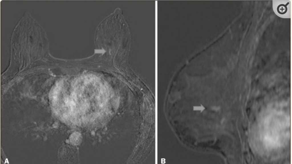
Contrast-enhanced, high-resolution MRI. Axial sequence, with digital subtraction (A) and sagittal MRI sequence (B), showing a linear area of enhancement (arrows) in the posterior third of the central region/junction of the medial quadrants of the left breast. The pathology study of the surgical specimen revealed DCIS, nuclear grade 2.
The use of catheter-based, accelerated partial-breast irradiation (APBI) offers many potential advantages over routine whole-breast radiation therapy, including decreased treatment time and decreased radiation dose delivered to uninvolved portions of the breast and adjacent organs. This has resulted in significant interest in APBI, with more than 32,000 women in the United States having had treatment with a breast brachytherapy catheter.
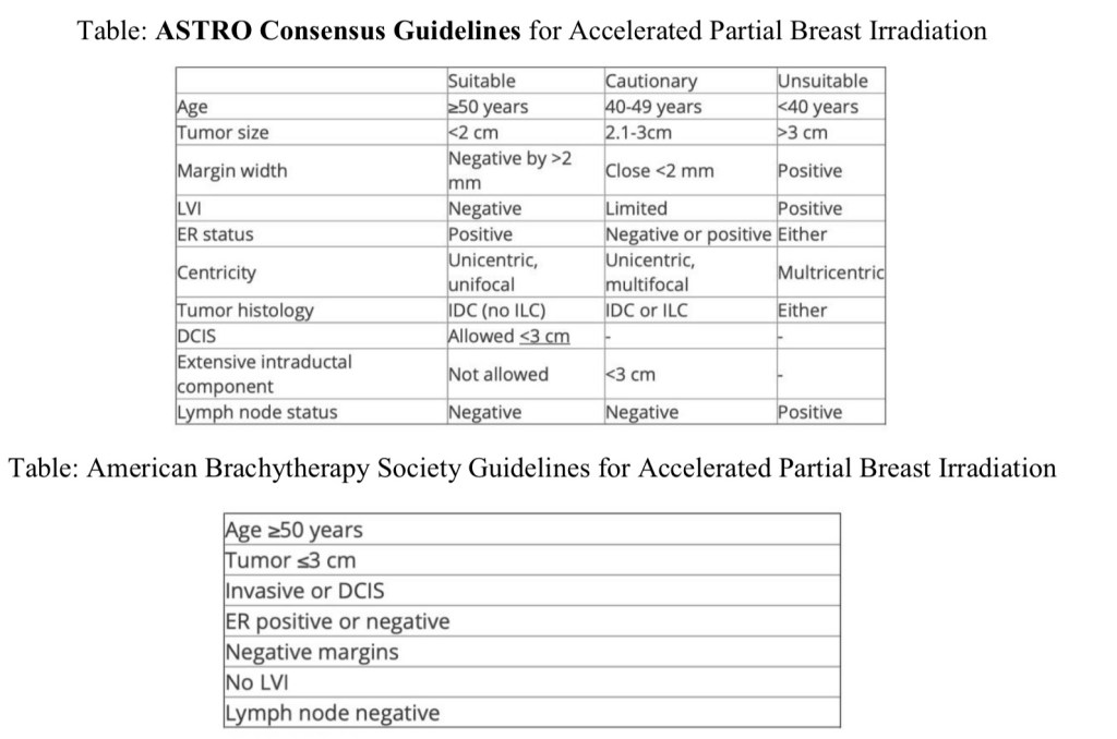
However, randomized clinical trials comparing APBI with conventional whole-breast radiation therapy have yet to be completed. In light of this, the American Society for Radiation Oncology (ASTRO) has issued a consensus statement regarding the appropriate use of APBI, categorizing potential patients for APBI into three groups: suitable, cautionary, and unsuitable. A similar, but somewhat more liberal guideline was put forth by The American Brachytherapy Society.
Accelerated partial breast irradiation includes multiple techniques such as: interstitial brachytherapy, applicator brachytherapy, external beam radiation therapy.
The Groupe Européen de Curie thérapie of European Society for Radiotherapy and Oncology (GEC-ESTRO) trial randomized 1184 patients with low-risk invasive carcinoma or ductal carcinoma in situ (DCIS) treated with breast-conserving surgery to either whole-breast irradiation (WBI) or APBI using multi catheter brachytherapy. The cumulative incidence of local recurrence at 5 years was 1.44% with APBI and 0.92% with WBI.
The Florence trial randomized 520 patients to either WBI or APBI with intensity-modulated radiation therapy, with a mean follow-up of 5 years. The rate of local recurrence was 1.5% in both arms.
The American Society of Breast Surgeons’ Mammo Site Registry found 5-year rates of local recurrence were less than 4%.
An older randomized trial from Hungary using interstitial APBI and electrons found that the 5-year local recurrence rate of 4.7%.
It is important to recognize that these outcomes are likely due to the highly selected, low-risk patient populations included in many of the APBI trials as evidenced by the tables / guidelines: patients tend to be postmenopausal, tumors less than 3 cm, without DCIS, node negative, ER-positive.
Arthur D W, Vicini F A, Kuske RR, Wazer DE, American Brachytherapy Society. Accelerated partial breast irradiation: an updated report from the American Brachytherapy Society. Brachytherapy. 2003;2:124-130.
Hughes KS, Schnaper LA, Berry D, et al. Cancer and Leukemia Group B; Radiation Therapy Oncology Group; Eastern Cooperative Oncology Group. Lumpectomy plus tamoxifen with or without irradiation in women 70 years of age or older with early breast cancer. N Engl J Med. 2004;351:971-977.
National Comprehensive Cancer Network. NCCN Clinical Practice Guidelines in Oncology. Breast Cancer. Available at http://www.nccn.org.
Smith BD, Arthur DW, Buchholz TA, et al. Accelerated partial breast irradiation consensus statement from the American Society for Radiation Oncology (ASTRO). J Am Coll Surg. 2009;209:269-277.
Giuliano AE, Ballman KV, McCall L, et al. Effect of Axillary Dissection vs No Axillary Dissection on 10-Year Overall Survival Among Women With Invasive Breast Cancer and Sentinel Node Metastasis: The ACOSOG Z0011 (Alliance) Randomized Clinical Trial. JAMA. 2017; 318(10): 918-926. doi: 10.1001/jama.2017.11470.
The data for preoperative MRI in the setting of either lobular or ductal carcinoma is controversial.
Routine preoperative MRI is not mandatory for either entity and is not anticipated to change the rate of repeat excision for positive margins or recurrence. It should be used on a case-by-case basis, taking into account other factors, such as breast density and additional risk factors.
Invasive lobular carcinoma is described pathologically as small cells that infiltrate the mammary stroma and adipose tissue in a single file pattern due to a lack of E-cadherin. The cells induce minimal reaction in the surrounding tissue, making them insidious both radiographically and pathologically. Classic-type invasive lobular carcinoma is usually positive for estrogen receptor and negative for HER2, but this is not universally true, as HER2 subtypes do exist. Lobular cancers respond less well to neoadjuvant chemotherapy than their ductal counterparts.
Orvieto E, Maiorano E, Bottiglieri L, et al. Clinicopathologic characteristics of invasive lobular carcinoma of the breast: results of an analysis of 530 cases from a single institution. Cancer. 2008;113:1511-1520.
Purushotham A, Pinder S, Cariati M, Harries M, Goldhirsch A. Neoadjuvant chemotherapy: not the best option in estrogen receptor-positive, HER2-negative, invasive classical lobular carcinoma of the breast? J Clin Oncol. 2010;28:3552-3554.
Turnbull L, Brown S, Harvey I, et al. Comparative effectiveness of MRI in breast cancer (COMICE) trial: a randomized controlled trial. Lancet. 2010;375:563-571.
Inflammatory breast cancer (IBC) is a clinical diagnosis made on the basis of a breast that rapidly enlarges; that is red, firm, and has skin edema (peau d’orange); and that involves 30% of the breast or more with a palpable border.
On evaluation of a specimen obtained by skin punch biopsy, the hallmark of this disease is dermal lymphatic invasion by tumor cells, but this pathologic finding is not necessary to make the diagnosis.
Nearly all women with IBC have lymph node involvement at the time of diagnosis, and sentinel lymph node biopsy has been shown to have a high false-negative rate in these patients. Hence, sentinel node biopsy is not indicated.
Approximately 30% of women with IBC will have distant metastatic disease; thus, all women with IBC should have a thorough staging work-up at the time of diagnosis.
As IBC has an American Joint Committee on Cancer (AJCC) stage of IIIb or higher (owing to its designation of T4d, N0-3, M0-1), neoadjuvant chemotherapy is the recommended first course of treatment.
Skin-sparing mastectomy is contraindicated.
Because the degree of pathologic response to neoadjuvant chemotherapy predicts prognosis, and the intensity of preoperative therapy predicts response, all chemotherapy up front instead of a so-called sandwich approach of chemotherapy/surgery/chemotherapy is preferred.
Surgery is indicated if disease responds well, as this gives the best chance for disease-free survival.
Post-mastectomy radiation therapy is required to reduce the risk of chest wall recurrence in these patients.
Cristofanilli M, Gonzalez-Angulo AM, Buzdar AU, Kau SW, Frye DK, Hortobagyi GN. Paclitaxel improves the prognosis in estrogen receptor negative inflammatory breast cancer: the M.D. Anderson Cancer Center experience. Clin Breast Cancer. 2004;4:415-419.
Dawood S, Cristofanilli M. What progress have we made in managing Inflammatory breast cancer? Oncology (Williston Park). 2007;21:673-687.
National Comprehensive Cancer Network. NCCN Clinical Practice Guidelines in Oncology. Breast Cancer. Available at http://www.nccn.org.
Panades M, Olivotto IA, Speers CH, et al. Evolving treatment strategies for inflammatory breast cancer: a population-based survival analysis. J Clin Oncol. 2005;23:1941-1950.
Local recurrence in the breast may be accompanied by distant recurrence. Therefore, all patients who are diagnosed with a local or regional recurrence should have staging scans to rule out distant disease. If these scans are negative, surgical intervention is recommended if the tumor is operable.
In patients who develop an ipsilateral breast recurrence or new primary tumor following prior history of breast conservation with whole breast radiation therapy, further radiation is generally contraindicated. Therefore, the standard surgical treatment of the breast is mastectomy. While there have been case reports of repeat lumpectomy followed by accelerated partial-breast radiation therapy, there are insufficient data to support the long-term safety of this approach. If the tumor is not operable by mastectomy at presentation, neoadjuvant therapy with chemotherapy or hormone therapy may be considered as an initial step.
The approach to the axillary lymph nodes has been an area of debate since the era of SLNB. Nodal staging provides valuable prognostic information, even in the case of a local recurrence. If a patient is clinically node-negative at the time she presents with a local recurrence in the breast, SLNB may be attempted. There is a small body of literature supporting the use of reoperative SLNB in clinically node-negative patients who have an ipsilateral recurrence or new primary following prior breast conservation with SLNB. These studies have found that successful lymphatic mapping is more likely to occur when fewer nodes were removed at the time of the treatment for the index cancer. While lymphoscintography is not strictly required, it may help to identify sites of extra-axillary drainage due to alternate drainage pathways. In patients who have had prior completion axillary lymph node dissection, reoperative SLNB is possible, but less likely to be associated with successful lymphatic mapping. In these cases, axillary exploration is less likely to yield any additional lymph nodes.
Cardosa F, Fallowfield L, Costa A, et al; and ESMO Guidelines Working Group. Locally recurrent or metastatic breast cancer: ESMO Clinical Practice Guidelines for diagnosis, treatment and follow-up. Ann Oncol. 2011;22(Suppl 6):vi25-vi30.
Cox CE, Furman BT, Kiluk JV, et al. Use of reoperative sentinel lymph node biopsy in breast cancer patients. J Am Coll Surg. 2008;207:57-61.
Karam A, Stempel M, Cody HS III, Port ER. Reoperative sentinel lymph node biopsy after previous mastectomy. J Am Coll Surg. 2008;207:543-548.
Kaur P, Kiluk JV, Meade T, et al. Sentinel lymph node biopsy in patients with previous ipsilateral complete axillary lymph node dissection. Ann Surg Oncol. 2011;18:727-732.
Port E, Garcia-Etienne C, Park J, Fey J, Borgen PI, Cody HS 3rd. Reoperative sentinel lymph node biopsy: a new frontier in the management of ipsilateral breast tumor recurrence. Ann Surg Oncol. 2007;14:2209-2214.
Breast cancer associated with pregnancy occurs anywhere in the first postpartum year, during pregnancy, or anytime during lactation.
Management of pregnant women newly diagnosed with breast cancer can be complex and depends on the trimester of pregnancy and desires for breast conservation.
Most obstetricians will recommend to deliver the baby at the earliest at 34 weeks to avoid morbidity of prematurity and treatment for the breast cancer should not be delayed until delivery. Studies have shown worse survival in those patients who delayed treatment 3 to 6 months.
It is safe to administer chemotherapy during pregnancy, but the risk of congenital malformations in the first trimester can be as high as 20% so it is generally recommended to start chemotherapy in the second or third trimester. The only agents that are contraindicated are taxanes and trastuzumab.
A study published in 2006 did not show any significant short term complications for children that were exposed to fluorouracil, doxorubicin, cyclophosphamide (FAC) chemotherapy in utero. It is not safe however to administer radiation or hormone therapy during pregnancy because of risks to the fetus.
Surgery is safe in the second and third trimester and is generally avoided in the first trimester due to risk of spontaneous abortion.
Surgical options include mastectomy and breast conservation, if applicable. If a patient has lumpectomy, radiation therapy must wait until after delivery. Sentinel node biopsy has been shown to be safe during pregnancy followed by axillary node dissection if the sentinel node is tumor positive. It is generally felt that preoperative lymphoscintigraphy is safe during pregnancy, although only a few small studies have examined sentinel node biopsy in pregnant patients. Injection of blue dye to identify a sentinel node is not recommended because of the small risk of allergic reaction and uncertain affects on the fetus.
Therapeutic abortion is no longer necessary or recommended.
Barnes DM, Newman LA. Pregnancy-associated breast cancer: a literature review. Surg Clin North Am. 2007:87:417-430.
Berry DL, Theriault RL, Holmes FA, et al. Management of breast cancer during pregnancy using a standardized protocol. J Clin Oncol. 1999;17:855-861.
Hahn KME, Johnson PH, Gordon N, et al. Treatment of pregnant breast cancer patients and outcomes of children exposed to chemotherapy in utero. Cancer. 2006;107:1219-1226.
Khera SY, Kiluk JV, Hasson DM, et al. Pregnancy-associated breast cancer patients can safely undergo lymphatic mapping. Breast J. 2008;14:250-254.
Mondi MM, Cuenca RE, Ollila DW, Stewart JH 4th, Levine EA. Sentinel lymph node biopsy during pregnancy: initial clinical experience. Ann Surg Oncol. 2007;14:218-221.









 Rodrigo Arrangoiz MS, MD, FACS, FSSO