
👉The primary application of ThyroSeq is to provide accurate cancer diagnosis in thyroid nodules with indeterminate FNA cytology
👉Indeterminate FNA cytology encompasses diagnostic categories III, IV, and V of the Bethesda System for Reporting Thyroid Cytopathology
👉Uncertain and variable risk of cancer in these nodules hampers clinical management of these patients
👉ThyroSeq stratifies thyroid nodules with indeterminate cytology into those that are most likely benign and can frequently be followed by observation and those that have a high probability of being cancer or pre-cancer, which in most cases need surgical management

👉ThyroSeq Clinical Validation Study Overview:
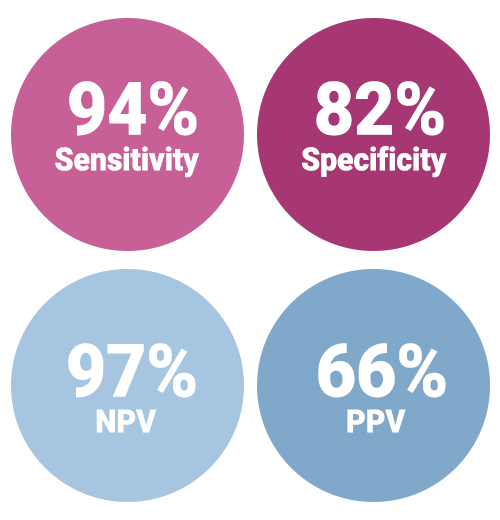
👉Clinical validation of ThyroSeq v3 performed in the recently completed prospective double-blind multicenter international study (ClinicalTrials.gov identifier NCT02352766)
👉Patients were enrolled in the study at 10 medical centers, nine located in the U.S. and one in Singapore
👉Totally, 782 patients with one or more thyroid nodules sampled by FNA ( 1013 samples ) were enrolled
👉Of those, 257 FNA samples from 234 patients had Bethesda III to V cytology, underwent surgery, and had FNA samples sufficient for molecular analysis
👉These 257 FNA samples comprised a final validation set that determined ThyroSeq v3 performance. The study had no post-unblinding sample exclusion

👉Performance of ThyroSeq v3 in the prospective double-blind multicenter study was recently published in JAMA Oncology. (Steward, DL et al. JAMA Oncol. 2018.)
👉In this multicenter study, clinical performance of ThyroSeq v3 was validated in all main types of thyroid cancer, including Hurthle cell (oncocytic) cancer
👉In fact, the study included 10 Hurthle cell carcinomas, 34 Hurthle cell adenomas, and 5 hyperplastic nodules with Hurthle cell predominance
👉The performance of ThyroSeq v3 allowed to detect all Hurthle cell carcinomas (sensitivity, 100%; 95%CI:69.2-100%), with all 5 hyperplastic nodules with Hurthle cell predominance classified as Negative, and overall test specificity of 66.7% (95%CI: 49.8-80.9%).
👉In the study, the majority of nodules with false-positive test results were clonal neoplasms and not hyperplastic nodules
👉There were five missed cancers that were all intrathyroidal and low stage
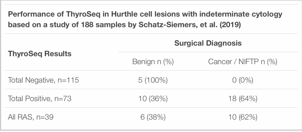
👉Additionally, ThyroSeq performance in Hurthle cell lesions was assessed in an independent study from NYU that included 188 indeterminate Hurthle cell predominant thyroid FNAs that had ThyroSeq v2/v3 results
👉Surgical follow up was available for 33 cases:
👉Detection of other lesions:

👉According to the ThyroSeq clinical validation study supplemental table above, reported in JAMA Oncology, ThyroSeq GC has a benign call rate of 61%, as compared to the Afirma GSC rate of 54%, which indicates that ThyroSeq allows more patients to safely avoid diagnostic surgeries
👉Additionally, ThyroSeq GC has a higher negative call rate for histologically benign nodules with indeterminate cytology (82%) than Afirma GSC (68%). This means that patients with nodules that have indeterminate cytology but are histologically benign would avoid diagnostic surgery with ThyroSeq GC more often than with Afirma GSC.
#Arrangoiz #ThyroidSurgeon #ThyroidExpert #CancerSurgeon #HeadandNeckSurgeon #ThyroidNodules #Thyroseq

The hallmarks of inflammatory breast cancer include warmth, edema, and erythema encompassing more than half of her breast.
Although inflammatory breast cancer is largely a clinical diagnosis, a skin punch biopsy revealing dermal lymphatic invasion confirms the diagnosis even in the absence of any obvious breast mass.
The standard treatment at this time involves neoadjuvant chemotherapy, modified radical mastectomy followed by postmastectomy radiation therapy.
Additional adjuvant systemic therapy is also administered based on the characteristics of the cancer (hormone receptor positive disease will receive hormonal therapy and HER2(+) disease anti-HER2-based therapy.
References
Sabiston Textbook of Surgery: The Biological Basis of Modern Surgical Practice, 20th ed. Ch. 34: Diseases of the Breast.




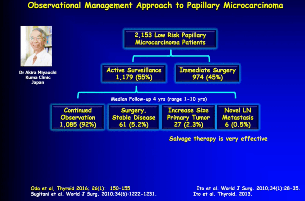
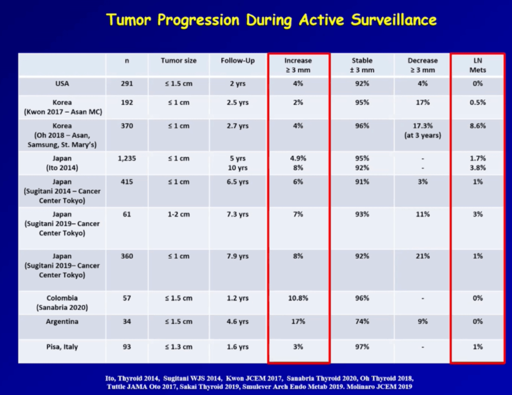
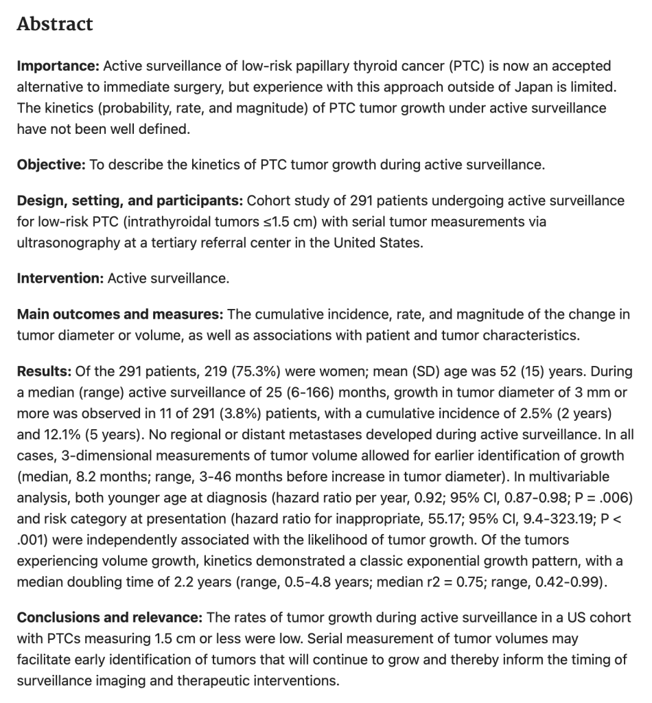
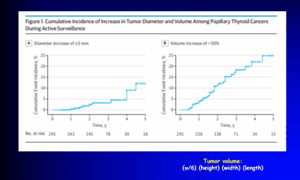








Both chemotherapy and aromatase inhibitors are indicated in high-risk HR+ breast cancer. Studies have shown aromatase inhibitors to be superior to tamoxifen in breast cancer recurrence and mortality in postmenopausal women.
References
Albain KS, Barlow WE, Shak S, et al. Prognostic and predictive value of the 21-gene recurrence score assay in postmenopausal women with node-positive, oestrogen-receptor-positive breast cancer on chemotherapy: a retrospective analysis of a randomised trial. Lancet Oncol. 2010; 11(1): 55-65.
Gnant M, Filipits M, Greil R, et al. Predicting distant recurrence in receptor-positive breast cancer patients with limited clinicopathological risk: using the PAM50 Risk of Recurrence score in 1478 postmenopausal patients of the ABCSG-8 trial treated with adjuvant endocrine therapy alone. Ann Oncol. 2014; 25(2): 339-45.
Early Breast Cancer Trialists’ Collaborative Group, Davies C, Godwin J, et al. Relevance of breast cancer hormone receptors and other factors to the efficacy of adjuvant tamoxifen: patient-level meta-analysis of randomised trials. Lancet. 2011; 378(9793): 771-84.
Francis PA, Pagani O, Fleming GF, et al. Tailoring Adjuvant Endocrine Therapy for Premenopausal Breast Cancer. N Engl J Med. 2018; 379(2): 122-137.
Early Breast Cancer Trialists’ Collaborative Group. Aromatase inhibitors versus tamoxifen in early breast cancer: patient-level meta-analysis of the randomised trials. Lancet. 2015; 386(10001): 1341-52.
Vogel VG, Costantino JP, Wickerham DL, et al. Update of the National Surgical Adjuvant Breast and Bowel Project Study of Tamoxifen and Raloxifene (STAR) P-2 Trial: Preventing breast cancer. Cancer Prev Res (Phila). 2010; 3(6): 696-706.