- Magnetic Resonance Imaging:
- Mammography:
- Remains the standard for radiographic evaluation of DCIS
- The cost and accessibility of magnetic resonance imaging (MRI):
- Make it less feasible as an effective screening method
- However, there is evidence that patients at high risk for breast cancer or those with very dense breasts:
- May benefit from screening with MRI
- Contrast-enhanced MRI:
- Is more sensitive than mammography:
- In the detection of both DCIS and invasive cancer
- Is more sensitive than mammography:
- However, fibrocystic changes and other benign findings:
- Can mimic DCIS on MRI:
- Leading to unnecessary biopsies
- Can mimic DCIS on MRI:
- MRI is increasingly being utilized after initial diagnosis in the preoperative evaluation:
- To identify multicentric and contralateral lesions:
- Because presence of either of these may change the surgical treatment strategy
- Hollingsworth et al. (2008):
- Reported that MRI detected multicentric disease:
- Defined as a separate focus of cancer more than 5 cm away from the index lesion or discontinuous growth to another breast quadrant:
- In 4.3% of 149 patients who presented with DCIS
- Reported that MRI detected multicentric disease:
- Lehman et al. (2007):
- Reported the utility of MRI in detecting contralateral breast cancer in a group of 969 patients with unilateral breast cancer:
- 196 of whom had DCIS
- Of the patients with DCIS:
- MRI prompted additional biopsies in 18 patients
- Contralateral breast cancer was detected in five patients:
- 28% of those biopsied and 2.6% of those with DCIS
- The sensitivity of detecting contralateral breast cancer was:
- 71%
- The specificity of detecting contralateral breast cancer was:
- 90%
- While MRI is associated with increased likelihood of change in the surgical plan for a patient with unilateral breast cancer:
- It is unclear whether these altered (and usually more extensive) surgical plans are actually treating clinically significant disease that might have otherwise decreased the patient’s disease-free or overall survival
- Reported the utility of MRI in detecting contralateral breast cancer in a group of 969 patients with unilateral breast cancer:
- To identify multicentric and contralateral lesions:
- Mammography:
- In a review of over 2,300 patients with breast-conserving therapy (BCT, i.e., lumpectomy and radiation) for DCIS at Memorial Sloan Kettering between 1997 and 2010:
- There was no association between receipt of preoperative MRI and risk of locoregional recurrence or contralateral breast cancer:
- Regardless of whether the patient received radiation (Pilewskie et al., 2014)
- There was no association between receipt of preoperative MRI and risk of locoregional recurrence or contralateral breast cancer:
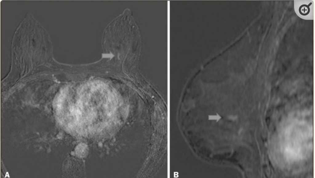
- The typical appearance of DCIS on MRI:
- Is non-mass enhancement
- Although mammography can be more sensitive than MRI for DCIS associated with calcifications:
- Uncalcified DCIS may be better visualized by MRI
- Kuhl and colleagues prospectively assessed 7,319 women who had undergone both preoperative mammography and MRI:
- Of 167 women with pure DCIS on final pathology:
- 92% (n = 153) were diagnosed by MRI and 56% (n = 93) were diagnosed by mammography:
- Of those diagnosed with high-grade DCIS:
- 48% were missed by mammography but diagnosed by MRI only
- Of those diagnosed with high-grade DCIS:
- 92% (n = 153) were diagnosed by MRI and 56% (n = 93) were diagnosed by mammography:
- Of 167 women with pure DCIS on final pathology:
- A meta-analysis looking at the association of preoperative MRI and surgical management of patients with DCIS:
- Showed no significant difference in the proportion of women with positive margins or in the need for re-excision after BCS
- Overall mastectomy rates did not differ significantly, whether or not preoperative MRI was performed (odds ratio [OR] 1.23; p = .34)
- Pilewskie and colleagues reported a large series of women undergoing BCS for DCIS:
- Found no difference in locoregional recurrence rates or contralateral breast cancer rates:
- In women who had perioperative MRI and those who did not
- Found no difference in locoregional recurrence rates or contralateral breast cancer rates:
- Although MRI can be useful in assessment of extent of disease and is an adjunct to traditional imaging in patients who have discordant results or mammographically occult disease:
- Routine use of MRI is not advocated for the perioperative management of DCIS
#Arrangoiz #CancerSurgeon #BreastSurgeon #SurgicalOncologist #BreastCancer #LCIS #DCIS #DuctalCarcinomaInsitu #LobularNeoplasia #LobularCarcinomaInsitu #Surgeon #Teacher #Miami #Mexico #MSMC #MountSinaiMedicalCenter
