Blog

Micro invasive Breast Cancer

  • In 1982, Lagios et al. introduced the term “microinvasion” in breast pathology as synonymous with invasion less than 1 mm.
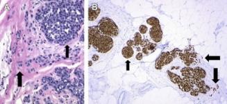
    • Microinvasive carcinoma is characterized by the extension of cancer cells beyond the in-situ component (ductal carcinoma in situ and lobular carcinoma in situ) into the adjacent breast tissue with no focus more than 0.1 cm in greatest dimension Figure.
      When there are multiple foci of microinvasion, only the size of the largest focus is used to classify the lesion, and the size of the individual foci should not be added together.
      Lesions that fulfill this definition are staged T1mic.
      The tumor focus/foci must invade into non-specialized interlobular or interductal stroma.
      The cells deemed to be invasive must be distributed in a non-organoid pattern that does not represent tangential sectioning of a duct or a lobular structure with in-situ carcinoma.
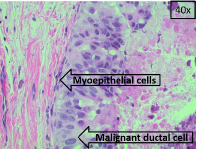
      Tangentially sectioned in-situ carcinoma foci that simulate microinvasion are distributed in the specialized intralobular and periductal stroma and usually occur as compact groups of tumor cells that have a smooth border surrounded by a circumferential layer of myoepithelial cells and stroma or a thickened basement membrane.
      At sites of microinvasion, tumor cells are distributed singly or as small groups that have irregular shapes reminiscent of conventional invasive carcinoma with no particular orientation.
  • Buccal Squamous Cell Carcinoma

    A. Background
    1. Carcinoma of the buccal mucosa is relatively uncommon in North America, compared with other oral cavity cancers such as carcinomas of the tongue or floor of the mouth.

    2. Squamous cell carcinoma is the most common pathology (more than 90% of the cases) and more prevalent in those who use tobacco and alcohol.

    B. Problem:
    1. As the orifice of the upper aerodigestive tract, the oral cavity plays a critical role in breathing, speech, and swallowing.

    2. The buccal region is particularly important in bolus formation, preventing food from spilling into the lateral oral gutters or extraorally during the oral preparatory phase of swallowing.

    3. Cancer of the buccal mucosa and subsequent treatment of the disease may interfere with these functions.

    4. Buccal carcinoma has the propensity to become aggressive, with high rates of locoregional recurrence.

    5. Diagnosis and treatment at an early stage leads to significantly improved prognosis and function over advanced disease.

    C. Epidemiology:
    1. Squamous cell carcinoma of the buccal mucosa accounts for 5% to 10% of all cancers of the oral cavity in North America and Western Europe.

    2. It occurs more often in men, with a male:female ratio of 3-4:1, and most commonly in the 7th or 8th decade of life.

    3. The incidence of buccal carcinoma is much higher in Asia.
    – In Southeast Asia, the disease is the most common form of oral cavity cancer.
    – In India, buccal carcinoma is the most common cancer in men and the third most common cancer in women.
    – The higher rate of buccal carcinoma in Asia is likely related to the widespread practice of betel nut chewing.
    – Betel nut, composed mainly of the fruit of the Areca Palm and often mixed with tobacco, is placed along the buccal mucosa to induce a feeling of euphoria.
    – Buccal carcinoma related to betel nut chewing tends to develop at an earlier age, with most cases occurring between the ages of 40 to 70.

    D. Etiology:
    1. Tobacco and alcohol use are the main etiologic agents associated with the development of buccal carcinoma.

    2. In North America, a history of using tobacco is documented in 70% of patients.

    3. Although alcohol by itself is not thought to be a significant risk, tobacco and alcohol have a well-recognized synergistic effect in the development of carcinoma.

    4. In Asia, betel nut is a significant etiologic agent, in addition to tobacco and alcohol.

    5. In India, over 90% of patients with buccal carcinoma have a history of using betel nut.

    6. Other suspected but not confirmed etiologic agents include human papilloma virus, poor oral hygiene, and chronic irritation.

    7. Premalignant conditions include oral submucosal fibrosis and lichen planus:
    – The latter has a reported transformation rate of 0.5% to 3%, whereas the former has a malignant transformation rate of 0.5%.

    E. Clinical Manifestations:
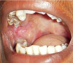
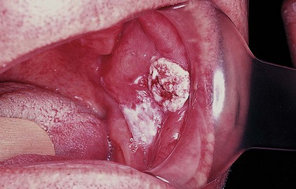
    1. Buccal carcinoma commonly presents as a slow-growing mass on the buccal mucosa.

    Unknown

    Philadelphia Illustration Dept./Elsevier
    Buccal SCC

    2. Small lesions tend to be asymptomatic and are often noted incidentally on dental examination.

    3. Pain commonly occurs as the lesion enlarges and ulceration develops.

    4. Oral intake may worsen the pain and lead to malnutrition and dehydration.

    5. Associated symptoms include bleeding, poor denture fit, facial weakness or sensory changes, dysphagia, odynophagia, and trismus.

    6. A detailed medical history is important to determine the patient’s candidacy for surgery or radiation therapy.

    7. The person often has a history of tobacco and alcohol use.

    8. A history of previous malignancies of the upper aerodigestive tract should be ascertained.

    9. Comprehensive examination of the head and neck should be conducted with a focus on the oral cavity.

    10. The mucosa of all the subsites of the oral cavity and oropharynx should be examined systematically.

    11. Palpation is important to determine the depth of invasion.

    12. Mandibular or maxillary alveolar invasion should be noted on inspection and palpation.

    13. Dentition must also be assessed, especially if irradiation is part of the planned management.

    14. The larynx and hypopharynx should be assessed by means of examination with a mirror or flexible endoscopy to rule out a second primary tumor of the upper aerodigestive tract.

    15. The ears should be examined in those patients with a history of otalgia because a lack of evidence of ear disease suggests referred pain due to malignancy.

    16. The neck and parotid gland should be carefully examined for adenopathy.

    17. Diaz et al found that 27% of patients presented with clinically positive nodes.

    18. The risk of nodal disease at presentation increases with advanced-stage disease.

    19. A meta-analysis of four studies with 223 cases of buccal carcinoma by Chhetri et al found that most presented with T2 or T3 disease (12% T1, 47% T2, 19% T3, 22% T4).

    20. The rate of nodal metastases at presentation was 40% for T2 disease and 52% for T3 disease.

    21. Signs of advanced disease on physical examination include bleeding, skin ulceration, facial swelling, neck mass, trismus, facial numbness, and paralysis of the facial musculature.

    22. The lesion often has 1 of 3 morphologic types:
    – Exophytic – The exophytic type is the most common, appearing as a papillary mass that becomes ulcerated when large.
    – Ulceroinfiltrative – The ulceroinfiltrative variety appears as an ulcer that penetrates deep into the underlying structures, with surrounding induration.
    – Verrucous – Verrucous carcinomas are uncommon variants of oral-cavity carcinomas; among these, the buccal mucosa is the most common site. These lesions appear as papillary masses, and keratinization gives them a whitish appearance.

    Rodrigo Arrangoiz MS, MD, FACS a head and neck surgeon and is amember of Sociedad Quirúrgica S.C at the America British Cowdray Medical Center.

    He is first author on some publications on oral cavity cancer:

    Training:

    • General surgery:

    • Michigan State University:

    • 2004 al 2010

    • Surgical Oncology / Head and Neck Surgery / Endocrine Surgery:

    • Fox Chase Cancer Center (Filadelfia):

    • 2010 al 2012

    • Masters in Science (Clinical research for health professionals):

    • Drexel University (Filadelfia):

    • 2010 al 2012

    • Surgical Oncology / Head and Neck Surgery / Endocrine Surgery:

    • IFHNOS / Memorial Sloan Kettering Cancer Center:

    • 2014 al 2016

    Breast Cancer Epidemiology

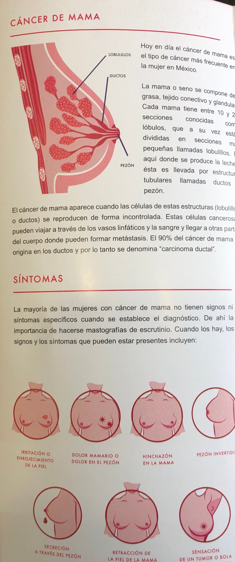
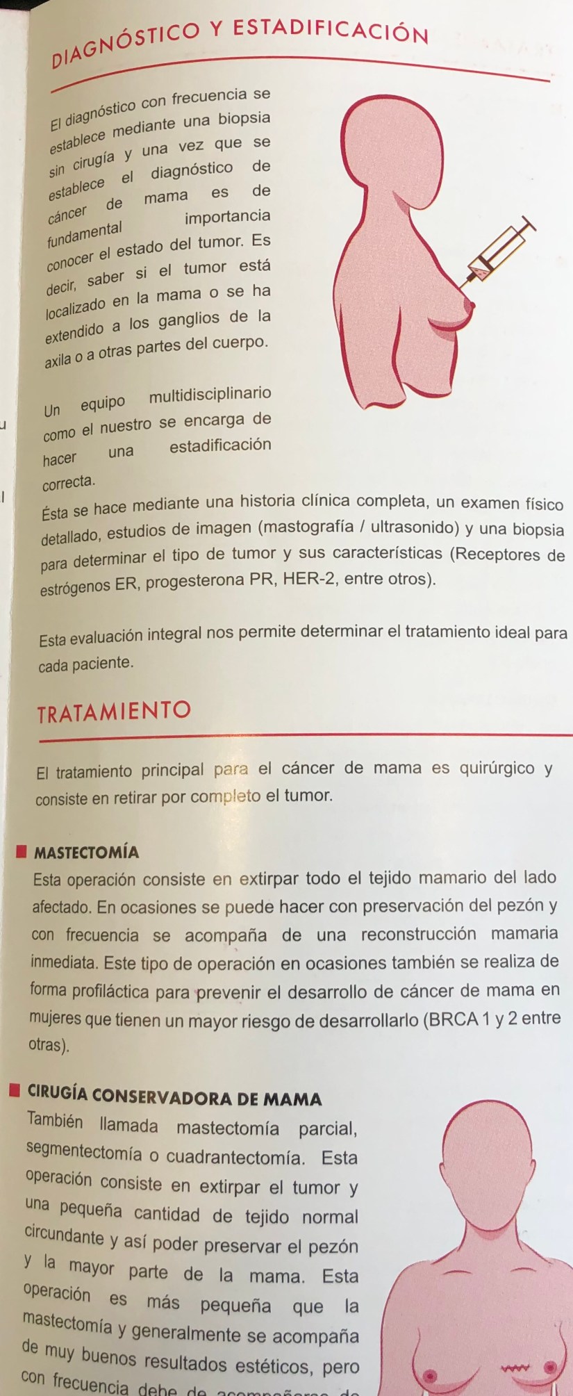
    Breast cancer remains the most common cancer diagnosed and the second most common cause of cancer-related mortality among women in the United States.

    • The American Cancer Society estimates that in 2018, approximately 268,670 new cases of invasive breast cancer will be diagnosed and nearly 41,400 breast cancer related deaths will occur.

    • Currently, the lifetime risk of breast cancer among women is 1 in 8 or 12% compared to 1 in 11 for women in the 1970s:

      • This increase in risk over the past four decades is attributed to:

        • Longer life expectancy

        • Changes in reproductive patterns

        • Hormone use

        • The rising prevalence of obesity

        • Increased detection through screening mammography.

    • Although the incidence of breast cancer has risen, breast cancer mortality has decreased:

      • Breast cancer death rates have decreased 36% from 1989 to 2012, after slowly increasing (0.4% per year) since 1975.

        • This likely reflects the increased use of screening mammography beginning in the early 1980s leading to detection of earlier stage disease, as well as continued improvements in systemic adjuvant therapy.

    • Breast cancer incidence rates are highest in non-Hispanic white women, followed by African American women and are lowest among Asian/Pacific Islander women.

    • Contrastingly, breast cancer death rates are highest for African American women, followed by non-Hispanic white women, and are lowest for Asian/Pacific Islander women:

      • Furthermore, the difference in long-term breast cancer mortality by race/ethnicity persists and is increasing with breast cancer death rates 42% higher in African American than Caucasian women in 2012:

        • This disparity reflects a combination of factors, including differences in stage at diagnosis, obesity, comorbidities, tumor characteristics, screening, access, adherence, and response to treatment.

    Rodrigo Arrangoiz MS, MD, FACS a surgical oncologist and is a member of Sociedad Quirúrgica S.C at the America British Cowdray Medical Center in Mexico City:

    • He is an expert in the management of breast cancer:

      • If you have any questions about breast cancer statistics please fill free to ask Dr. Arrangoiz

    Training:

    • General surgery:

    • Michigan State University:

    • 2004 al 2010

    • Surgical Oncology / Head and Neck Surgery / Endocrine Surgery:

    • Fox Chase Cancer Center (Filadelfia):

    • 2010 al 2012

    • Masters in Science (Clinical research for health professionals):

    • Drexel University (Filadelfia):

    • 2010 al 2012

    • Surgical Oncology / Head and Neck Surgery / Endocrine Surgery:

    • IFHNOS / Memorial Sloan Kettering Cancer Center:

    • 2014 al 2016


    img_0832-2img_0834-2img_0833-2img_0835-2img_0836-2img_0837-2

    Intraductal Papilloma (IDP)

    Introduction

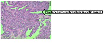
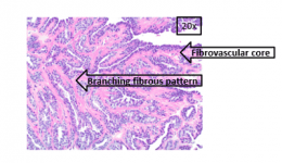
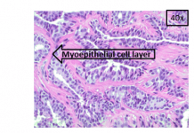
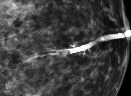
    • In the breast, intraductal papilloma (IDP) is a benign lesion that consists of branching fibrovascular cores occurring within a cystic cavity with overlying layers of epithelial and myoepithelial cells:
      • The classic pathologic features of intraductal papilloma (IDP) include an encysted solid mass with a branching fibrovascular pattern.

    image.phpimage-1.phpimage-2.php

    • The large/central subtype (L/C ST) specifically refers to an IDP arising from a large duct.
      • IDP (L/C ST) is generally grossly apparent, solitary, and centrally located in the breast; it has accordingly been referred to as “solitary papilloma” and “central papilloma.”


    • IDP (L/C ST) stands in contrast to IDP small/peripheral subtype (S/P ST), which originates at the terminal duct lobular unit (TDLU) and is usually located peripherally in the breast, is not grossly apparent, and generally occurs in multiples.

    17093

    Epidemiology

    • IDP (L/C ST) is primarily found in middle-aged women.
      • In a study of 179 women with solitary / central  papilloma:
        • The mean age at diagnosis was 48 years, and occurrence substantially decreased after age 75 years.
        • Younger women were also identified in this series:
          • The youngest was aged 18 years.
    • IDPs are relatively common and are found in 1% to 5% of breast biopsies.
    • Current evidence suggests that IDP (L/C ST) is more common that IDP (S/P ST).

    Etiology

    • Molecular evidence has shown that IDPs frequently demonstrate loss of heterozygosity (LOH):
      • Involving specific loci on chromosome 16:
        • This suggests they are clonal neoplasms.
          • However, the studies showing this do not distinguish between IDP (L/C ST) and IDP (S/P ST):
            • Therefore, they are unable to evaluate genetic differences between these lesions.

    Presentation

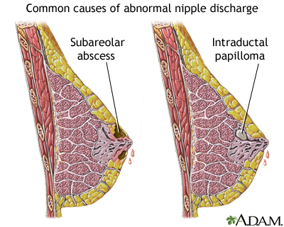
    • IDP (L/C ST) frequently presents as a unilateral serous or bloody nipple discharge:
      • It may also present as a palpable breast mass and may on occasion present as breast pain.
    • Mammography reveals no abnormality in most cases but may show duct ectasia, microcalcifications, or a mass.
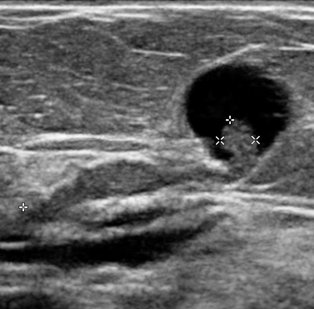
    • Ultrasonography may be more sensitive than mammography for detecting IDP (L/C ST) and can reveal duct ectasia, nodules, or a cyst with or without a polyp.

    64db50775d6c438409d5c3562b3f6a_big_gallery

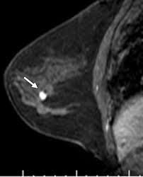
    • On MRI, IDP (L/C ST) appears as dilated ducts with an associated enhancing, well-circumscribed mass:
      • MRI is currently the most sensitive imaging modality for detecting IDP (L/C ST).

    a94bfb04a3c884ade36a8218e71c0f_galleryUnknown


    Rodrigo Arrangoiz MS, MD, FACS a surgical oncologist and is a member of Sociedad Quirúrgica S.C at the America British Cowdray Medical Center in Mexico City:

    • He is an expert in the management of breast cancer:

      • If you have any questions about intraductal papilloma and bloody nipple discharge please fill free to ask Dr. Arrangoiz

    Training:

    • General surgery:

    • Michigan State University:

    • 2004 al 2010

    • Surgical Oncology / Head and Neck Surgery / Endocrine Surgery:

    • Fox Chase Cancer Center (Filadelfia):

    • 2010 al 2012

    • Masters in Science (Clinical research for health professionals):

    • Drexel University (Filadelfia):

    • 2010 al 2012

    • Surgical Oncology / Head and Neck Surgery / Endocrine Surgery:

    • IFHNOS / Memorial Sloan Kettering Cancer Center:

    • 2014 al 2016


    Sociedad Quirúrgica S.C. multidisciplinary breast cancer clinic:


    img_0832-2img_0833-2img_0834-2img_0835-2img_0836-2img_0837-2


    Ductal Carcinoma In Situ (DCIS)

    Mammographic detection of calcification remains the mainstay of detection of nonpalpable malignancy.

    • The term ductal carcinoma in situ (DCIS) encompasses a heterogeneous group of lesions that differ in their clinical presentation, histologic appearance, and biological potential.

    • DCIS is characterized by proliferation of presumably malignant epithelial cells within the mammary ductal system, with no evidence of invasion into the surrounding stroma on routine light microscopic examination.

    • Classification schemes that divide DCIS histologically into a variety of subtypes emphasize architectural features or growth pattern of the neoplastic cells, cytologic features of ductal cells, and cell necrosis, both singly and in combination:

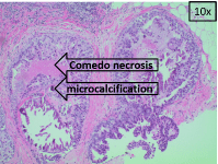
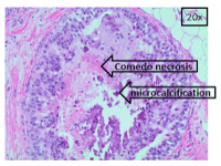
      • Comedo-type necrosis is characterized by prominent necrosis (eosin positive) in the center of the involved spaces.

      • The necrotic material frequently becomes calcified.

      • The tumor cells are large and show nuclear pleomorphism.

      • Mitotic activity may be prominent.

    image.phpimage-1.phpimage-2.php


    Rodrigo Arrangoiz MS, MD, FACS a surgical oncologist and is a member of Sociedad Quirúrgica S.C at the America British Cowdray Medical Center in Mexico City:

    • He is an expert in the management of breast cancer.

    Training:

    • General surgery:

    • Michigan State University:

    • 2004 al 2010

    • Surgical Oncology / Head and Neck Surgery / Endocrine Surgery:

    • Fox Chase Cancer Center (Filadelfia):

    • 2010 al 2012

    • Masters in Science (Clinical research for health professionals):

    • Drexel University (Filadelfia):

    • 2010 al 2012

    • Surgical Oncology / Head and Neck Surgery / Endocrine Surgery:

    • IFHNOS / Memorial Sloan Kettering Cancer Center:

    • 2014 al 2016

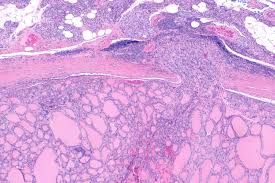
    Follicular Thyroid Carcinoma (FTC)

    Unknown

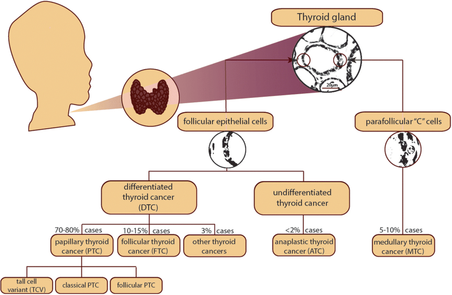
    Follicular thyroid carcinoma (FTC) is the second most common cancer of the thyroid, after papillary thyroid carcinoma (PTC).

    • Follicular and papillary thyroid cancers are considered to be differentiated thyroid cancers:
      • Together they make up 95% of thyroid cancer cases.
    12943_2018_866_Fig1_HTML

    FTC and other thyroid neoplasms arising from follicular cells (adenomas, papillary/follicular carcinoma, and noninvasive follicular thyroid neoplasm with papillary-like nuclear features [NIFTP]) show a broad range of overlapping clinical and cytologic features.

    • FTC resembles the normal microscopic pattern of the thyroid:
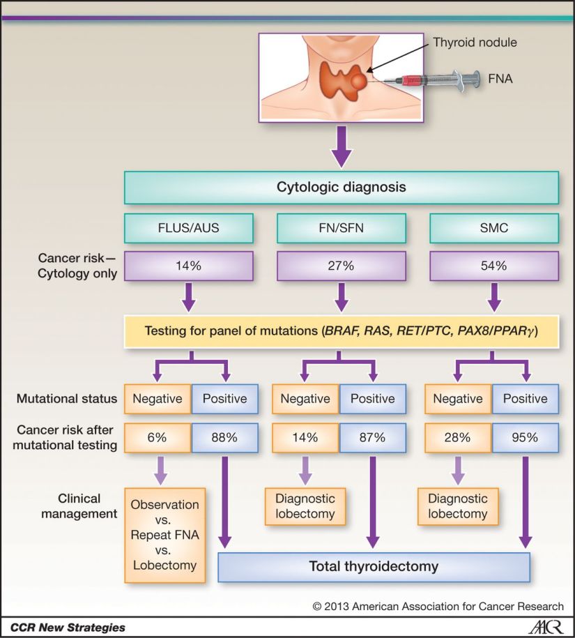
      • A clear distinction between benign and malignant disease based solely on cytological examination of a needle biopsy specimen may be difficult:
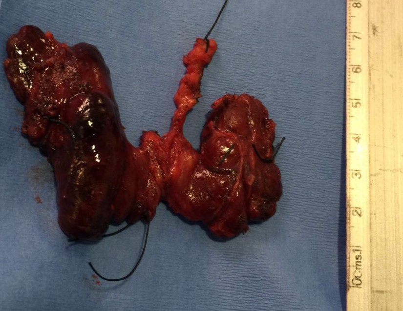
        • For this reason, a surgical procedure to remove all or a large portion of the thyroid gland may be necessary to obtain sufficient tissue for a definitive diagnosis of FT:
          • Pathological examination showing capsular or vascular invasion may be required for this determination.

    Hurthle cell carcinoma should be considered a variant of FTC.

    Pathophysiology

    • Activating point mutations in the ras oncogene are well known in patients with follicular adenoma and carcinoma, especially in poorly differentiated (55%) and anaplastic carcinoma (52%):
      • As a result of such mutations, p21-RAS becomes locked in its active conformation, leading to the constitutive activation of the protein and tumor development.
        • The biochemical pathways that this process follows may be therapeutic targets for FTC. 
          • Accidental (not diagnostic) x-ray exposure may influence both occurrence and pattern of ras mutation.


    • A study of differential gene expression profiling of aggressive and nonaggressive follicular carcinomas identified 94 genes that distinguish follicular carcinomas from follicular adenomas (including PBP and CKS2) and 4 genes that distinguish aggressive follicular carcinomas from nonaggressive follicular carcinomas (NID2, TM7SF2, TRIM2, and GLTSCR2).

    Epidemiology

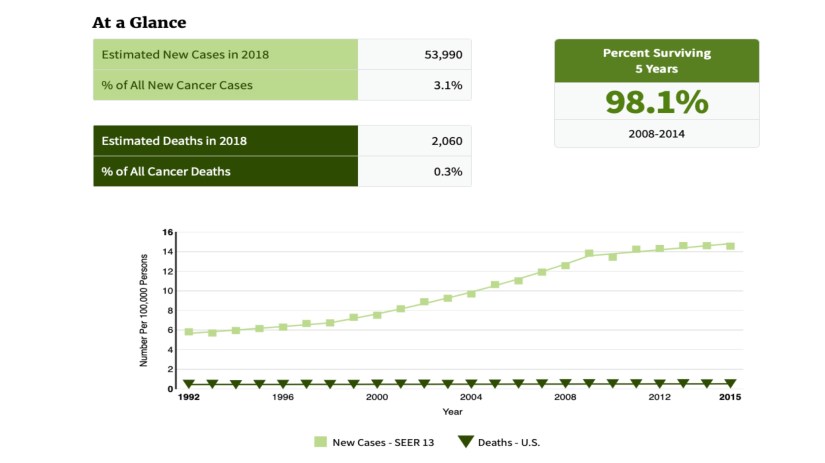
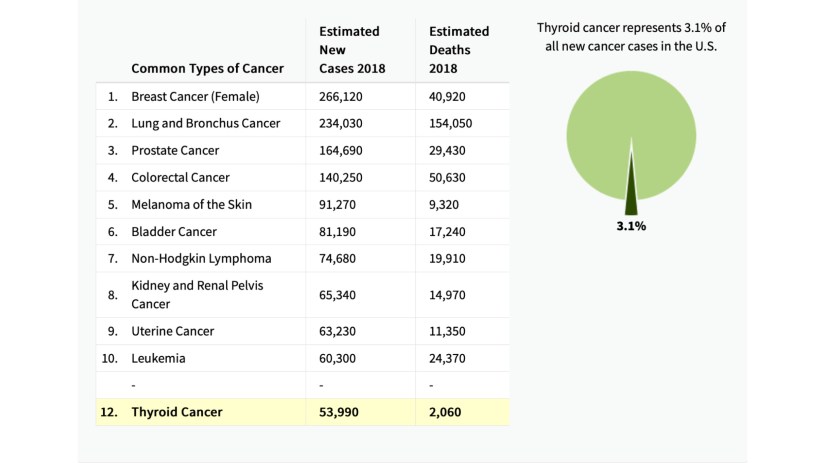
    • The American Cancer Society (ACS) estimates that 53,990 new thyroid cancers will occur in 2018 (13,090 in men and 40,900 in women)
    • The ACS estimates 2060 deaths from thyroid cancer in 2018, (960 in men and 1100 in women).
    • In women, thyroid cancer is the fifth most common cancer:
      • Accounting for approximately 5% of all new cases.
    • In the United States, about 10% to 15% of all thyroid cancers are follicular.
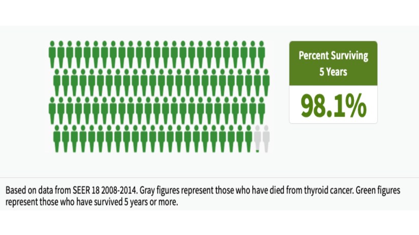
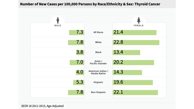
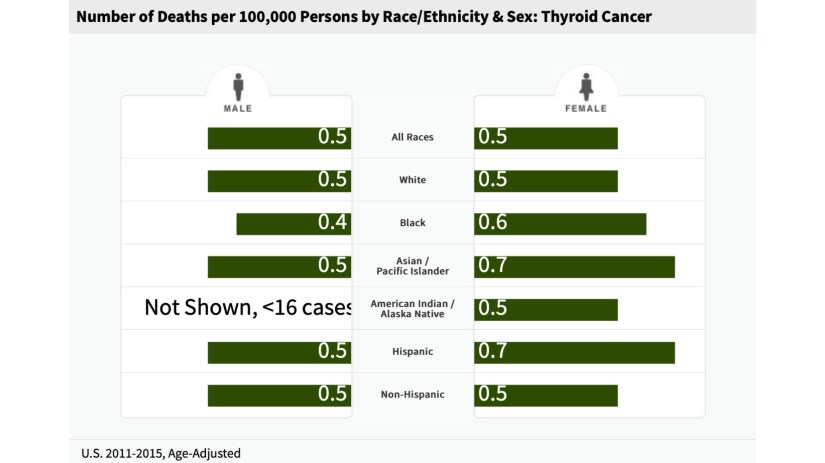
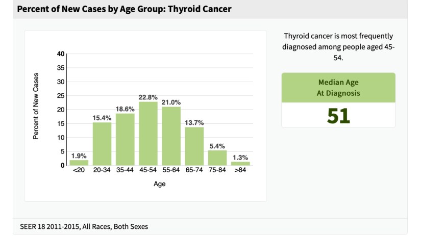
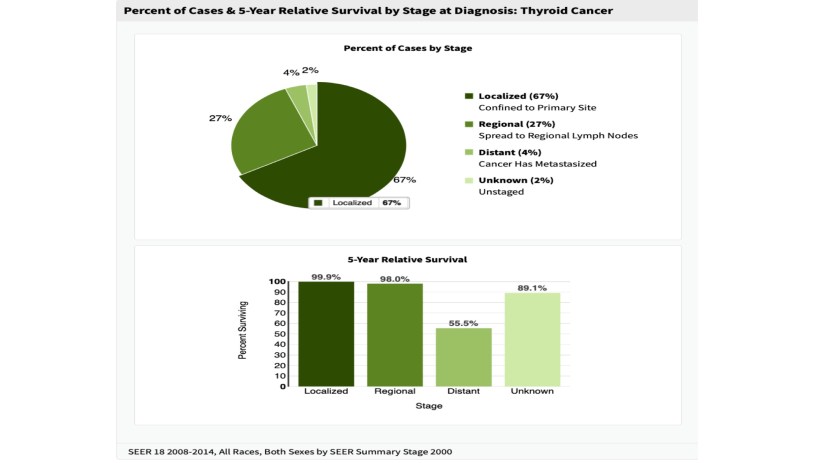
    Thyroid Cancer StatisticsSurvival Statistics at 5 Years for Thyroid CancerHow Common is Thyroid Cancer Compared to other CancersNumber of New Cases of Thyroid Cancer Based on Age, RaceNumber of Deaths for Thyroid CancerDeath from Thyroid Cancer by Age GroupNew Cases of Thyroid Cancer by AgeThyroid Cancer 5 Year Survival by Stage
    • Thyroid cancers are quite rare, accounting for only 1.5% of all cancers in adults and 3% in children.
    • The highest incidence of thyroid carcinomas in the world is among female Chinese residents of Hawaii:
      • In Hawaii, the incidence of FTC ranges from 10 to 30 new cases a year per million inhabitants.
      • In recent years, the frequency of FTC has appeared to increase; however, this increase is related to improvement in diagnostic techniques and a successful campaign of information about this carcinoma.

    Rodrigo Arrangoiz MS, MD, FACS cirujano de tumores de cabeza y cuello / cirugia endocrina miembro de Sociedad Quirúrgica S.C. experto en el manejo del cáncer de tiroides.

    Cumple con los requisitos determinados por el Dr. Saha para realizar cirugía de tiroides de manera efectiva y segura:

    • Cirugia general y gastrointestinal:

    • Michigan State University:

    • 2004 al 2010image-48

    • Cirugia oncológica / tumores de cabeza y cuello / cirugia endocrina:

    • Fox Chase Cancer Center (Filadelfia):

    • 2010 al 2012

    image-39

    • Maestria en ciencias (Clinical research for healthprofessionals):

    • Drexel University (Filadelfia):

    • 2010 al 2012image-50

    • Cirugia de tumores de cabeza y cuello / cirugiaendocrina
    • IFHNOS / Memorial Sloan Kettering Cancer Center:

    • 2014 al 2016

    image-51

    http://www.sociedadquirurgica.com

    http://www.hiperparatiroidismo.info

    http://www.cirugiatiroides.com

    #Arrangoiz

    #CirugiadeTumoresdeCabezayCuello

    #CirugiaEndocrina

    #CirugiaOncologica

    #HeadandNeckSurgery

    #EndocrineSurgery

    #SurgicalOncology

    ¿Quién debe de realizar su cirugía de tiroides?

    • El entrenamiento y el volumen del cirujano son factores críticos para obtener el mejor resultado con las menores complicaciones en la cirugía de tiroides.

    Presentation1


    • Ashok Shaha MD, FACS cirujano de cabeza y cuello de Memorial Sloan Kettering Cancer Center experto en tiroides durante su plática inaugural de la American Head and Neck Society nos habla de quien debe estar realizando cirugía de tiroides.


    Rodrigo Arrangoiz MS, MD, FACS cirujano de tumores de cabeza y cuello / cirugia endocrina miembro de Sociedad Quirúrgica S.C. cumple con los requisitos determinados por el Dr. Saha para realizar cirugía de tiroides .

    Su entrenamiento fue el siguiente:

    • Cirugia general y gastrointestinal:
    • Michigan State University:
    • 2004 al 2010image-48• Cirugia oncológica / tumores de cabeza y cuello / cirugia endocrina:
    • Fox Chase Cancer Center (Filadelfia):
    • 2010 al 2012image-39• Maestria en ciencias (Clinical research for healthprofessionals):
    • Drexel University (Filadelfia):
    • 2010 al 2012image-50• Cirugia de tumores de cabeza y cuello / cirugiaendocrina
    • IFHNOS / Memorial Sloan Kettering Cancer Center:
    • 2014 al 2016image-51 

    http://www.sociedadquirurgica.com

    http://www.hiperparatiroidismo.info

    http://www.cirugiatiroides.com

    #Arrangoiz

    #CirugiadeTumoresdeCabezayCuello

    #CirugiaEndocrina

    #CirugiaOncologica

    #HeadandNeckSurgery

    #EndocrineSurgery

    #SurgicalOncology

    Tratamiento del Hiperparatiroidismo Primario

    Rodrigo Arrangoiz MS, MD, FACS cirujano de tumores de cabeza y cuello / cirugia endocrina es experto en el manejo del hiperparatiroidismo primario.

    • Video del Dr. Arrangoiz, pionero en México de la técnica de paratiroidectomia radioguiada mínimamente invasiva descrita por el Dr. James Norman, sobre el hiperparatiroidismo:


    Introdujo a nuestro país la técnica de exploración bilateral de cuello con valoración de la funcionalidad de las glándulas paratiroides con paratiroidectomia radioguiada mínimamente invasiva:

    Su entrenamiento fue el siguiente:

    • Cirugia general y gastrointestinal:
    • Michigan State University:
    • 2004 al 2010image-48• Cirugia oncológica / tumores de cabeza y cuello / cirugia endocrina:
    • Fox Chase Cancer Center (Filadelfia):
    • 2010 al 2012image-39• Maestria en ciencias (Clinical research for healthprofessionals):
    • Drexel University (Filadelfia):
    • 2010 al 2012image-50• Cirugia de tumores de cabeza y cuello / cirugiaendocrina
    • IFHNOS / Memorial Sloan Kettering Cancer Center:
    • 2014 al 2016image-51 

    http://www.sociedadquirurgica.com

    http://www.hiperparatiroidismo.info

    http://www.cirugiatiroides.com

    #Arrangoiz

    #CirugiadeTumoresdeCabezayCuello

    #CirugiaEndocrina

    #CirugiaOncologica

    #HeadandNeckSurgery

    #EndocrineSurgery

    #SurgicalOncology

    #Hyperparathyroidism

    #Hiperparatiroidismo

    Experto en Tiroides

    Rodrigo Arrangoiz MS, MD, FACS cirujano de tumores de cabeza y cuello / cirugia endocrina / cirugía oncológica es experto en el manejo de patología  de la glándula tiroides y paratiroides.

    Su entrenamiento fue el siguiente:

    • Cirugia general y gastrointestinal:
    • Michigan State University:
    • 2004 al 2010image-48• Cirugia oncológica / tumores de cabeza y cuello / cirugia endocrina:
    • Fox Chase Cancer Center (Filadelfia):
    • 2010 al 2012image-39• Maestria en ciencias (Clinical research for healthprofessionals):
    • Drexel University (Filadelfia):
    • 2010 al 2012image-50

    • Cirugia de tumores de cabeza y cuello / cirugiaendocrina
    • IFHNOS / Memorial Sloan Kettering Cancer Center:
    • 2014 al 2016image-51

    https://www.cirugiatiroides.com

    https://hiperparatiroidismo.info


    bed1cf48db8f6c6d39001781c7725a8e.jpgcropped-18403652_10206829497335208_5004404657991480104_n1.jpgF1.large20d943b1-6f20-49c3-9754-2f3756051c9312fc1993-1232-40ac-91a0-f394c7a8de2ebd310df2-281c-45ee-84ef-1877498e710a5b081828-c940-488d-bd45-21f33bd5f84f2131cd57-aa86-4187-801d-bb4d2ab2d6ef


    Can Low-Dose Tamoxifen Prevent Recurrences With Reduced Toxicity?

    For patients with intraepithelial neoplasia (also known as noninvasive breast cancers) treatment with tamoxifen at a dose of only 5 mg/day for 3 years, rather than the standard 20 mg/day, can reduce recurrence at a similar rate to the higher dose, according to the results of the randomized, phase 3 TAM-01 study.

    • The regimen did not significantly worsen menopausal symptoms or major adverse events compared with placebo.

      • Tamoxifen was developed in the 1960s, but the minimal effective dose has never been established, Rodrigo Arrangoiz MS, MD, FACS

    Intraepithelial neoplasia represents 15% to 25% of all breast cancers, and includes a heterogeneous spectrum of disorders including atypical ductal hyperplasia (ADH), ductal carcinoma in situ (DCIS), and lobular carcinoma in situ (LCIS). 

    • Tamoxifen is very effective in prevention (risk reducing), but its potential side effects, including:

    • Endometrial cancer, deep vein thrombosis (DVT), and menopausal symptoms, act as a barrier to its use.

    De Censi and colleagues hypothesized that a reduced dose and a shorter duration would be effective.

    • That idea was based on a 2003 study from this same Italian group, which showed that 5 mg of tamoxifen was comparable to 20 mg in reducing breast cancer proliferation, as measured by Ki-67 level.

    At San Antonio Breast Cancer Symposium (SABCS) 2018, the TAM-01 study which included 500 women aged less than 75 years with intraepithelial neoplasia was presented:

    • They were randomized to receive either tamoxifen 5 mg/day for 3 years, or to placebo

    • The median follow-up in the study was 5.1 years.

    • Patients had a mean age of 54 years, and just under half in both groups were premenopausal.

    • Most of the cohort had DCIS (69% of tamoxifen patients, 70% of placebo patients), followed by ADH (20% in both groups), and LCIS (11% and 10%, respectively).

    In total, there were 42 breast cancer or DCIS events:

    • There were 28 such events in the placebo group compared with 14 in the tamoxifen group, for a hazard ratio (HR) of 0.48 (95% CI, 0.26–0.92; P = .024).

      • The placebo patients had a rate of 23.9 events per 1,000 person-years, compared with 11.6 events per 1,000 person-years with tamoxifen.

    • There were 12 cases of contralateral breast cancer in the placebo group, compared with 3 cases in the tamoxifen group, for an HR of 0.24 (95% CI, 0.07–0.87; P = .018).

    • There was one case of endometrial cancer in the tamoxifen group, and none in the placebo patients.

    • There were four other neoplasms with tamoxifen and six with placebo.

    • Both groups had one case of DVT or pulmonary embolism, and both had two cases of coronary heart disease.

      • With 20 mg/day of tamoxifen:

        • 2.7 cases of endometrial cancer and 2.4 cases of DVT/pulmonary embolism would be expected.

    • Notably, the regimen did not appear to significantly worsen menopausal symptoms.

      • There was a significant increase in the frequency of daily hot flashes (P = .05), but the absolute increase was less than one.

      • There was no difference between tamoxifen and placebo with regard to vaginal dryness or pain during intercourse (P = .57), or with regard to musculoskeletal pain/arthralgia (P = .84).

    img_0832-2img_0833-2img_0836-2img_0835-2img_0834-2img_0837-2

    Rodrigo Arrangoiz MS, MD, FACS a surgical oncologist and is amember of Sociedad Quirúrgica S.C at the America British Cowdray Medical Center in Mexico City:

    • He is an expert in the management of breast cancer.

      • If you have any questions about risk reducing therapies for breast cancer please fill free to ask him.

    Training:

    • General surgery:

    • Michigan State University:

    • 2004 al 2010

    • Surgical Oncology / Head and Neck Surgery / Endocrine Surgery:

    • Fox Chase Cancer Center (Filadelfia):

    • 2010 al 2012

    • Masters in Science (Clinical research for health professionals):

    • Drexel University (Filadelfia):

    • 2010 al 2012

    • Surgical Oncology / Head and Neck Surgery / Endocrine Surgery:

    • IFHNOS / Memorial Sloan Kettering Cancer Center:

    • 2014 al 2016