Blog

Mucoepidermoid Carcinoma (MEC)

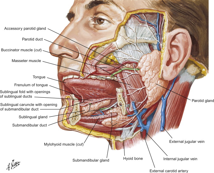
3-s2.0-b9780123813619000081-f08-03-9780123813619

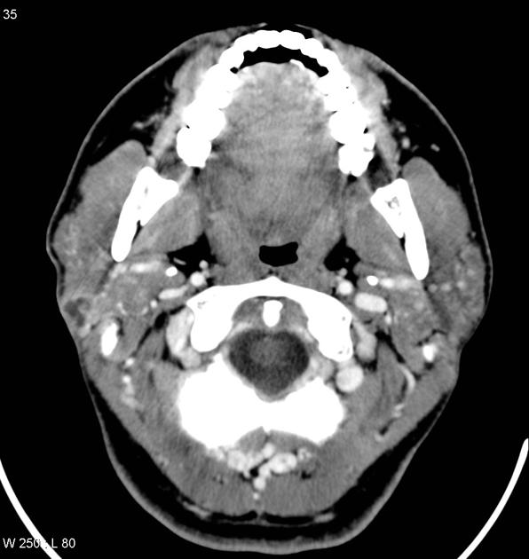
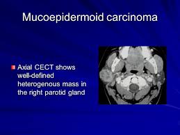
  • Mucoepidermoid carcinoma (MEC) is the most common malignant neoplasm of the mayor and minor salivary glands:
    • They encompass between 2.8% to 15.5% of all salivary gland tumors
    • They represent 12% to 35% of malignant salivary gland tumors
    • They encompass 6.5% to 41% of all minor salivary gland tumors:
      • Representing the most common type of malignant minor salivary gland tumor in most series:
        • Other series mention that adenoid cystic carcinoma is the most common malignant minor salivary gland tumor
  • Approximately half of the cases of MEC occur in the major salivary glands:
    • 65% to 80% of these occur in the parotid
    • 8% to 13% occur in the submandibular gland
    • 2% to 4% involve the sublingual gland
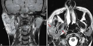
24f1f9d76f99646bc7ec674d51a9de_big_galleryunknown-1images

  • MEC of the minor salivary glands ordinarily arises on the palate:
    • But a number may also be found in the:
      • Retro molar area, floor of the mouth, buccal mucosa, lip, and tongue
  • Its prevalence is highest in the fourth to fifth decade of life:
    • 35 to 65 years of age:
      • With a female preponderance as high as 4:1
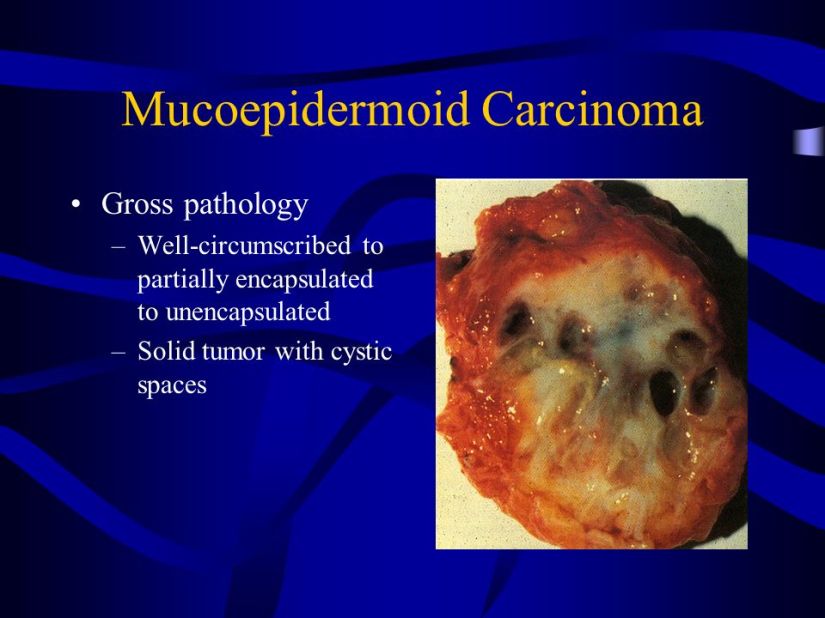
  • Grossly:
    • The tumor is poorly circumscribed
    • Measures from 3 to 5 cm

mucoepidermoid+carcinoma-1

  • Histologically:
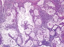
    • They are characterized by a mixed population of cells:
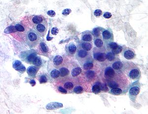
      • Including, mucin-producing cells, epidermoid cells with squamoid differentiation, clear cells, and intermediate cells that may predominate in numbers and are believed to be the progenitor of the other types of cells:
        • No myoepithelial cells are present

300px-mucoepidermoid_carcinoma_2_-_very_high_magunknown

  • The clinical behavior of MEC has proved to be difficult to predict but correlations to tumor grade and stage have been reported:
    • The histologic features that are most useful in predicting the aggressive nature of these tumors are:
      • A minor cystic component (less than 20%)
      • Tumor necrosis
      • Neural invasion
      • Cellular anaplasia
      • Brisk mitotic activity
    • Based on the presence or absence of these features and the clinical behavior, MEC are classified as:
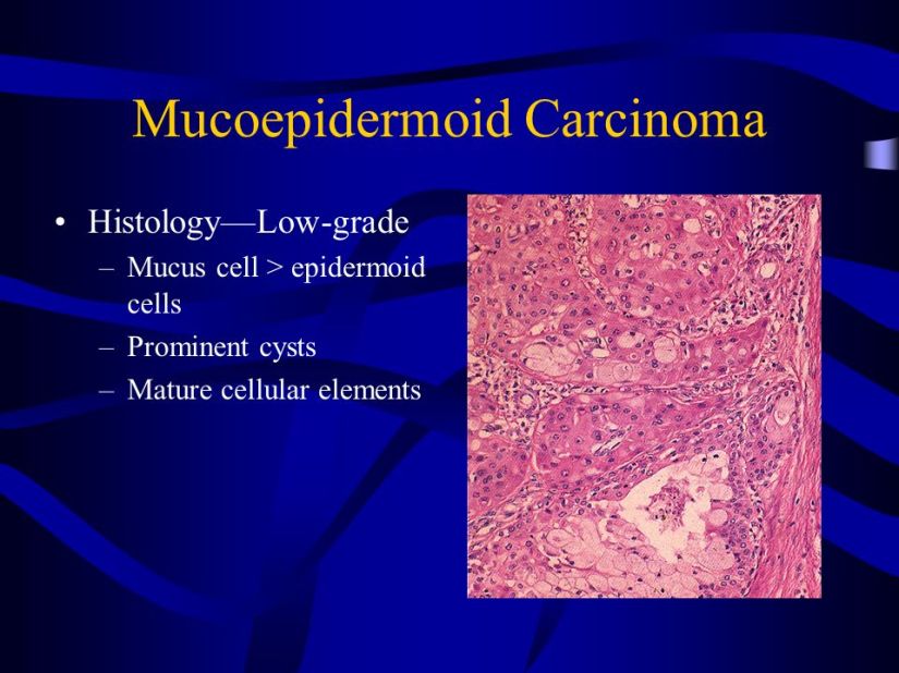
      • Low, intermediate, and high grade:
        • Low-grade MEC are well circumscribed, with pushing margins and dilated cystic areas containing mucin:
          • Mucin- producing, intermediate, or epidermoid cells make up the lining of these cystic structures.

mucoepidermoid+carcinoma

  • As the grade worsens:
    • The tumors become more infiltrative, poorly circumscribed, cystic formations are lost, and nests of tumor become more solid and irregular with intermediate or epidermoid cells dominating.
  • High-grade MEC are characterized by:
    • The invasion of adjacent structures
    • Atypical, brisk mitoses
    • Tumor necrosis
    • Perineural invasion
    • Cellular anaplasia
    • Lymph node (40% to 50%) metastasis
    • Distant metastases (33%).
      • These high-grade lesions are differentiated from primary of metastatic squamous cell carcinoma by the presence of intracellular mucin:
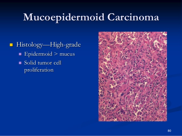
        • Sebaceous and clear cell carcinomas are additional differential diagnosis to consider.

salivary-glands-disorders-ii-80-638

Rodrigo Arrangoiz MS, MD, FACS a head and neck surgeon / surgical oncologist and is a member of Sociedad Quirúrgica S.C at the America British CowdrayMedical Center in Mexico City:

  • He is an expert in the management of salivary gland neoplasms:

    • If you have any questions about salivary gland neoplasms  please fill free to ask Dr. Arrangoiz

Training:

• General surgery:

• Michigan State University:

• 2004 al 2010

• Surgical Oncology / Head and Neck Surgery / Endocrine Surgery:

• Fox Chase Cancer Center (Filadelfia):

• 2010 al 2012

• Masters in Science (Clinical research for health professionals):

• Drexel University (Filadelfia):

• 2010 al 2012

• Surgical Oncology / Head and Neck Surgery / Endocrine Surgery:

• IFHNOS / Memorial Sloan Kettering Cancer Center:

• 2014 al 2016


#Arrangoiz

#HeadandNeckSurgeon

#SurgicalOncologist

#Surgeon

#CancerSurgeon

#CirujanodeCabezayCuello

#CirujanoOncologo
http://www.sociedadquirurgica.com

Diabetic Mastopathy

  • Diabetic mastopathy, also known as “lymphocytic mastopathy” or “sclerosing lymphocytic lobulitis”:
    • Is an uncommon mass-forming lesion seen in patients with insulin-dependent (type 1) diabetes mellitus:
      • Particularly in those who have long-standing disease with microvascular complications.
    • Diabetic mastopathy most often occurs in premenopausal women.
    • The typical presentation is a palpable unilateral mass.
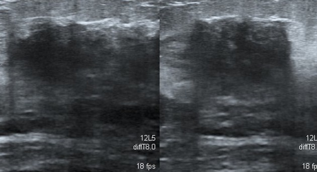
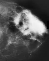
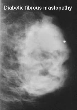
    • The mammographic and sonographic features of diabetic mastopathy may be suspicious for malignancy:
      • Mammography may reveal an ill-defined mass, distortion, or dense glandular breast tissue
      • Ultrasound may show an irregular hypoechoic mass with posterior shadowing.

unknown-1diabetic-fibrous-mastopathy-breast-xray72004d613f0fdcb31dd3b59df60c34_big_gallery

  • The characteristic constellation of findings in diabetic mastopathy includes:
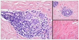
    • Lymphocytic lobulitis and ductitis
    • Lymphocytic perivasculitis
    • Stromal fibrosis with epithelioid fibroblasts.
    • Lymphocytic infiltrates, which can be fairly dense:
      • Surround ducts, lobules, and small vessels
      • May sometimes be associated with plasma cells
    • Immunohistochemical characterization of these infiltrates reveals mature B-lymphocytes with a small population of T cells.
    • Germinal centers are not typically seen here.
    • Involved lobules may be atrophic or unremarkable.
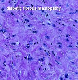
    • The stroma in diabetic mastopathy is dense and has a keloidal appearance.
    • Intrastromal epithelioid fibroblasts appear as plump cells with eosinophilic cytoplasm.
    • Nuclei are oval to round with vesicular nuclei:
      • Neither significant nuclear atypia nor mitotic figures are seen.
    • The distribution of fibroblasts within the stroma can be heterogeneous, and show a whorled or nodular growth pattern.

diabetic-fibrous-mastopathy-blueunknown


Rodrigo Arrangoiz MS, MD, FACS a surgical oncologist and is a member of Sociedad Quirúrgica S.C at the America British Cowdray Medical Center in Mexico City:

  • He is an expert in the management of breast cancer.

    • If you have any questions about diabetic mastopathy please fill free to contact Dr. Arrangoiz.

Training:

• General surgery:

• Michigan State University:

• 2004 al 2010

• Surgical Oncology / Head and Neck Surgery / Endocrine Surgery:

• Fox Chase Cancer Center (Filadelfia):

• 2010 al 2012

• Masters in Science (Clinical research for health professionals):

• Drexel University (Filadelfia):

• 2010 al 2012

• Surgical Oncology / Head and Neck Surgery / Endocrine Surgery:

• IFHNOS / Memorial Sloan Kettering Cancer Center:

• 2014 al 2016

Anaplastic Thyroid Carcinoma (ATC)

Clinical Manifestations 

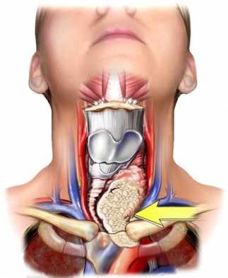
  • The typical patient with ATC has a long-standing neck mass:
    • Which starts to rapidly enlarge and may be painful.
  • Associated symptoms such as dysphonia, dysphagia, and dyspnea are common:
    • Due to local invasion of the recurrent laryngeal nerve, esophagus, and trachea.
  • The tumor is often large and may be fixed to surrounding structures:
    • May ulcerate through the skin with areas of necrosis.
  • Lymph nodes usually are palpable at presentation.
  • Evidence of metastatic spread also may be present:
    • Sites of metastases include:
      • Lung (25%)
      • Mediastinum (25%)
      • Liver (10%)
      • Bone (6%)
      • Kidney/adrenals (5%)
      • Heart (5%)
      • Brain (3%).

Diagnostic Workup

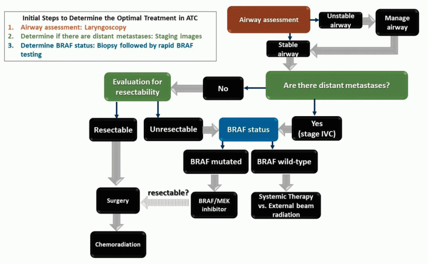
  • The most important consideration in the evaluation of patients with suspected ATC is to complete the assessment quickly with a focus on establishing disease burden and the status of the airway, as tumors often grow rapidly.
  • Diagnosis is confirmed by fine-needle aspiration (FNA) biopsy:
    • Revealing characteristic giant and multinucleated cells.
  • Differential diagnoses on FNA can include:
    • Poorly differentiated cancer
    • Lymphoma
    • Medullary carcinoma
    • Direct extension from a laryngeal carcinoma
    • Primary thyroid squamous cell carcinoma
    • Other metastatic carcinomas
    • Melanoma.
  • Immunohistochemistry is often needed to distinguish between these diagnostic possibilities:
    • ATCs often lack markers of thyroid and epithelial differentiation:
      • Such as thyroid transcription factor-1 (TTF-1) and thyroglobulin (Tg)
    •  ATC is positive for p53 (50% to 80% of the cases).
  • Core or incisional biopsy is occasionally needed to confirm the diagnosis, especially when there is predominantly necrotic material on the FNA.
  • Hyperthyroidism, hypocalcemia, and leucocytosis have been reported in ATC patients:
    • Therefore, laboratory evaluation should include a CBC, electrolytes, creatinine, liver profile, coagulation factors, and thyroid function tests.
    • Measurement of albumin and prealbumin levels provides an assessment of nutrition levels.
  • Anatomic imaging with contrast-enhanced neck CT or MRI helps to evaluate the extent of locoregional disease.
    • Neck ultrasonography is also used for this purpose. 
  • In patients with symptoms suggestive of advanced locoregional spread,:
    • Esophagoscopy and/or bronchoscopy can assess for esophageal or airway involvement.
  • Vocal cord paralysis is common in these patients and hence laryngoscopy in indicated:
    • It also allows for assessment of direct laryngeal or subglottic involvement.
  • PET-CT is useful to assess the extent of distant disease and may be more accurate than conventional neck, chest, abdomen, and pelvis CT.
  • Preoperative radiologic imaging and diagnostic biopsies of suspected tumors at distant sites should be conducted expeditiously, so as to not delay therapeutic interventions.

Staging

  • Due to its inherent aggressive nature:
    • ATC is automatically classified as stage IV (A, B, or C) at presentation.
  • According to AJCC 7th edition staging criteria:
    • T4a referred to ATC limited to the thyroid
    • T4b referred to tumors with gross extrathyroidal extension.
  • The AJCC 8th edition has changed the T classification so that rather than automatically being T4 disease, anaplastic cancers will now follow the same T definitions as differentiated thyroid cancer:
    • Intrathyroidal disease (T1 to T3a) is stage IVA
    • Gross extrathyroidal extension or cervical lymph node metastases is stage IVB,
    • Distant metastases are stage IVC.
  • Approximately:
    • 10% of patients with ATC present with only an intrathyroidal tumor
    • 40% have extrathyroidal invasion and/or lymph node disease.
    • 50% of patients present with diffusely metastatic disease.

Rodrigo Arrangoiz MS, MD, FACS cirujano de tumores de cabeza y cuello / cirugia endocrina miembro de Sociedad Quirúrgica S.C. experto en el manejo del cáncer de tiroides.

  • Cumple con los requisitos determinados por el Dr. Ashok Saha para realizar cirugía de tiroides de manera efectiva y segura:


Rodrigo Arrangoiz MS, MD, FACS es miembro de la American Thyroid Association:

2019 membership certificate arrangoiz, rodrigo2019 membership certificate arrangoiz, rodrigo

Entrenamiento:

  • Cirugia general y gastrointestinal:

• Michigan State University:

• 2004 al 2010image-48

• Cirugia oncológica / tumores de cabeza y cuello / cirugia endocrina:

• Fox Chase Cancer Center (Filadelfia):

• 2010 al 2012

image-39

• Maestria en ciencias (Clinical research for healthprofessionals):

• Drexel University (Filadelfia):

• 2010 al 2012image-50

• Cirugia de tumores de cabeza y cuello / cirugiaendocrina

• IFHNOS / Memorial Sloan Kettering Cancer Center:

• 2014 al 2016

image-51

http://www.sociedadquirurgica.com

http://www.hiperparatiroidismo.info

http://www.cirugiatiroides.com

#Arrangoiz

#CirugiadeTumoresdeCabezayCuello

#CirugiaEndocrina

#CirugiaOncologica

#HeadandNeckSurgery

#EndocrineSurgery

#SurgicalOncology

Anaplastic Thyroid Carcinoma (ATC)

anaplastic thyroid cancer

Etiology and Pathogenesis

  • Anaplastic thyroid carcinoma (ATC) accounts for 1.7% of all thyroid malignancies in the United States. 
  • Women are more commonly affected than men.
  • The majority of tumors are present in the seventh and eighth decades of life.
  • It has a very poor prognosis:
    • Median survival of 5 months.
    • 1-year survival of 20%.

8fb25e2b-e511-4643-a84b-fe9b1b6180fd_figure3

  • ATC is typically thought to originate due to dedifferentiation of more differentiated thyroid cancers of follicular cell origin:
    • Up to 80% of ATCs occur in patients with preexisting goiters:
      • The process of dedifferentiation is complex and often involves changes in various chromosomal regions affecting cell cycle and other signal transduction pathways:
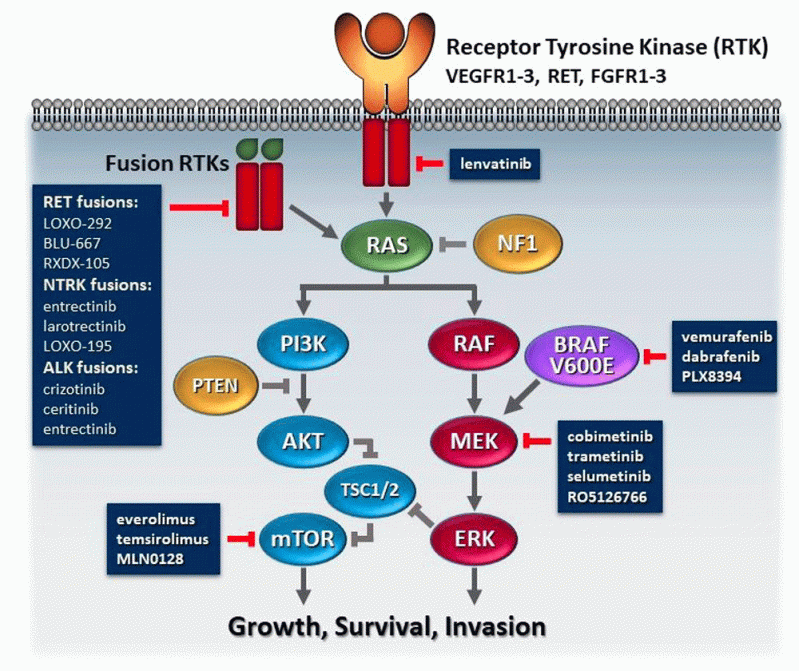
        • Several genes are known to be mutated in ATCs and include:
          • p53 (50% to 80%)
          • BRAF (20% to 40%)
          • RAS (20% to 40%)
          • PI3KCA (10% to 20%)
          • PTEN (5% to 15%)
          • AKT1 (5% to 10%)
          • CTNNB1 (5% to 60%). 

8fb25e2b-e511-4643-a84b-fe9b1b6180fd_figure2

  • Although some gene mutations are also commonly found in differentiated thyroid cancers (BRAF and RAS):
    • p53 tumor suppressor gene mutations are almost exclusively found in ATC.
      • Suggesting that they occur late in the dedifferentiation process.
  • BRAF V600E mutations lead to constitutive activation of the MAPK pathway:
    • Resulting in tumor growth and reduction in the expression of the sodium-iodide symporter, which correlates with resistance to radioactive iodine (RAI) therapy.
  • PIK3CA mutations result in aberrant activation of the PI3K/Akt/mTOR pathway and are found in more than 50% of ATCs and lead to increased growth.
  • AKT1 (5% to 10%) mutations in particular also lead to resistance to conventional chemotherapeutic agents:
    • PTEN mutations also affect this pathway
  • CTTNB1 encodes β-catenin:
    • A key member of the Wnt signaling pathway:
      • Causes tumor progression.
  • Two ATC-specific mutations in the anaplastic lymphoma kinase gene lead to dual activation of the MAPK and PI3K/Akt pathways:
    • Which leads to multilayer and anchorage-independent tumor growth.
  • Several microRNAs have also been reported to be dysregulated in ATC:
    • These include both upregulation (miRNA 222, 221, 146B, 106, 17 to 92) and downregulation (miRNA 618, 138, 125B, 30d, 26a)


Rodrigo Arrangoiz MS, MD, FACS cirujano de tumores de cabeza y cuello / cirugia endocrina miembro de Sociedad Quirúrgica S.C. experto en el manejo del cáncer de tiroides.

  • Cumple con los requisitos determinados por el Dr. Ashok Saha para realizar cirugía de tiroides de manera efectiva y segura:

Entrenamiento:

  • Cirugia general y gastrointestinal:

• Michigan State University:

• 2004 al 2010image-48

• Cirugia oncológica / tumores de cabeza y cuello / cirugia endocrina:

• Fox Chase Cancer Center (Filadelfia):

• 2010 al 2012

image-39

• Maestria en ciencias (Clinical research for healthprofessionals):

• Drexel University (Filadelfia):

• 2010 al 2012image-50

• Cirugia de tumores de cabeza y cuello / cirugiaendocrina

• IFHNOS / Memorial Sloan Kettering Cancer Center:

• 2014 al 2016

image-51

http://www.sociedadquirurgica.com

http://www.hiperparatiroidismo.info

http://www.cirugiatiroides.com

#Arrangoiz

#CirugiadeTumoresdeCabezayCuello

#CirugiaEndocrina

#CirugiaOncologica

#HeadandNeckSurgery

#EndocrineSurgery

#SurgicalOncology

Oncocytoma of Salivary Gland Origen

  •  Oncocytomas typically occur in older persons and are relatively rare:
    • Its a benign tumor composed of oncocytes:
      • It is also called oxyphilic adenoma
    • Represents 0.1% to 2% of salivary gland neoplasms:
      • They are more frequent than oncocytosis and oncocytic carcinoma:
        • They are classified according to the World Health Organization (WHO) classification, and histologically there are three distinct types:  oncocytosis, oncocytoma and oncocytic carcinoma


    • These tumors are characterized by a high mitochondrial content:
      • Which accounts for fluorodeoxyglucose avidity on positron emission tomography scans, similar to Warthin’s tumors. 
  • Clinical Features of Oncocytomas:
    • Usually there are identified in the parotid gland (78% to 84% cases), but the submandibular gland or minor salivary glands can be involved.
      • This tumor might be seen in other organs such as:
        • The nasal and thoracic cavities, ovaries, breast, kidney, thyroid, parathyroid, pituitary, larynx and pancreas
  • They often present as solitary slow growing painless masses:
    • Which are firm, multilobulated and mobile entities upon clinical examination.


  • They are diagnosed between the sixth through the eighth decades of life with a slightly higher incidence in women:
    • Mean age at presentation is 60 years.
    • 20% associated with radiation therapy or radiation exposure.
    • Rarely bilateral.
    • May be multiple.
    • Rarely synchronous with Warthin tumor or carcinoma ex pleomorphic adenoma
    • May occur in trisomy 7 or in BHD syndrome
  • Radiology Description:
    • Computed tomography (CT) and magnetic resonance imaging (MRI) are the image modalities of choice,

      • Specific MRI features:
        • T1 hypointense
        • Isointense to normal gland on fat saturated T2 and postcontrast T1
      • CT features:
        • Enhancing tumor and nonenhancing cystic component

f2.large

  • Pathology:
    • Well circumscribed with fibrous capsule, solid, tan-red-brown, lobulated, often small, may have cystic spaces
    • Microscopic description:
      • Eosinophilic or clear cell (glycogen) with sheets, trabeculae, acini or follicular patterns of monotonous large polygonal cells with well defined cell borders, deeply eosinophilic, granular cytoplasm, small round nuclei:
        • Oncocytic cells are thought to originate from the transformation of epithelial cells of salivary gland ducts or acini

      • Vascular stroma, may have clear cell change, background of oncocytic nodular hyperplasia, psammoma bodies, tyrosine rich crystals
      • No mitotic figures, no elastosis

unknownhistomorphological-features-of-clear-cell-variant-of-oncocytoma-of-the-parotid-gland-theoncocytoma+gross+histology+encapsulated

  • Complete surgical excision with a superficial parotidectomy is the treatments of choice.

Rodrigo Arrangoiz MS, MD, FACS a head and neck surgeon / surgical oncologist and is a member of Sociedad Quirúrgica S.C at the America British CowdrayMedical Center in Mexico City:

  • He is an expert in the management of salivary gland neoplasms:

    • If you have any questions about salivary gland neoplasms  please fill free to ask Dr. Arrangoiz


Training:

• General surgery:

• Michigan State University:

• 2005 al 2010

• Surgical Oncology / Head and Neck Surgery / Endocrine Surgery:

• Fox Chase Cancer Center (Filadelfia):

• 2010 al 2012

• Masters in Science (Clinical research for health professionals):

• Drexel University (Filadelfia):

• 2010 al 2012

• Surgical Oncology / Head and Neck Surgery / Endocrine Surgery:

• IFHNOS / Memorial Sloan Kettering Cancer Center:

• 2014 al 2016

#Arrangoiz

#HeadandNeckSurgeon

#SurgicalOncologist

#Surgeon

#CancerSurgeon

#CirujanodeCabezayCuello

#CirujanoOncologo
http://www.sociedadquirurgica.com

The American Thyroid Association (ATA) Guidelines for Assessment of Thyroid Nodules

The American Thyroid Association (ATA) guidelines for assessment of thyroid nodules are meant to improve inter- and intra-reader consistency during assessment of thyroid nodules on ultrasound, and to facilitate communication with referring endocrinologists.

  • The 2015 guidelines stress the importance of the ultrasonographic pattern of the nodule for risk stratification:

    • This, as well as the size of the nodule, are the two main criteria for FNA.

Initial evaluation

  • Serum thyrotropin (TSH) should be ordered

    • If the TSH is below normal limits, thyroid scintigraphy should be pursued

  • An incidental finding of focal FDG uptake in a greater than 1 cm thyroid nodule is concerning and FNA is warranted:

    • If less than 1 cm the nodule may be monitored similarly to a sub-centimeter thyroid nodule with a high risk sonographic pattern

    • If the thyroid demonstrates diffuse uptake compatible with chronic lymphocytic thyroiditis, further imaging or FNA is not warranted

On a thyroid ultrasound, a nodule is classified into one of five categories:

  • Benign pattern (0% risk):

    • No biopsy

  • Very low suspicion pattern (< 3% risk):

    • Biopsy if ≥ 2 cm (or ultrasound observation)

  • Low suspicion pattern (5% to 10% risk):

    • Biopsy if ≥ 1.5 cm

  • Intermediate suspicion pattern (10% to 20% risk):

    • Biopsy if ≥ 1 cm

  • High suspicion pattern (> 70% to 90% risk):

    • Biopsy if ≥ 1 cm

  • Completely cystic nodules with well-defined walls

  • Spongiform nodules and nodules with interspersed cystic spaces, without any of the features in more suspicious patterns

  • Isoechoic or hyperechoic nodule

  • Partially cystic nodule with a peripheral solid component

  • None of the following features:

    • Microcalcifications (see other points below)

    • Irregular margins

    • Extra thyroidal extension

    • Taller than wide

  • Hypoechoic solid nodule with smooth margins

  • None of the following features:

    • Microcalcifications (see other points below)

    • Irregular margins

    • Extra thyroidal extension

    • Taller than wide

  • Solid hypoechoic nodule (or solid hypoechoic component of a partially cystic nodule), with at least one of these features:

    • Microcalcifications

    • Irregular margins (infiltrative, microlobulated)

    • Extrathyroidal extension

    • Taller than wide

    • Rim calcifications with an extrusive soft tissue component

    • Lymphadenopathy

  • Dystrophic calcifications other than microcalcifications (e.g. coarse macrocalcification, rim calcifications) increase risk, but to a lesser degree than microcalcifications

  • A survey of cervical lymph nodes should be performed in all neck ultrasound studies

Rodrigo Arrangoiz MS, MD, FACS cirujano de tumores de cabeza y cuello / cirugia endocrina miembro de Sociedad Quirúrgica S.C. experto en el manejo del cáncer de tiroides.

Cumple con los requisitos determinados por el Dr. Saha para realizar cirugía de tiroides de manera efectiva y segura:

  • Cirugia general y gastrointestinal:

• Michigan State University:

• 2004 al 2010image-48

• Cirugia oncológica / tumores de cabeza y cuello / cirugia endocrina:

• Fox Chase Cancer Center (Filadelfia):

• 2010 al 2012

image-39

• Maestria en ciencias (Clinical research for healthprofessionals):

• Drexel University (Filadelfia):

• 2010 al 2012image-50

• Cirugia de tumores de cabeza y cuello / cirugiaendocrina

• IFHNOS / Memorial Sloan Kettering Cancer Center:

• 2014 al 2016

image-51

http://www.sociedadquirurgica.com

http://www.hiperparatiroidismo.info

http://www.cirugiatiroides.com

#Arrangoiz

#CirugiadeTumoresdeCabezayCuello

#CirugiaEndocrina

#CirugiaOncologica

#HeadandNeckSurgery

#EndocrineSurgery

#SurgicalOncology

Genetics in Breast Cancer

unknown

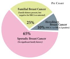
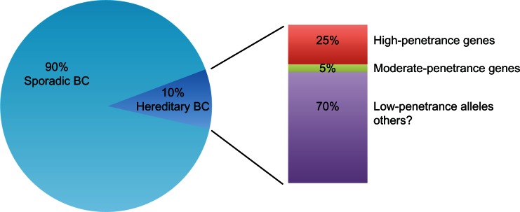
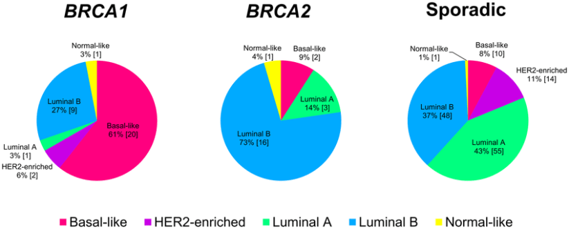
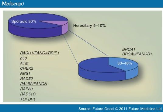
  • When talking about genetics and breast cancer, physicians typically first discuss risk factors associated with mutations in the tumor-suppressor genes BRCA1 and BRCA2, as these mutations represent most of the hereditary types of breast cancer.

  • Among families with a history of multiple breast cancers, 7% have BRCA-1 mutations.

  • For families with a history of breast and ovarian cancers, BRCA-1 mutations are present in 40%.

  • BRCA mutations greatly increase a patient’s risk of developing ovarian cancer and are associated with a lifetime risk of 40%.

genetic-susceptibility-in-hereditary-breast-cancer-up-to-10-of-all-breast-cancers-are.png

  • Similarly, BRCA2 mutations confer an increased risk of both breast and ovarian cancer:

    • In families at high risk for these cancers, 10% to 20% carry a BRCA-2 mutation.

association-between-hereditary-breast-cancers-and-molecular-subtypes-distribution-of

  • In addition to BRCA-1 and BRCA-2 gene mutations, other genes and syndromes are associated with an increased risk for breast cancer:

    • The HER2 gene, also referred to as neu or ERBB2 (receptor tyrosine-protein kinase erbB-2), codes for a transmembrane tyrosine kinase receptor, a member of the epidermal growth factor receptor (EGFR) family:

      • When activated, this receptor family causes an increase in activity of molecular pathways associated with tumor growth and progression:

        • Gene amplification at chromosome 17q (Chr17q) involves the HER gene and is observed in about 10% to 15% of breast cancers:

          • This percentage is lower than the rate (20% to 25%) reported in historical studies, in which different diagnostic thresholds were used:

            • Chr17 polysomy could also have led to discordant interpretations between high signals due to polysomy and those due to an absolute increase of HER2 gene copy number.

            • HER2 amplification is an adverse prognostic factor associated with:

              • An increased metastatic potential and reduced survival and is a predictive factor of response to anti-HER2 therapy.

    • Other genes at this locus on CHr17q (MED1, STARD3, GRB7, THRA, RARA, IGFBP4, CCR7, KRT20, KRT19, and GAST) might also be amplified and could play functional roles in breast cancer development and progression.

738249-fig1

  • A 2007 study showed that US women of Ashkenazi Jewish descent have the highest prevalence of the BRCA1 mutation, at 8.3%.

  • The variable prevalence of BRCA1 mutation in other racial and ethnic groups in the United States is as follows:

    • Hispanic American women: 3.5%

    • Non-Hispanic American white women: 2.2%

    • African American women: 1.3%

    • Asian American women: 0.5%

  • Independent of BRCA status, Ashkenazi Jewish women have double the baseline risk for breast cancer:

  • Li-Fraumeni syndrome (LFS) is a rare genetic cancer syndrome associated with mutations of tumor-suppressor gene TP53:

    • Which is inherited in an autosomal dominant pattern.

    • Together, male and female patients with LFS have a 50% risk of developing cancer by age 40 years and a 90% risk by age 60 years.

    • Female patients with LFS have almost a 100% lifetime risk of developing cancer:

      • They also have a 56% risk of developing breast cancer by age 45 years, and a more than 90% risk by age 60 years:

        • Most breast cancers are diagnosed in patients younger than age 40 years.

      • Patients with LFS also have rates of bilateral breast cancer approaching 25%.

    • LFS is also associated with cancers of other sites:

      • Including brain cancer, leukemia, soft-tissue sarcomas, and adrenocortical cancer.

  • Cowden disease (CD) is a rare genetic syndrome caused by mutations in the PTEN gene (phosphatase and tensin homolog):

    • Patients with CD have a lifetime risk of developing breast cancer that ranges from 25% to 50%.

    • CD is commonly associated with several benign breast abnormalities,:

      • Fibroadenomas, fibrocystic breast disease, ductal epithelial hyperplasia, and nipple malformations.

    • This syndrome is also associated with:

      • Intestinal hamartoma, cutaneous lesions, thyroid cancer (usually follicular thyroid carcinoma), ovarian cancer, colon cancer.

  • In addition to their link to breast cancer, BRCA-2 mutations are associated with an increased risk for the following types of cancer:

    • Malignant melanoma:

      • Elevated risk for melanomas of both the skin and eye

    • Ovarian (10% lifetime risk):

    • Prostate – 20% risk

    • Pancreatic -7%, or higher if there is a family history of pancreatic cancer

    • Gallbladder and bile duct

    • Stomach

  • While a small percentage of patients with early-onset breast cancer have a BRCA2 mutation:

    • It is present in 10% to 20% of families at high risk for breast cancer and ovarian cancer.

    • BRCA2 mutations are associated with an increased incidence of:

      • High-grade estrogen-receptor-positive (ER+), progesterone-receptor positive (PR+), HER2-negative breast cancers (luminal type).

Rodrigo Arrangoiz MS, MD, FACS a surgical oncologist and is a member of Sociedad Quirúrgica S.C at the America British Cowdray Medical Center in Mexico City:

  • He is an expert in the management of breast cancer.

    • If you have any questions about hereditary breast cancer please fill free to contact Dr. Arrangoiz.

Training:

• General surgery:

• Michigan State University:

• 2004 al 2010

• Surgical Oncology / Head and Neck Surgery / Endocrine Surgery:

• Fox Chase Cancer Center (Filadelfia):

• 2010 al 2012

• Masters in Science (Clinical research for health professionals):

• Drexel University (Filadelfia):

• 2010 al 2012

• Surgical Oncology / Head and Neck Surgery / Endocrine Surgery:

• IFHNOS / Memorial Sloan Kettering Cancer Center:

• 2014 al 2016

Pathology, Immunohistochemistry, and SLN Biopsy

  • The prognostic significance of sentinel lymph node (SLN) disease detected on immunohistochemistry (IHC) has been widely debated and results from two  studies indicate that SLN micrometastases detected by IHC staining are clinically insignificant and routine use of IHC can be abandoned.

    • In the ACOSOG Z0010 and National Surgical Adjuvant Breast and Bowel Protocol (NSABP) B-32 trials:

      • All patients were treated on the basis of H&E SLN stains only.

        • To control for treatment bias, clinicians and patients were blinded to the results of IHC staining.

      • In ACOSOG Z0010:

        • A prospective observational study of SLN biopsy

        • Occult metastases were found by IHC in 8.9% of 3945 patients who were SLN-negative by H&E:

          • 5-year survival was not different between those patients who were H&E negative and IHC negative, and those who were H&E negative and IHC positive:

            • 95.8% vs 95.1% (P = 0.53)

      • In NSABP B-32:

        • A prospective randomized study of SLN biopsy plus ALND vs SLN biopsy alone (with ALND limited to SLN-positive patients)

        • Occult metastases were found by IHC in 15.9% of 3887 H&E-negative patients:

          • Although overall, disease-free and distant disease–free survival were significantly worse for IHC-positive than for IHC-negative patients:

            • The absolute difference in overall survival was only 1.2%:

              • 94.6% vs 95.8% (P = 0.03).

  • The results of the ACOSOG Z0010 and NSABP B-32 trials suggest that SLN micrometastases found only by IHC are clinically insignificant and that IHC staining of SLNs is unnecessary:

    • Therefore, routine use of IHC staining of SLNs is not recommended and should be limited to selective use at the discretion of the pathologist.

    • In addition, intraoperative frozen-section analysis of the SLN can be avoided if clinical suspicion of nodal involvement is low and the patient otherwise meets the entry criteria for the Z-11 trial.

      • The data is fairly clear for the above recommendations.

Rodrigo Arrangoiz MS, MD, FACS a surgical oncologist and is a member of Sociedad Quirúrgica S.C at the America British CowdrayMedical Center in Mexico City:

  • He is an expert in the management of breast cancer

Training:

• General surgery:

• Michigan State University:

• 2004 al 2010

• Surgical Oncology / Head and Neck Surgery / Endocrine Surgery:

• Fox Chase Cancer Center (Filadelfia):

• 2010 al 2012

• Masters in Science (Clinical research for health professionals):

• Drexel University (Filadelfia):

• 2010 al 2012

• Surgical Oncology / Head and Neck Surgery / Endocrine Surgery:

• IFHNOS / Memorial Sloan Kettering Cancer Center:

• 2014 al 2016

Management of the Axilla in Patients With Invasive Breast Cancer I

showimage.ashx

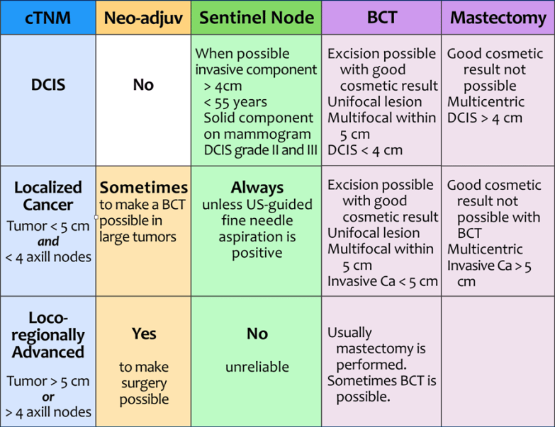
  • Sentinel lymph node biopsy (SLNB) has replaced axillary lymph node dissection (ALND) for the staging of clinically node-negative breast cancer patients, demonstrating equivalent survival to ALND for lymph node–negative patients while resulting in reduced morbidity.

    • For the majority of patients with pathologically positive SLNs, completion ALND was recommended by the American Society of Clinical Oncology Guidelines and the National Comprehensive Cancer Network (NCCN):

      • However, based on data from the American College of Surgeons Oncology Group (ACOSOG) Z0011 trial  these guidelines have changed:

        • ALND may be omitted in selected patients with 1 or 2 positive SLNs.


  • The ACOSOG Z0011 trial:

    • 813 patients

    • Clinical T1 to T2 node-negative tumors (N0) who were found to have hematoxylin and eosin (H&E)-positive SLNs were randomized to ALND vs no further axillary surgery:

      • Patients with palpable lymph nodes or clinical T3 tumors were not eligible for this study.

    • The protocol mandated the use of standard whole-breast radiation without an axillary field.

    • Patients with > 3 positive SLNs were excluded from the study.

    • The trial was closed early due to poor accrual with an enrollment of only 47% of the targeted 1900 patients.

    • It still showed equivalent results between the two treatment arms for loco-regional failure and survival.:

      • At 6.3 years’ follow-up, no differences were found between the two groups in the rates of:

        • Axillary recurrence:

          • 0.5% vs 0.9%

        • In-breast recurrence:

          • 3.6% vs 1.9%

        • Overall locoregional recurrence:

          • 4.1% vs 2.8% (P = 0.53)

      • Disease-free were similar:

        • 82.2% vs 83.8%

      • Overall survival were similar:

        • 91.9% vs 92.5%

    • The majority of women in this trial were:

      • Older than 50 years (64%)

      • Had clinical T1 tumors (68%)

      • Had ER-positive tumors (77%)

      • Had only one positive SLN (60%),

      • Received whole-breast radiation (89%)

      • Received systemic therapy (96%: 58% adjuvant chemotherapy and 46% adjuvant hormonal therapy).

    • Forty percent of patients had micrometastases or isolated tumor cells and 60% had macrometastases in the sentinel nodes.

    • Additional positive axillary nodes were found in 27.3% of the ALND patients.

    • This study excluded patients:

      • Undergoing mastectomy

      • Patients receiving neoadjuvant chemotherapy.


  • The results from ACOSOG Z0011 were practice changing and ALND is no longer the standard of care for patients who meet all of the following criteria:

    • T1 to T2 tumors

    • One to two positive SLNs without extracapsular extension


    • Patient acceptance and completion of whole-breast radiation therapy without extended fields of therapy

    • Patient acceptance and completion of adjuvant therapy (hormonal, cytotoxic, or both)


  • The results from ACOSOG Z0011 are not directly applicable to patients who:

    • Have T3 tumors

    • Have more than 2 positive nodes

    • Are undergoing mastectomy

    • Are undergoing partial breast radiation

    • Have been identified as having matted axillary nodes or preoperative palpable nodes

    • Are receiving neoadjuvant chemotherapy

a58284a1f10a0e__tab-staging-1

Rodrigo Arrangoiz MS, MD, FACS a surgical oncologist and is a member of Sociedad Quirúrgica S.C at the America British CowdrayMedical Center in Mexico City:

  • He is an expert in the management of breast cancer:

    • If you have any questions about breast cancer AND the management of the axilla in patients with invasive cancer Dr. Arrangoiz

Training:

• General surgery:

• Michigan State University:

• 2004 al 2010

• Surgical Oncology / Head and Neck Surgery / Endocrine Surgery:

• Fox Chase Cancer Center (Filadelfia):

• 2010 al 2012

• Masters in Science (Clinical research for health professionals):

• Drexel University (Filadelfia):

• 2010 al 2012

• Surgical Oncology / Head and Neck Surgery / Endocrine Surgery:

• IFHNOS / Memorial Sloan Kettering Cancer Center:

• 2014 al 2016

Thyroid Nodule Size Irrelevant When It Comes to Malignancy

Large thyroid nodules in asymptomatic patients show malignancy and false-negative rates on fine needle aspiration (FNA) that are similar to those of small nodules, suggesting that nodule size alone should not be a reason for surgical resection, according to findings from a new meta-analysis.

      • “Based on these data, surgical resection of large cytologically benign nodules is not recommended in the absence of other clinical indications,” for thyroidectomy, say Nicole A. Cipriani, MD, assistant professor of pathology, University of Chicago, Illinois, and coauthors of the study, recently published in Thyroid.

With conflicting data on whether larger nodule size is associated with a greater risk of malignancy and false-negative FNA rates, some surgeons choose to err on the side of caution and resect larger thyroid nodules, regardless of FNA results.

But the issue is controversial. Surgery itself is associated with potential risks, including increased physical and psychological morbidity, as well as a heavier financial burden, compared with the alternative approach of close clinical follow-up of cytologically benign nodules.

To assess the existing evidence, Cipriani and a team of multiple reviewers analyzed findings from 35 studies that stratified thyroid nodules by size and included data on benign and other cytology.

      • The analysis included more than 20,000 nodules, of which more than 7000 had benign cytology.
      • Of 21 datasets that allowed for comparison of malignancy rates by thyroid nodule size:
          • 81% showed malignancy rates of larger nodules to be similar to or lower than rates of smaller nodules:
              • The overall malignancy rate of large nodules (3 cm or greater) was 13.1% compared with 19.6% among nodules 3 cm or smaller (odds ratio [OR], 0.72).

              • And in studies stratifying nodules as 4 cm or greater, the malignancy rate was similar, at 20.9%, to that of nodules 4 cm or smaller, at 19.9% (OR, 0.85).

False-negative FNA rates according to nodule size were available in 17 datasets, with only one study linking higher false-negative rates with larger nodules.

      • In those studies, the overall false-negative rate of nodules 3 cm or greater was 7.2% compared with 5.7% of those smaller than 3 cm (OR, 1.47; CI, 0.80 – 2.69).
      • And the overall false-negative rate of nodules 4 cm or greater was only slightly higher than that of nodules smaller than 4 cm, at 6.7% vs 4.5% (OR, 1.38; CI, 1.06 – 1.80), which was again not statistically significant.

  • Although some studies have attempted to correlate larger thyroid nodule size with the risk of malignancy or false-negative FNA, the overall inconsistency of evidence is reflected in the American Thyroid Association (ATA) guidelines, which state that:
        • “Based on the evidence, it is still unclear if patients with thyroid nodules 4 cm or larger and benign cytology carry a higher risk of malignancy and should be managed differently than those with smaller nodules.”

  • It is surprising that multiple authors still recommend surgical resection of large cytologically benign nodules due to the perceived (but not actual) high false-negative rates:
      • Based on the data provided by Cipriani et al I have (Rodrigo Arrangoiz) stopped recommending thyroidectomy for large cytologically being nodules based on size alone.

              • The study by Cipriani et al showed no significant differences in the rate of malignancies between nodules smaller than 4 cm (140 of 546; 26.5%) compared with those 4 cm or larger (33 of 106; 31.1%).
              • It also indicated that most malignancies among indeterminate thyroid nodules were low-risk regardless of tumor size, with the researchers concluding size should not be driving more aggressive diagnostic surgeries for cytologically indeterminate thyroid nodules.

ATA guidelines recommend resection primarily for nodules assessed on FNA to be malignant:

      • But there are some exceptions for cytologically benign nodules, including those that are symptomatic, growing in size, of cosmetic concern, or substernal, with symptoms of concern including difficulty swallowing or breathing, or tracheal deviation.
            • Close follow-up (including repeat ultrasound and/or FNA) can identify patients who ultimately require resection, and those with indolent disease may avoid surgery for cytologically benign but large thyroid
  • Rodrigo Arrangoiz MS, MD, FACS a head and neck surgeon / endocrine surgery specialist member of Sociedad Quirúrgica S.C at the America British Cowdray Medical Center in Mexico City:
                • He is an expert in the management of thyroid cancer:

                • If you have any questions about indications for surgery for thyroid nodules Dr. Arrangoiz:

    Training:

    • General surgery:

    • Michigan State University:

    • 2004 al 2010

    • Surgical Oncology / Head and Neck Surgery / Endocrine Surgery:

    • Fox Chase Cancer Center (Filadelfia):

    • 2010 al 2012

    • Masters in Science (Clinical research for health professionals):

    • Drexel University (Filadelfia):

    • 2010 al 2012

    • Surgical Oncology / Head and Neck Surgery / Endocrine Surgery:

    • IFHNOS / Memorial Sloan Kettering Cancer Center:

    • 2014 al 2016

    http://www.cirugiatiroides.com