COMPLICATIONS OF NASOPHARYNGEAL CARCINOMA (NPC) TREAMENT WITH RADIATION OR CHEMORADIOTHERAPY

  • Although treatment of NPC with radiotherapy or concurrent chemoradiation yields good response:
    • There are many complications:
      • That can adversely affect:
        • The quality of life of these patients
  • Complications of NPC treatment:
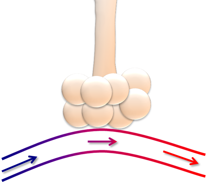
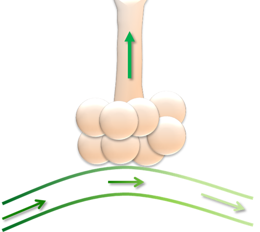
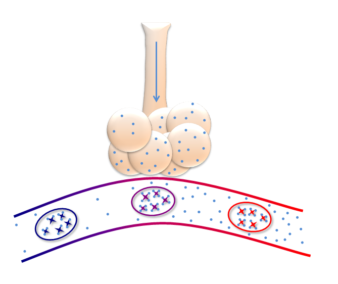
    • Xerostomia:
      • Is almost universal:
        • After conventional radiotherapy:
          • This leads to:
            • Dry mouth
            • Poor oral hygiene
            • Dental caries
    • Hearing impairment:
      • Is also frequently seen:
        • May be related to the combined effects of:
          • Direct radiation insult to the hearing apparatus
          • Persistent disturbance of Eustachian tube function
          • Chemotherapy-induced ototoxicity
    • Soft tissue fibrosis following radiotherapy:
      • May lead to:
        • Restriction of:
          • Neck movement or mouth opening:
            • Often accompanied by discomfort
    • Cranial nerve palsies:
      • Are usually due to:
        • Incomplete healing of damage caused by the tumor:
          • Although cranial nerves (especially CN IX, X, XI and XII):
            • Can also be damaged by radiation
    • Dysphagia:
      • Can be due to:
        • Cranial nerve palsies or
        • Pharyngeal stricture
    • Hormonal insufficiency:
      • Can develop due to:
        • Damage to:
          • The hypothalamic-pituitary axis or
          • End organs such as:
            • The thyroid gland
    • Carotid artery stenosis:
      • Can develop following neck irradiation:
        • And may result in cerebral ischaemia
    • The more serious sequelae are:
      • Damage of higher functions:
        • That lead to memory loss
        • Cognitive dysfunction, and
        • Neuropsychological dysfunction:
          • This can occur with or without radiological evidence:
            • Of temporal lobe necrosis

#Arrangoiz #Teacher #CancerSurgeon #HeadandNeckSurgeon #HeadandNeckCancer #SurgicalOncologist

Nasopharyngeal Papillary Adenocarcinoma

  • Nasopharyngeal Papillary Adenocarcinoma (NPAC):
    • Epidemiology:
      • Patients ranged in age from 11 to 64 years:
        • Median = 37 years
      • The ratio of male to female patients:
        • Is 5:4
    • They most frequently present with:
      • Airway obstruction
    • Primary NPACs can be classified into two main categories with different morphological features and clinical behavior:
      • The conventional or mucosal surface origin type
      • The salivary gland type:
        • Two main pathological subtypes:
          • Adenoid cystic carcinoma and
          • Mucoepidermoid carcinoma
    • Histomorphologic evaluation show:
      • An invasive carcinoma:
        • With papillary and glandular features:
          • The tumor display transition from normal surface epithelium to neoplasm
      • Cytologically this tumors are:
        • Composed of moderately pleomorphic, columnar, and pseudostratified cells with eosinophilic cytoplasm:
          • The nuclei are round to oval
          • They have vesicular or optically clear chromatin
        • Histochemical analysis confirms:
          • The production of epithelial mucin
        • Immunohistochemical study demonstrated:
          • Diffuse keratin and epithelial membrane antigen reactivity
          • Focal carcinoembryonic antigen reactivity
        • No immunoreactivity was seen with:
          • S-100 protein
          • Glial fibrillary acidic protein, or
          • Thyroglobulin:
            • Taken together:
              • These findings indicate an origen:
                • From the nasopharyngeal surface epithelium
              • Help to differentiate these tumors from:
                • Seromucous gland lesions and
                • Metastatic papillary thyroid carcinoma
      • The biologic potential:
        • Is that of a low-grade malignant tumor
      • Simple and complete surgical excision:
        • Is the treatment of choice

#Arrangoiz #HeadandNeckSurgeon #CancerSurgeon #NasopharyngealCancer #Surgeon #Teacher

The NSABP B-14 and the NSABP B-20 Trial were used for the development and validation of the 21-gene reverse transcription polymerase chain reaction (RT-PCR) recurrence score assay to predict recurrence of estrogen receptor (ER)-positive, node-negative breast cancer.

  • A multistep approach was used for the synthesis and development of the:
    • Oncotype DX 21-gene RT-PCR assay
  • The developers selected:
    • 250 candidate genes from:
      • Published literature
      • Genomic databases, and
      • Experiments based on:
        • DNA array:
          • Performed on fresh frozen tissue
  • Data were then analyzed from:
    • Three independent studies:
      • Of breast cancer patients:
        • Involving a total of 447 patients:
          • Including the tamoxifen-only group of:
            • The NSABP B-20
    • This data was used to test the relationship between:
      • Expression of the 250 genes:
        • And the recurrence of breast cancer
    • These data was used to select:
      • The final 21-gene panel and
      • To develop an algorithm:
        • To determine the recurrence score (RS):
          • For each tumor sample
  • Finally:
    • This assay was validated by:
      • Calculating RS on cancer cells contained in paraffin blocks:
        • From patients in the NSABP B-14 trial
    • In multivariable Cox model:
      • The RS provided significant predictive power:
        • For recurrence:
          • That was independent of:
            • Age and
            • Tumor size
      • The RS was also:
        • Predictive of:
          • Overall survival (OS) and
            • Could be used as a continuous function to:
              • Predict distant recurrence in individual patients
  • The NSABP B-14 trial:
    • Was designed to assess the effect of tamoxifen on disease-free survival and OS:
      • In patients with breast cancer that are:
        • ER-positive
        • Lymph node-negative
  • The NSABP B-20 trial:
    • Evaluated the benefit of two chemotherapy regimens:
      • Methotrexate and fluorouracil followed by leucovorin vs
      • Cyclophosphamide, methotrexate, and fluorouracil) and
      • Tamoxifen over tamoxifen alone:
        • In patients with:
          • ER-positive
          • Lymph node-negative breast cancer
  • Further evaluation by the Oncotype DX, 21-gene RT-PCR assay:
    • Of 651 tissue blocks from the NSABP B-20 trial:
      • Showed additional clinical value of this score:
        • For weighing the benefit of chemotherapy for different ER-positive, lymph-node-negative breast cancer patients:
          • Treated with surgery and tamoxifen
      • Patients with a high RS (≥31):
        • Had significant benefit from chemotherapy:
          • With a decrease in 10-year distant recurrence rate
      • While patients with a low RS (<18):
        • Had minimal benefit from chemotherapy
      • The results of patients with intermediate RS scores (18 to 30):
        • Were uncertain
      • The clinical utility of this assay to stratify patients into low- (RS less than 18) , intermediate- (RS 18 to 30), and high-risk (RS ≥31) groups:
        • Can be applied not only to the prediction of recurrence:
          • But also to decision making with regard to chemotherapy:
            • In patients with ER-positive, lymph-node-negative breast cancer treated with tamoxifen
  • The additional analysis of 895 samples:
    • From patients treated with tamoxifen in both the B-14 and B-20 trials:
      • 355 patients given placebo from B-14 and
      • 424 patients treated with chemotherapy and tamoxifen from B-20:
        • Has provided further evidence for the clinical use of the the Oncotype DX assay recurrence score
    • In addition to predicting chemotherapy benefit and distant recurrence:
      • These data have also shown significant correlation with:
        • Locoregional recurrence:
          • Based on RS
      • In the patients given tamoxifen:
        • 10-year estimates of locoregional recurrence were:
          • 4.3% for patients with a low RS (<18) and
          • 15.8% for patients with a high RS (>30)
            • This trend was also evident in the patients who received placebo or chemotherapy and tamoxifen
  • The NSABP B-17 trial:
    • Compared lumpectomy alone with lumpectomy plus breast radiation in:
      • 818 patients:
        • Who had localized ductal carcinoma in situ (DCIS)
  • The NSABP B-18 trial:
    • Evaluated if preoperative versus postoperative chemotherapy with:
      • Adriamycin and cyclophosphamide:
        • Was more effective in prolonging:
          • Disease-free survival and OS in patients with operable breast cancer
  • The NSABP B-24 trial:
    • Examined the benefit of tamoxifen after lumpectomy and radiotherapy in patients with DCIS in:
      • Decreasing the occurrence of cancer in both breasts
  • The NSABP B-27 trial:
    • Compared three chemotherapy regimens:
      • Preoperative adriamycin and cyclophosphamide alone versus
      • The addition of taxotere preoperatively or postoperatively and
        • The effect on survival
  • The NSABP B-35 trial:
    • Compared the use of tamoxifen versus anastrozole after lumpectomy and radiotherapy to treat ER- and progesterone-positive DCIS in both pre- and postmenopausal women:
      • With regard to prevention of breast cancer occurrences

REFERENCES

  1. Mamounas EP, Tang G, Fisher B, et al. Association between the 21-gene recurrence score assay and risk of locoregional recurrence in node-negative, estrogen receptor-positive breast cancer: results from NSABP B-14 and NSABP B-20. J Clin Onc. 2010;28:1677-1683.
  2. Paik S, Shak S, Tang G, et al. A multigene assay to predict recurrence of tamoxifen-treated, node-negative breast cancer. New Engl J Med. 2004;351:2817-2826.
  3. Paik S, Tang G, Shak S, et al. Gene expression and benefit of chemotherapy in women with node-negative, estrogen receptor-positive breast cancer. J Clin Oncol. 2006;24:3726-3734.

#Arrangoiz #BreastSurgeon #BreastCancer #CancerSurgeon #SurgicalOncologist #Teacher #Surgeon

Recently published thyroid-related articles!


ETV5 overexpression contributes to tumor growth and progression of thyroid cancer through PIK3CA. Meng D, Li Z, Ma X, Wu L, Fu L, Qin G. Life Sci. 2020 Apr 20:117693. PMID: 32325133 https://www.ncbi.nlm.nih.gov/pubmed/32325133

Mid-Term and Long-Term Impact of Permanent Hypoparathyroidism After Total Thyroidectomy. Ponce de León-Ballesteros G, Bonilla-Ramírez C, Hernández-Calderón FJ, Pantoja-Millán JP, Sierra-Salazar M, Velázquez-Fernández D, Herrera MF. World J Surg. 2020 Apr 22. PMID: 32322939 https://www.ncbi.nlm.nih.gov/pubmed/32322939

Prediction mode of more than 5 central lymph nodes metastases in clinically node-negative ipsilateral papillary thyroid carcinoma with tumor size 1 to 4 cm. Jin L, Sun HL, Zhou L, Xie L, Zhuang YY, Wang JB. Medicine (Baltimore). 2020 Apr;99(16):e19809. PMID: 32311998 https://www.ncbi.nlm.nih.gov/pubmed/32311998

Low-risk papillary thyroid microcarcinoma: Optimal management toward a more conservative approach. Ramundo V, Sponziello M, Falcone R, Verrienti A, Filetti S, Durante C, Grani G. J Surg Oncol. 2020 May;121(6):958-963. PMID: 32189352 https://www.ncbi.nlm.nih.gov/pubmed/32189352
American Association of Clinical Endocrinologists American Thyroid Association ThyCa, Inc. Endocrine Society Graves’ Disease and Thyroid Foundation

Gas Exchange

  • This diagram represents a normal cluster of alveoli with a normal capillary:
    • Delivering carbon dioxide (CO2) and picking up oxygen (O2)
A normal cluster of alveoli with a normal capillary, delivering carbon dioxide (CO2) and picking up oxygen (O2).
  • Carbon dioxide:
    • Travels dissolved in the blood
  • Approaching the alveolus:
    • The CO2:
      • Easily crosses through the blood:
        • Across the capillary wall, and into the alveolus
CO2 crosses through the blood, across the capillary wall, and into the alveolus.
  • Because CO2 crosses so readily into the alveolus from the serum:
    • Ventilation occurs readily:
      • This means that the major determinants of CO2 in the blood are:
        • The rate of production:
          • Increased with:
            • Elevated metabolic demand:
              • Such as in:
                • Sepsis
                • Exercise
        • The rate of elimination:
          • Largely determined by:
            • The minute ventilation:
              • Which is the amount of air moving through the lungs in one minute:
                • Quantified as:
                  • The tidal volume:
                    • Green shaded arrows and
                  • The respiratory rate:
                    • Represented by the back and forth arrows
              • The higher the minute ventilation:
                • The lower the CO2
              • Normal minute ventilation is about:
                • 6 to 8 L/min
              • In times of stress:
                • With increased CO2 production:
                  • The minute ventilation may be:
                    • 10 to 15 L/min
lungs with arrows depicting minute ventilation and respiratory rate
  • Conversely:
    • The pathway for oxygen is less simple:
      • Oxygen is transported largely bound to:
        • Hemoglobin inside the red blood cells:
          • The hemoglobin in this schematic:
            • Demonstrates the four binding sites per hemoglobin molecule inside the red blood cells
          • Oxygen is represented by small blue dots
          • The concentration of oxygen is high in the alveoli, and:
            • It diffuses down the concentration gradient:
              • Into the capillary:
                • Into the RBC, and
                  • Binds with Hgb
 The hemoglobin in this schematic demonstrates the four binding sites per hemoglobin molecule inside the red blood cells. Oxygen is represented by small blue dots. The concentration of oxygen is high in the alveoli, and it diffuses down the concentration gradient, into the capillary, into the RBC, and binds with Hgb.
  • While this binding allows for great efficiency in carrying oxygen:
    • The multiple steps for oxygen transport:
      • As compared to the simplicity of CO2:
        • Explains some of the differential clinical effects seen with ventilation and oxygenation
  • A small amount of oxygen is carried dissolved in the plasma, but compared to the amount bound to hemoglobin, this amount is trivial:
    • The oxygen-carrying capacity of the blood is described by the equation:
      • Delivery of Oxygen = Cardiac Output x (Hgb x 1.39 x Oxygen Saturation) + (PaO2 x 0.003):
        • This equation intuitively makes sense:
          • As the more Hgb available to carry oxygen:
            • The more oxygen that can be delivered

#Arrangoiz #Surgeon #Teacher

NSABP B-18 Trial

  • The NSABP B-18 trial:
    • Was initiated in 1988 to determine:
      • Whether four cycles of doxorubicin and cyclophosphamide given preoperatively:
        • Improved survival and DFS:
          • When compared with the same chemotherapy given postoperatively
        • Secondary aims included:
          • The evaluation of preoperative chemotherapy:
            • In downstaging the primary breast tumor and involved axillary lymph nodes
    • Women aged 50 years or older:
      • Also received tamoxifen for 5 years
    • The results of the trial showed that:
      • With preoperative chemotherapy:
        • 36% of patients:
          • Achieved a clinical complete response
        • 43% of patients:
          • Achieved a clinical partial response
        • 13% of patients:
          • Achieved a pathologic complete response
      • Patients who received preoperative chemotherapy:
        • Were more likely to receive breast-conserving surgery:
          • 67% vs 60%:
            • Than patients receiving postoperative chemotherapy
      • There was no difference in:
        • DFS
        • Distant DFS
        • Overall survival:
          • Between the two groups
    • At 9 years:
      • The rate of ipsilateral breast tumor recurrence:
        • Was numerically higher in the preoperative chemotherapy group than the postoperative chemotherapy group (10.7% vs 7.6%):
          • Although this difference was not statistically significant (P=0.12)

REFERENCES

  1. Fisher ER, Wang J, Bryant J, Fisher, Mamounas E, Wolmark N. Pathobiology of preoperative chemotherapy: findings from the National Surgical Adjuvant Breast and Bowel (NSABP) protocol B-18. Cancer. 2002;95:681-695.
  2. Wolmark N, Wang J, Mamounas E, Bryant J, Fisher B. Preoperative chemotherapy in patients with operable breast cancer: nine-year results from National Surgical Adjuvant Breast and Bowel Project B-18. J Natl Cancer Inst Monogr. 2001:96-102.

#Arrangoiz #BreastSurgeon #CancerSurgeon #SurgicalOncologist #BreastCancer #Surgeon #Teacher

American Joint Committee on Cancer (AJCC) Staging of Cutaneous Squamous Cell Carcinoma of the Head and Neck

  • Primary Tumor
    • TX – Primary tumor cannot be assessed
    • Tis – Carcinoma in situ
    • T1 – Tumor smaller than or equal to 2 cm in greatest dimension
    • T2 – Tumor larger than 2 cm, but smaller than or equal to 4 cm in greatest dimension
    • T3- Tumor larger than 4 cm in maximum dimension or minor bone erosion or perineural invasion or deep invasion*
    • T4 – Tumor with gross cortical bone / marrow, invasion, skull base invasion and/or skull base foramen invasion
      • T4a – Tumor with gross cortical bone / marrow invasion
      • T4b – Tumor with skull base invasion and/or skull base foramen involvement
        • *Deep invasion is defined as invasion beyond the subcutaneous fat or greater than 6 mm:
          • As measured from the granular layer of adjacent normal epidermis to the base of the tumor
        • *Perineural invasion for T3 classification is defined as:
          • Tumor cells within the nerve sheath of a nerve lying deeper than the dermis or
          • Measuring 0.1 mm or larger in caliber, or
          • Presenting with clinical or radiographic involvement of named nerves without skull base invasion or transgression
  • Regional Lymph Nodes
    • cNX- Regional lymph nodes cannot be assessed
    • cN0 – No regional lymph node metastasis
    • cN1 – Metastasis in a single ipsilateral lymph node, 3 cm or smaller in greatest dimension and ENE(−)
    • cN2 – Metastasis in a single ipsilateral node larger than 3 cm but not larger than 6 cm in greatest dimension and ENE(−); or metastases in multiple ipsilateral lymph nodes, none larger than 6 cm in greatest dimension and ENE(−); or in bilateral or contralateral lymph nodes, none larger than 6 cm in greatest dimension and ENE(−)
      • cN2a – Metastasis in a single ipsilateral node larger than 3 cm but not larger than 6 cm in greatest dimension and ENE(−)
      • cN2b – Metastases in multiple ipsilateral nodes, none larger than 6 cm in greatest dimension and ENE(−)
      • cN2c – Metastases in bilateral or contralateral lymph nodes, none larger than 6 cm in greatest dimension and ENE(−)
    • cN3 – Metastasis in a lymph node larger than 6 cm in greatest dimension and ENE(−); or metastasis in any node(s) and clinically overt ENE [ENE(+)]
      • cN3a – Metastasis in a lymph node larger than 6 cm in greatest dimension and ENE(−)
      • cN3b – Metastasis in any node(s) and ENE (+)
        • Note:
          • A designation of “U” or “L” may be used for any N category:
            • To indicate metastasis above the lower border of the cricoid (U) or below the lower border of the cricoid (L)
            • Similarly, clinical and pathological extranodal extension (ENE):
              • Should be recorded as ENE(−) or ENE(+)
  • pNX – Regional lymph nodes cannot be assessed
  • pN0 – No regional lymph node metastasis
  • pN1 – Metastasis in a single ipsilateral lymph node, 3 cm or smaller in greatest dimension and ENE(−)
  • pN2 – Metastasis in a single ipsilateral lymph node, 3 cm or smaller in greatest dimension and ENE(+); or larger than 3 cm but not larger than 6 cm in greatest dimension and ENE(−); or metastases in multiple ipsilateral lymph nodes, none larger than 6 cm in greatest dimension and ENE(−); or in bilateral or contralateral lymph node(s), none larger than 6 cm in greatest dimension, ENE(−)
    • pN2a – Metastasis in single ipsilateral node 3 cm or smaller in greatest dimension and ENE(+); or a single ipsilateral node larger than 3 cm but not larger than 6 cm in greatest dimension and ENE(−)
    • pN2b – Metastases in multiple ipsilateral nodes, none larger than 6 cm in greatest dimension and ENE(−)
    • pN2c – Metastases in bilateral or contralateral lymph node(s), none larger than 6 cm in greatest dimension and ENE(−)
  • pN3 – Metastasis in a lymph node larger than 6 cm in greatest dimension and ENE(−); or in a single ipsilateral node larger than 3 cm in greatest dimension and ENE(+); or multiple ipsilateral, contralateral, or bilateral nodes, any with ENE(+); or a single contralateral node of any size and ENE(+)
    • pN3a – Metastasis in a lymph node larger than 6 cm in greatest dimension and ENE(−)
    • pN3b – Metastasis in a single ipsilateral node larger than 3 cm in greatest dimension and ENE(+); or multiple ipsilateral, contralateral, or bilateral nodes, any with ENE(+); or a single contralateral node of any size and ENE(+)
      • Note:
        • A designation of “U” or “L” may be used for any N category to indicate metastasis above the lower border of the cricoid (U) or below the lower border of the cricoid (L)
        • Similarly, clinical and pathological extranodal extension (ENE) should be recorded as ENE(−) or ENE(+)
  • Distant Metastasis
    • M0 – No distant metastasis
    • M1 – Distant metastasis
  • G Histologic Grade
    • GX – Grade cannot be assessed
    • G1 – Well differentiated
    • G2 – Moderately differentiated
    • G3 – Poorly differentiated
    • G4 – Undifferentiated

#Arrangoiz #HeadandNeckSurgeon #CancerSurgeon #SurgicalOncologist #SkinCancer

ACOSOG Z0010: A multicenter prognostic study of sentinel node (SN) and bone marrow (BM) micrometastases in women with clinical T1/T2 N0 M0 breast cancer

  • Background:
    • Sentinel node (SN) biopsy (SNB) with immunohistochemistry (IHC) of histologically negative SN:
      • Identifies metastases not seen by standard histology
    • The impact of IHC-detected BM metastasis:
      • Has been reported in several large single-institution studies
    • 5,539 patients were entered into this prospective multicenter observational study:
      • To determine the clinical significance of SN and BM mets
  • Methods:
    • Patients underwent lumpectomy and SNB with bilateral iliac crest BM aspiration
    • BM and histologically negative SN were evaluated:
      • With IHC in a central laboratory (results not clinically reported)
    • Overall survival (OS), disease-free survival, and locoregional recurrence were determined
    • Results with OS (the primary endpoint) are shown here
  • Results:
    • SN were successfully identified:
      • In 5,184 of 5,485 pts:
        • 94.5%
    • Histologic SN mets were found:
      • In 1,239 pts:
        • 23.9%
    • IHC detected:
      • An additional 350 pts (10.5%) with SN metastasis
      • BM metastasis were identified by IHC:
        • In 105 of 3491 examined (3.0%)
    • 5-yr overall survival is shown in the Table
    • BM IHC positivity:
      • Significantly predicted:
        • Decreased OS (p=0.015)
    • A multivariable analysis that included:
      • SN and BM status, ER, PR, grade, size, and age:
        • Showed that neither IHC detected metastasis in SN (p=0.66) or BM (p=0.08):
          • Were independent predictors of OS:
            • Although BM status showed a strong trend
  • Conclusions:
    • The detection of BM metastasis by IHC in patients with clinical T1 to T2, N0, M0 breast cancer:
      • Identifies those patients at significantly increased risk for death
    • The impact of BM metastasis on outcome:
      • Supports and confirms prior studies
    • In this study:
      • SN IHC-detected metastasis:
        • Appear to have no significant impact on OS
      • The routine examination of SN by IHC:
        • Is not supported in this patient population by this study
5-year OS by SN and BM status

#Arrangoiz #BreastSurgeon #BreastCancer #CancerSurgeon #SurgicalOncologist

What where the surgical side effect of sentinel lymph node dissection as reported by the American College of Surgeons Oncology Group (ACOSOG) Z0010 trial?

  • The overall significant complication rate from sentinel lymph node dissection:
    • Reported from the ACOSOG Z0010 trial:
      • Was low:
        • With 32% of patients:
          • Reporting at least one surgical side effect:
            • Less than 1% of patients:
              • Requiring hospitalization
    • The most common surgical side effect:
      • Was axillary paresthesias:
        • Affecting 307 (8.6%) of 3573 patients
    • Allergic reaction:
      • Was reported in less than 1% of the cases
      • There was only one:
        • Anaphylactic reaction reported
    • Lymphedema:
      • Was reported in 7% of the cases
    • Brachial plexus injury:
      • Was reported in less than 1% of patients

REFERENCES

  1. Hunt KK, Ballman KV, McCall LM, et al. Factors associated with local-regional recurrence after a negative sentinel node dissection: results of the ACOSOG Z0010 trial. Ann Surg. 2012;256:428-436.
  2. Boughey JC, Hunt KK. The ACOSOG experience. In: Kuerer HM, ed. Kuerer’s Breast Surgical Oncology. New York, NY: McGraw-Hill Companies; 2010:517-530.

#Arrangoiz #BreastSurgeon #BreastCancer #CancerSurgeon #SurgicalOncologist

How to Treat Hot Flashed in Women with Breast Cancer Treated with Endocrine Therapy?

  • The Arimidex, Tamoxifen, Alone or in Combination trial (ATAC):
    • Found hot flashes are a frequent side effect of anti-estrogen therapy:
      • However:
        • Anastrozole was associated with significantly fewer hot flashes than tamoxifen:
          • 34% vs 40%:
            • p<0.0001
    • Because of their negative influence on overall quality of life:
      • Hot flashes:
        • May decrease medication compliance
    • Hot flashes may be decreased or alleviated:
      • With non-hormonal therapies:
        • A recent systematic review of 13 randomized controlled trials compared possible non-hormonal agents for hot flash treatments:
          • Hot flash reduction was equivalent regardless of patient use of tamoxifen or aromatase inhibitor
        • Citalopram 10 mg, 20 mg, and 30 mg daily:
          • Had comparable outcomes
        • Venlafaxine 75 mg daily:
          • Improved hot flashes and alleviated hot flash symptoms:
            • Faster than clonidine
        • Gabapentin 900 mg daily was more effective than 300 mg
        • Paroxetine 10 mg daily had fewer side effects than 20 mg
        • Interestingly:
          • Acupuncture had similar efficacy to venlafaxine and gabapentin and:
            • Was suggested to have longer durability with fewer side effects
        • Vitamin E has been found to have modest effectiveness:
          • At best in small clinical trials

REFERENCES

  1. Baum M, Budzar AU, Cuzick J, et al. Anastrozole alone or in combination with tamoxifen versus tamoxifen alone for adjuvant treatment of postmenopausal women with early breast cancer: first results of the ATAC randomised trial. Lancet. 2002;359(9324):2131-2139.
  2. Johns C, Seav SM, Dominick SA, et al. Informing hot flash treatment decisions for breast cancer survivors: a systematic review of randomized trials comparing active interventions. Breast Cancer Res Treat. 2016;156(3):415-426.
  3. Bordeleau L, Pritchard K, Goodwin P, Loprinzi C. Therapeutic options for the management of hot flashes in breast cancer survivors: an evidence-based review. Clin Ther. 2007;29(2):230-241.
  4. Loprinzi CL, Wolf SL, Barton DL, Laack NN. Symptom management in premenopausal patients with breast cancer. Lancet Oncol. 2008;9(10):993-1001.

#Arrangoiz #Surgeon #BreastSurgeon #SurgicalOncology #CancerSurgeon #Teacher