Blog

Embryogenesis of the Parathyroid Glands

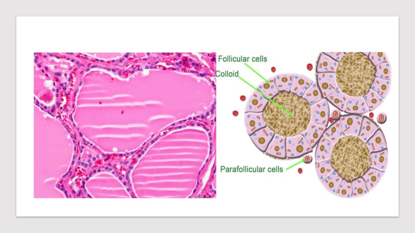
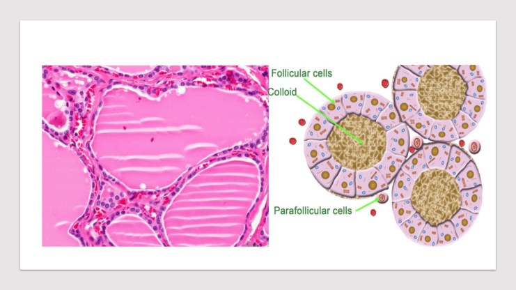
Presentation1Presentation2

  • Based on the classic paper by Norris and others:
    • The development of the parathyroid glands in humans can be divided into five stages:
      • Preprimordial stage
      • Early primordial stage
      • Branchial complex stage
      • Isolation stage
      • Definitive form stage
  • The preprimordial stage:
    • Indicates the period between the formation of the pharynx and the earliest appearance of a recognizable parathyroid anlage:
      • During this stage, at 4 mm to 8 mm in length:
        • The third and fourth pharyngeal pouches show a slight dorsal extension.
      • The third pouch:
        • Which has the form of a tubelike lateral expansion of the primitive pharynx:
          • Makes contact with the ectoderm of the pharyngeal cleft and then continues its growth in a downward and ventral direction.
  • The early primordial stage:
    • When the embryo is about 9 mm in length:
      • The parathyroid tissue can be recognized
    • Proliferation and differentiation of large, clear cells occur in the third and fourth pouches:
      • Resulting in:
        • A thickening of the third and fourth pouches
        • Formation of a budlike nodule of the fourth pouch
  • The branchial complex stage:
    • The derivatives of the third and fourth pharyngeal pouches become separated from each other to reach independent positions. 
    • During the early phase of this stage:
      • The pharyngeal pouches are still joined to the primitive pharynx by pharyngobranchial ducts:
        • These latter, subsequently, narrow and finally divide:
          • Which determines the definitive separation of the third and caudal pharyngeal complexes from the primitive pharynx.
      • At the beginning of this stage:
        • The primordial thymus and PIII are intimately joined:
          • Subsequently, the thymus begins a period of rapid ventral growth:
            • Until the lower pole comes in contact with the pericardium.
          • On the other hand, the growth of the PIII is not as rapid, and it remains a budlike projection from the superior end of the thymus cord.
          • Finally, it takes a sphere shape, intimately attaching to the upper pole of the thymus cord.
        • The position of the caudal pharyngeal complex in relation to the median anlage of the thyroid depends on:
          • Changes in form, size, and position of the rapidly growing lateral lobe of the median thyroid.
          • During this stage, the PIV rudiment is still attached to lateral thyroid body. 
          • When the embryo is 13 mm to 14 mm long, the PIII and PIV migrate together with the thymus and ultimobranchial bodies, respectively.
          • Because of the extension of the cervical spine and the descent of the heart and great vessels:
            • The complex derived from the third branchial (parathymus) is drawn toward the superior mediastinum and, thus:
              • Migrates in a medial and caudal direction through the entire length of the embryonic neck to reach its final position, and separation of the PIII from the thymus begins.
          • The PIV follows the thyroid migration of the ultimobranchial bodies, which travel toward the lateral part of the main median thyroid rudiment:
            • Their descent in the neck is thus relatively limited. 
            • They remain in contact with the posterior part of the middle third of the thyroid lobes.
      • The complex branchial stage ends when the embryo is approximately 18 to 20 mm in length.

Presentation2Presentation1

  • The isolation stage:
    • Is characterized by the separation of the parathyroid rudiments (PIII and PIV) from the other elements of the third (the thymus) and of the caudal pharyngeal complexes (the ultimobranchial bodies), respectively.
    • The isolation of the parathyroid glands is usually accomplished when the embryo is 20 mm in length. 
    • After completing the descent through the neck:
      • The PIII increases in size and separation from the thymus occurs, because of cephalic regression of the last.
      • PIII is thus abandoned at the level of the anterior or posterolateral region of the inferior poles of the thyroid lobes, or at the level of the thyrothymic ligaments, vestigial structures indicative of their former connections.
    • The two elements of the caudal pharyngeal complex also grow separately and are conjoined by a connecting stalk:
      • The interruption of this stalk, determining the isolation of the PIV, occurs once the lateral and the median thyroid become incorporated. 
      • The final position of the PIV in relation to the thyroid gland is determined by the place at which the inclusion of the ultimobranchial body (lateral thyroid element) occurs.
  • The definitive form stage:
    • Indicates the period from the end of the isolation to the time when the parathyroids assume their definitive form.
 

20d943b1-6f20-49c3-9754-2f3756051c93.jpg

Rodrigo Arrangoiz MS, MD, FACS a head and neck surgeon / endocrine surgeon / surgical oncologist and is a member of Sociedad Quirúrgica S.C at the America British Cowdray Medical Center in Mexico City:

  • He is an expert in the management parathyroid diseases.

  • Publication on parathyroid embryology and anatomy:

Training:

• General surgery:

• Michigan State University:

• 2004 al 2010

• Surgical Oncology / Head and Neck Surgery / Endocrine Surgery:

• Fox Chase Cancer Center (Filadelfia):

• 2010 al 2012

• Masters in Science (Clinical research for health professionals):

• Drexel University (Filadelfia):

• 2010 al 2012

• Surgical Oncology / Head and Neck Surgery / Endocrine Surgery:

• IFHNOS / Memorial Sloan Kettering Cancer Center:

• 2014 al 2016

#Arrangoiz#Teacher

#Surgeon

#Cirujano


#ThyroidExpert

#ThyroidSurgeon

#CirujanodeTiroides

#ExpertoenTiroides

#ExpertoenParatiroides

#Paratiroides

#Hiperparatiroidismo

#CancerdeTiroides

#ThyroidCancer

#PapillaryThyroidCancer


#SurgicalOncologist

#CirujanoOncologo

#CancerSurgeon

#CirujanodeCancer

#HeadandNeckSurgeon

#CirugiaEndocrina

#EndocrineSurgery

#CirujanodeCabezayCuello

http://www.cirugiatiroides.com

http://www.sociedadquirurigca.com

http://www.hiperparatiroidismo.info
 

Embriología de la Glándula Tiroides

Presentation3

  • El cirujano moderno de cabeza y cuello / cirujano endocrino / cirujano de tiroides deben tener un conocimiento completo de la ciencias básicas detrás del desarrollo de la glándula tiroides y paratiroides, así como de las posibles anomalías congénitas que surgen de estas glándulas, ya que pueden afectar a la integridad de la cirugía, así como las complicaciones de la cirugía:

    • La glándula tiroides es la primera de las glándulas endocrinas del cuerpo que se desarrolla y aparece como una evaginación de los primitivos primarios alrededor de la tercera semana de gestación (aproximadamente el día 24):

      • La glándula tiroides se forma como una proliferación (engrosamiento) de las células epiteliales endodérmicas que se encuentran en la superficie media del suelo faríngeo en desarrollo:

        • El sitio de este desarrollo se encuentra entre dos estructuras clave:

          • El tubérculo impar y la cópula, y es reconocido como el foramen ciego.

Presentation1

  • El foramen ciego ubicado en la base de la lengua es el lugar de origen de la glándula tiroides en la unión entre la primera y la segunda bolsa branquial (faríngea), inmediatamente dorsal al saco aórtico:

    • La glándula tiroides tiene un doble origen embriológico:

      • La faringe primitiva y la cresta neuroectodérmica / neural.

    • La glándula tiroides inicialmente surge caudal al tubérculo impar, que también se conoce como la yema media de la lengua:

      • Esta inflamación embrionaria se desarrolla a partir del primer arco branquial y se produce en la línea media del piso de la faringe en desarrollo:

        • Finalmente, ayudando a formar la lengua a medida que las dos hinchazones linguales laterales la sobre-crecen.

      • El foramen ciego comienza rostral a la cópula:

        • También conocida como la eminencia hipobranquial (hipofaríngea):

          • Esta hinchazón embriológica mediana consiste en un mesodermo que surge de la segunda bolsa branquial (aunque también están involucradas la tercera y cuarta bolsas branquiales):

            • La glándula tiroides, por lo tanto, se origina entre la primera y la segunda bolsas branquiales.

        • El precursor de la tiroides inicial, el primordio de la tiroides, comienza como un engrosamiento simple de la línea media (endodermo) y se desarrolla para formar el divertículo de la tiroides o anlage de la tiroides (este anlage mediano forma la mayor parte de la glándula tiroides):

          • Esta estructura al principio es inicialmente hueca:

            • Aunque luego se solidifica (formando los elementos foliculares de la glándula tiroides) y se vuelve bilobulado.

        • La división de la glándula tiroidea en lóbulos laterales, si no está presente desde el principio, se produce tan temprano que es imposible establecer si la tiroides surge como una sola unidad o como un órgano pareado.

        • El tallo generalmente tiene un lumen, el conducto tirogloso, que no desciende a los lóbulos laterales.

        • Los dos lóbulos están ubicados a ambos lados de la línea media y están conectados a través de un istmo.

        • Este anlage tiroideo sigue el corazón primitivo a medida que desciende caudalmente.

        • A principios de la quinta semana de gestación, el conducto atenuado pierde su luz y posteriormente se rompe en fragmentos:

          • La parte proximal se retrae y desaparece, dejando solo el foramen ciego en la base de la lengua para marcar su origen, y el extremo caudal se desarrolla como la glándula tiroides encapsulada bilobulada y alcanza su posición adulta final en la séptima semana de gestación.

  • Los anlages laterales apareados se originan en las porciones ventrales de la cuarta y quinta bolsas branquiales y se fusionan con el anlage medular de la tiroides aproximadamente a la quinta semana de gestación:

    • Aportando hasta el 30% del peso de la glándula tiroides.

    • Los anlages laterales son de origen neuroectodérmico / neural (cuerpos ultimobranquiales) y producen las células parafoliculares o C que producen calcitonina (desde la cresta neural, estas células C migran a los cuerpos ultimobranquios), que se encuentran en la región posterior superior de la glándula tiroides.

    • La fusión del anlage medular de la tiroides y los cuerpos ultimobranquiales explica por qué las células parafoliculares o las células C no están dispersas en toda la tiroides, sino que están limitadas a una región profunda entre los tercios medio y superior de los lóbulos laterales a lo largo de un eje lobular central imaginario:

      • Las bases para la fusión de la mediana y la anidación lateral de la tiroides no están claras, pero se afirma que el lugar de la fusión de estas dos estructuras ocurre en el tubérculo de Zuckerkandl.

      • Las células parafoliculares o C pertenecen a un grupo de derivados de la cresta neural conocidos como células de captación y descarboxilación del precursor de aminas (APUD).

Presentation2

  • Las células foliculares de la tiroides se desarrollan a partir del anlage medular de la tiroides y se hacen evidentes alrededor de la octava semana de gestación y comienzan a producir coloides y absorben yodo radioactivo alrededor de la semana 11 de gestación:

    • La evidencia de tiroxina viene con la aparición de coloides.

    • El desarrollo del folículo tiroideo se anuncia por la aparición de un material intracelular periódico ácido-Schiff (PAS) positivo:

    • Lo que conduce a la formación de folículos por brotes o división de los folículos primarios:

      • La diferenciación histológica de la glándula tiroides se puede conceptualizar en tres etapas:

        • Pre-coloide (7 a 13 semanas)

        • Coloide (13 a 14 semanas)

        • Folicular (después de 14 semanas)


20d943b1-6f20-49c3-9754-2f3756051c93.jpg

Rodrigo Arrangoiz MS, MD, FACS es cirujano de cabeza y cuello / cirugía endocrina / cirugía oncológica miembro de Sociedad Quirúrgica S.C en el hospital ABC en Santa Fé en la ciudad de México:

  • Es experto en patología de la glándula tiroides:

  • Tiene publicación sobre la embriología y anatomía de la glándula tiroides:

Entrenamiento:

• General surgery:

• Michigan State University:

• 2004 al 2010

• Surgical Oncology / Head and Neck Surgery / Endocrine Surgery:

• Fox Chase Cancer Center (Filadelfia):

• 2010 al 2012

• Masters in Science (Clinical research for health professionals):

• Drexel University (Filadelfia):

• 2010 al 2012

• Surgical Oncology / Head and Neck Surgery / Endocrine Surgery:

• IFHNOS / Memorial Sloan Kettering Cancer Center:

• 2014 al 2016

#Arrangoiz#Teacher

#Surgeon

#Cirujano


#ThyroidExpert

#ThyroidSurgeon

#CirujanodeTiroides

#ExpertoenTiroides

#ExpertoenParatiroides

#Paratiroides

#Hiperparatiroidismo

#CancerdeTiroides

#ThyroidCancer

#PapillaryThyroidCancer


#SurgicalOncologist

#CirujanoOncologo

#CancerSurgeon

#CirujanodeCancer

#HeadandNeckSurgeon

#CirugiaEndocrina

#EndocrineSurgery

#CirujanodeCabezayCuello

http://www.cirugiatiroides.com

http://www.sociedadquirurigca.com

Embryology of the Thyroid Gland

  • The modern head and neck surgeon / endocrine surgeon / thyroid surgeon should have a complete understanding of the basic science behind the development of the thyroid and parathyroid glands as well as knowledge of the possible congenital abnormalities arising out of these glands, as they may impact the completeness of surgery as well as the complications of surgery:

  • The thyroid gland is the first of the body’s endocrine glands to develop appearing as an outpouching of the primitive foregut around the third week of gestation (roughly the 24th day):
    • The thyroid gland forms as a proliferation (thickening) of the endodermal epithelial cells found on the median surface of the developing pharyngeal floor:
      • The site of this development lies between two key structures:
        • The tuberculum impar and the copula, and is recognized as the foramen cecum.

Presentation1

  • The foramen cecum located at the base of the tongue is the site of origin of the thyroid gland at the junction between the first and second branchial (pharyngeal) pouches, immediately dorsal to the aortic sac:
    • The thyroid gland has a double embryologic origin:
      • The primitive pharynx and the neuroectodermal / neural crest.
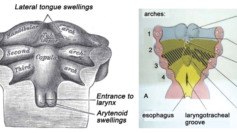
        • The thyroid gland initially arises caudal to the tuberculum impar, which is also known as the median tongue bud:
          • This embryonic swelling develops from the first branchial arch and occurs on the mid-line of the floor of the developing pharynx:
            • Eventually helping form the tongue as the two lateral lingual swellings overgrow it.
        • The foramen cecum begins rostral to the copula:
          • Also known as the hypobranchial (hypopharyngeal) emi- nence:
            • This median embryologic swelling consists of mesoderm that arises from the second branchial pouch (although the third and fourth branchial pouches are also involved):
              • The thyroid gland, therefore, originates between the first and second branchial pouches. 
        • The initial thyroid precursor, the thyroid primordium, starts as a simple mid-line thickening (endoderm) and develops to form the thyroid diverticulum or thyroid anlage (this median anlage forms the bulk of the thyroid gland):
          • This structure at the outset is initially hollow:
            • Though it later solidifies (forming the follicular elements of the thyroid gland) and becomes bilobed.
        • Division of the thyroid gland into lateral lobes, if not present from the beginning, takes place so early that it is impossible to establish whether the thyroid arises as a single unit or as a paired organ.
        • The stalk usually has a lumen, the thyroglossal duct, that does not descend into the lateral lobes.
        • The two lobes are located on either side of the midline and are connected via an isthmus.
        • This thyroid anlage follows the primitive heart as it descends caudally.
        • Early during the fifth week of gestation, the attenuated duct loses its lumen and subsequently breaks into fragments:
          • The proximal part retracts and vanishes, leaving only the foramen cecum at the base of the tongue to mark its origin, and the caudal end develops as the bilobed encapsulated thyroid gland and reaches its final adult position by the 7th week of gestation.

Presentation2

  • The paired lateral anlages originate from the ventral portions of the fourth and fifth branchial pouches and fuse with the median thyroid anlage at approximately the fifth week of gestation:
    • Contributing up to 30% of the thyroid gland weight.
  • The lateral anlages are neuroectodermal / neural crest in origin (ultimobranchial bodies) and produce the calcitonin producing parafolicullar or C cells (from the neural crest these the C-cells migrate to the ultimobranchial bodies), which come to lie in the superior posterior region of the thyroid gland.
    • The fusion of the median thyroid anlage and the ultimobranchial bodies explains why the parafollicular cells or C cells are not scattered throughout the entire thyroid but are limited to a region deep within the middle to upper thirds of the lateral lobes along an imaginary central lobar axis:
      • The bases for the fusion of the median and lateral thyroid anlages is unclear, but the site of the fusion of these two structures is stated to occur at the tubercle of Zuckerkandl.
    • The parafolicullar or C cells belong to a group of neural crest derivatives known as the amine precursor uptake and decarboxylation cells (APUD) .

Presentation3

  • The thyroid follicular cells develop from the median thyroid anlage and become apparent around the 8th week of gestation and start producing colloid and take up radioactive iodine around the 11th week of gestation:

    • Evidence of thyroxine comes with the appearance of colloid.

    • The development of the thyroid follicle is heralded by the appearance of an intracellular periodic acid-Schiff (PAS)-positive material:

      • Which leads to follicle formation by budding or division of the primary follicles:

        • The histologic differentiation of the thyroid gland can be conceptualized into three stages:

          • Pre-colloid (7 to 13 weeks)

          • Colloid (13 to 14 weeks)

          • Follicular (after 14 weeks)


20d943b1-6f20-49c3-9754-2f3756051c93.jpg

Rodrigo Arrangoiz MS, MD, FACS a head and neck surgeon / endocrine surgeon / surgical oncologist and is a member of Sociedad Quirúrgica S.C at the America British Cowdray Medical Center in Mexico City:

  • He is an expert in the management thyroid diseases.

  • Publication on thyroid embryology and anatomy:

Training:

• General surgery:

• Michigan State University:

• 2004 al 2010

• Surgical Oncology / Head and Neck Surgery / Endocrine Surgery:

• Fox Chase Cancer Center (Filadelfia):

• 2010 al 2012

• Masters in Science (Clinical research for health professionals):

• Drexel University (Filadelfia):

• 2010 al 2012

• Surgical Oncology / Head and Neck Surgery / Endocrine Surgery:

• IFHNOS / Memorial Sloan Kettering Cancer Center:

• 2014 al 2016

#Arrangoiz#Teacher

#Surgeon

#Cirujano


#ThyroidExpert

#ThyroidSurgeon

#CirujanodeTiroides

#ExpertoenTiroides

#ExpertoenParatiroides

#Paratiroides

#Hiperparatiroidismo

#CancerdeTiroides

#ThyroidCancer

#PapillaryThyroidCancer


#SurgicalOncologist

#CirujanoOncologo

#CancerSurgeon

#CirujanodeCancer

#HeadandNeckSurgeon

#CirugiaEndocrina

#EndocrineSurgery

#CirujanodeCabezayCuello

http://www.cirugiatiroides.com

http://www.sociedadquirurigca.com

Sebaceous Gland Carcinoma

  • Sebaceous gland carcinoma is an aggressive, uncommon, cutaneous tumor:

    • First well-described by Allaire in 1891.

  • This tumor is thought to arise from sebaceous glands in the skin and, thus, may arise anywhere on the body where these glands exist, including the genitalia:

    • Approximately 75% of these tumors arise in the periocular region:

      • In this region, sebaceous gland carcinoma represents 1.0% to 5.5% of eyelid malignancies:

        • Fourth after basal cell carcinoma, squamous cell carcinoma, and melanoma.

      • Women tend to be affected somewhat more often than men:

        • With 57% to 77% of patients being women in several large series.

      • Most patients present in their sixth or seventh decade of life:

        • Although the range is from early childhood through the nineties:

          • The youngest reported case arose in a 3-year-old child.

      • An area rich in a variety of types of sebaceous glands:

        • Sebaceous gland carcinoma resembles normal sebaceous glands.

        • One may reasonably speculate that sebaceous gland carcinoma arises from mature sebaceous glands.

        • Histologic studies have suggested that periocular sebaceous gland carcinomas arise from the sebaceous glands in this region:

          • The following five types of sebaceous glands are seen in the periocular region:

            • Meibomian glands of the tarsal plate

            • Glands of Zeis of the cilia

            • Sebaceous glands of the eyebrows

            • Glands of the caruncle

            • Glands of the fine hair follicles of the eyelid surface

  • This tumor exhibits an aggressive clinical course:

    • With a significant tendency for both local recurrence and distant metastasis:

      • Reported local recurrence rates range from 9% to 36%, with larger series reporting recurrence rates in the 30% range:

        • Local recurrence tends to occur within 5 years.

      • The rate of metastasis in extraocular and ocular sebaceous gland carcinoma is thought to be similar, occurring in 14% to 25% of cases:

        • First to the draining lymph nodes and then to distant sites:

          • Sites of distant metastasis include the:

            • Liver, lungs, bones, and brain

  • Diagnosis and therapy tend to be delayed because sebaceous gland carcinoma is frequently mistaken for more common benign entities, further complicating treatment of this aggressive malignancy:

    • In addition, a varied histologic appearance may occur, and delayed diagnosis or misdiagnosis following a biopsy is not uncommon.

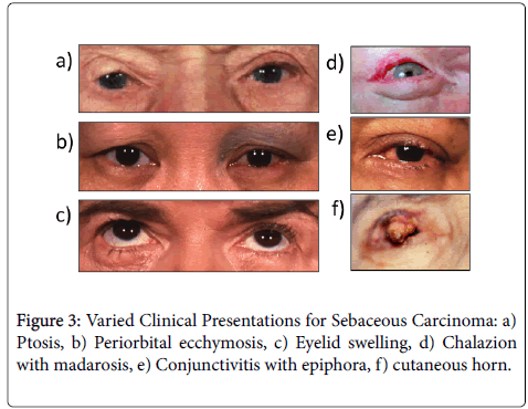
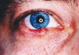
  • When arising in the periocular region, the clinical presentation is often variable, and sebaceous gland carcinoma is often not initially suspected:

    • Instead, patients may receive multiple courses of incision and drainage for chalazion before a definitive biopsy is performed.

  • Most sebaceous gland carcinomas have no obvious etiology:

    • Only a few are associated with Muir-Torre syndrome:

      • Although sebaceous adenoma and epithelioma are more specific markers for Muir-Torre syndrome:

        • An evaluation for this syndrome is advisable once sebaceous gland carcinoma is diagnosed:

          • In approximately 40% of cases, patients with Muir-Torre syndrome develop some type of sebaceous tumor before or concurrent with visceral malignancy.

clinical-ophthalmology-eyelid-swelling-8-635-g003Unknown


20d943b1-6f20-49c3-9754-2f3756051c93

Training:

• General surgery:

• Michigan State University:

• 2004 al 2010

• Surgical Oncology / Head and Neck Surgery / Endocrine Surgery:

• Fox Chase Cancer Center (Filadelfia):

• 2010 al 2012

• Masters in Science (Clinical research for health professionals):

• Drexel University (Filadelfia):

• 2010 al 2012

• Surgical Oncology / Head and Neck Surgery / Endocrine Surgery:

• IFHNOS / Memorial Sloan Kettering Cancer Center:

• 2014 al 2016

#Arrangoiz

#Surgeon

#Cirujano

#SurgicalOncologist

#CirujanoOncologo

#CancerSurgeon

#CirujanodeCancer

http://www.sociedadquirurigca.com

Merkel Cell Carcinoma

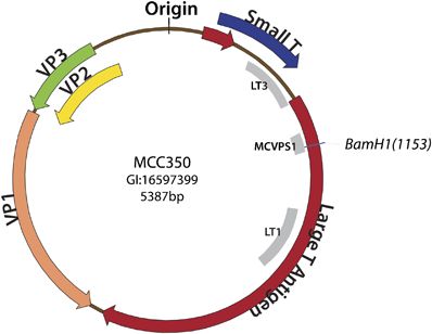
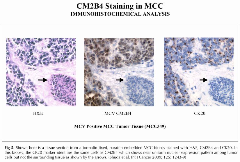
  • Merkel Cell Polyomavirus (MCV) is one of seven currently known human oncoviruses:

    • It is suspected to cause the majority of cases of Merkel cell carcinoma, a rare but aggressive form of skin cancer:

      • Approximately 80% of Merkel cell carcinoma (MCC) tumors have been found to be infected with MCV:

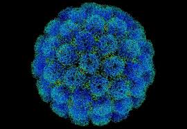
        • Polyomaviruses are small (~5400 base pair), non-enveloped, double-stranded DNA viruses:

          • MCV is the fifth polyomavirus that infects humans to be discovered.


  • El poliomavirus de células de Merkel (MCV) es uno de los siete oncovirus humanos conocidos actualmente:

    • Se sospecha que causa la mayoría de los casos de carcinoma de células de Merkel, una forma rara pero agresiva de cáncer de piel:

      • Aproximadamente el 80% de los tumores de carcinoma de células de Merkel (MCC) se han encontrado infectados con el MCV:

        • Los poliomavirus son virus de ADN de doble cadena, pequeños (~ 5400 pares de bases), sin envoltura:

          • MCV es el quinto poliomavirus que infecta a los humanos por descubrir.


modpathol20093f3images

Untitled


20d943b1-6f20-49c3-9754-2f3756051c93

Training:

• General surgery:

• Michigan State University:

• 2004 al 2010

• Surgical Oncology / Head and Neck Surgery / Endocrine Surgery:

• Fox Chase Cancer Center (Filadelfia):

• 2010 al 2012

• Masters in Science (Clinical research for health professionals):

• Drexel University (Filadelfia):

• 2010 al 2012

• Surgical Oncology / Head and Neck Surgery / Endocrine Surgery:

• IFHNOS / Memorial Sloan Kettering Cancer Center:

• 2014 al 2016

#Arrangoiz

#Surgeon

#Cirujano

#SurgicalOncologist

#CirujanoOncologo

#CancerSurgeon

#CirujanodeCancer

http://www.sociedadquirurigca.com

Cáncer de Mama Inflamatorio

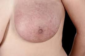
  • El cáncer de mama inflamatorio es una forma rara pero altamente agresiva de cáncer de mama:
    • Se considera una entidad distinta con características clínico-patológicas únicas.
  • Los síntomas de eritema y aumento del tamaño de los senos generalmente se desarrollan en el transcurso de unas pocas semanas:
    • Los signos clínicos son el resultado de una embolia tumoral linfovascular, que es patognomónica para el cáncer de mama inflamatorio.
  • El diagnóstico oportuno puede ser difícil, ya que el cáncer de mama inflamatorio puede simular una enfermedad infecciosa como la mastitis o un absceso mamario:
    • Sin embargo, el diagnóstico y el tratamiento oportuno son muy importantes para proporcionar una intervención multidisciplinaria lo antes posible.
    • Se debe realizar estudios de estratificación para descartar metástasis a distancia en el momento del diagnóstico.
  • Una combinación de terapia sistémica neoadyuvante, mastectomía radical modificada y radioterapia adyuvante es un tratamiento estándar para el cáncer de mama inflamatorio.

Unknown

symptoms-signs-of-inflammatory-breast-cancer

Rodrigo Arrangoiz MS, MD, FACS cirujano oncology y miembro de Sociedad Quirúrgica S.C en el America British Cowdray Medical Center en la ciudad de Mexico:

  • Es experto en el manejo del cáncer de mama.

20d943b1-6f20-49c3-9754-2f3756051c93

Training:

• General surgery:

• Michigan State University:

• 2004 al 2010

• Surgical Oncology / Head and Neck Surgery / Endocrine Surgery:

• Fox Chase Cancer Center (Filadelfia):

• 2010 al 2012

• Masters in Science (Clinical research for health professionals):

• Drexel University (Filadelfia):

• 2010 al 2012

• Surgical Oncology / Head and Neck Surgery / Endocrine Surgery:

• IFHNOS / Memorial Sloan Kettering Cancer Center:

• 2014 al 2016

#Arrangoiz

#Surgeon

#Cirujano

#SurgicalOncologist

#CirujanoOncologo

#BreastSurgeon

#CirujanodeMama

#CancerSurgeon

#CirujanodeCancer

http://www.sociedadquirurigca.com


img_0832-2img_0833-2img_0834-2img_0835-2img_0836-2img_0837-2


The Impact of Hashimoto Thyroiditis on Thyroid Nodule Cytology and Risk of Thyroid Cancer

Abstract

Context: The impact of Hashimoto thyroiditis (HT) on the risk of thyroid cancer and its accurate detection remains unclear. The presence of a chronic lymphocytic infiltration imparts a logical mechanism potentially altering neoplastic transformation, while also influencing the accuracy of diagnostic evaluation.

Methods: We performed a prospective, cohort analysis of 9851 consecutive patients with 21,397 nodules ≥1 cm who underwent nodule evaluation between 1995 and 2017. The definition of HT included (i) elevated thyroid peroxidase antibody (TPOAb) level and/or (ii) findings of diffuse heterogeneity on ultrasound, and/or (iii) the finding of diffuse lymphocytic thyroiditis on histopathology. The impact of HT on the distribution of cytology and, ultimately, on malignancy risk was determined.

Results: A total of 2651 patients (27%) were diagnosed with HT, and 3895 HT nodules and 10,168 non-HT nodules were biopsied. The prevalence of indeterminate and malignant cytology was higher in the HT vs non-HT group (indeterminate: 26.3% vs 21.8%, respectively, P < 0.001; malignant: 10.0% vs 6.4%, respectively, P < 0.001). Ultimately, the risk of any nodule proving malignant was significantly elevated in the setting of HT (relative risk, 1.6; 95% CI, 1.44 to 1.79; P < 0.001), and was maintained when patients with solitary or multiple nodules were analyzed separately (HT vs non-HT: 24.5% vs 16.3% solitary; 22.1% vs 15.4% multinodular; P < 0.01).

Conclusion: HT increases the risk of thyroid malignancy in any patient presenting for nodule evaluation. Diffuse sonographic heterogeneity and/or TPOAb positivity should be used for risk assessment at time of evaluation.


20d943b1-6f20-49c3-9754-2f3756051c93.jpg

Rodrigo Arrangoiz MS, MD, FACS a head and neck surgeon / endocrine surgeon / surgical oncologist and is a member of Sociedad Quirúrgica S.C at the America British Cowdray Medical Center in Mexico City:

  • He is an expert in the management thyroid carcinoma.

Training:

• General surgery:

• Michigan State University:

• 2004 al 2010

• Surgical Oncology / Head and Neck Surgery / Endocrine Surgery:

• Fox Chase Cancer Center (Filadelfia):

• 2010 al 2012

• Masters in Science (Clinical research for health professionals):

• Drexel University (Filadelfia):

• 2010 al 2012

• Surgical Oncology / Head and Neck Surgery / Endocrine Surgery:

• IFHNOS / Memorial Sloan Kettering Cancer Center:

• 2014 al 2016

#Arrangoiz

#Teacher

#Surgeon

#Cirujano

#SurgicalOncologist

#CirujanoOncologo

#CancerSurgeon

#CirujanodeCancer

#HeadandNeckSurgeon

#CirugiaEndocrina

#EndocrineSurgery

#CirujanodeCabezayCuello

http://www.cirugiatiroides.com

http://www.sociedadquirurigca.com


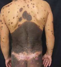
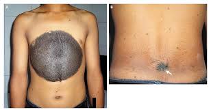
Nevos Congénitos Gigantes

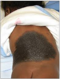
  • Un nevó congénito gigante se define como:
    • Un nevó que mide al menos 15 cm de diámetro o al menos el doble del tamaño de la palma de la mano de la persona afectada.
  • Los pacientes con nevos congénitos gigantes tienen un riesgo de por vida estimado del:
    • 4% a 10% de desarrollar un melanoma.
  • Aproximadamente la mitad de los melanomas que se desarrollan en nevos congénitos gigantes se desarrollan dentro de los primeros 5 años de vida.
  • Las decisiones sobre el manejo de los nevos congénitos gigantes son difíciles porque tales lesiones son a menudo tan extensas que la excisión quirúrgica profiláctica es imposible:
    • Cuando la ubicación y el tamaño de una lesión permiten la excisión profiláctica:
      • La excisión debe considerarse antes de los 2 años de edad.
    • Aunque la evidencia es limitada:
      • Los posibles indicadores de pronóstico negativo que pueden ayudar en la toma de decisiones incluyen:
        • El tamaño de la lesión
        • La presencia de lesiones satelitales


UnknownUnknown-1images


20d943b1-6f20-49c3-9754-2f3756051c93

Training:

• General surgery:

• Michigan State University:

• 2004 al 2010

• Surgical Oncology / Head and Neck Surgery / Endocrine Surgery:

• Fox Chase Cancer Center (Filadelfia):

• 2010 al 2012

• Masters in Science (Clinical research for health professionals):

• Drexel University (Filadelfia):

• 2010 al 2012

• Surgical Oncology / Head and Neck Surgery / Endocrine Surgery:

• IFHNOS / Memorial Sloan Kettering Cancer Center:

• 2014 al 2016

#Arrangoiz

#Surgeon

#Cirujano

#SurgicalOncologist

#CirujanoOncologo

#CancerSurgeon

#CirujanodeCancer

http://www.sociedadquirurigca.com

Thyroid Physiology

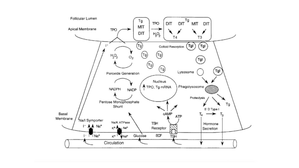
  • The basic functional unit of the thyroid gland is the:

    • Thyroid follicle:

      • The thyroid follicle is made up of a:

        • Single layer of cells that forms a sphere that surrounds a protein aggregate called colloid.

      • The thyroid follicular cells are polarized:

        • With the side toward the colloid called the:

          • Apical membrane

        • And the outer side of the cell in contact with capillaries called the:

          • Basal membrane (Figure)

  • The synthesis of thyroid hormone is activated after binding of:

    • Thyrotropin-stimulating hormone (TSH) to the basal membrane surface receptor:

      • The TSH receptor

    • TSH stimulates all the steps of thyroid hormone synthesis and secretion, including:

      • Iodide transport

      • Synthesis of thyroglobulin

      • Iodination of thyroglobulin

      • Secretion of thyroid hormones

    • TSH binding of the TSH receptor on the basal membrane of the thyroid follicular cell activates adenylate cyclase:

      • To increase intracellular cAMP:

        • Which activates a cascade of numerous steps in the thyroid hormone synthetic pathway

  • The first step is transport of iodide across the basal membrane into the follicular cell in an energy-dependent manner by the Na+/I symporter:

    • The iodide becomes covalently attached to the precursor thyroid hormone glycoprotein:

      • Thyroglobulin:

        • This occurs at the interface between the apical membrane and the colloid by the enzyme thyroperoxidase (TPO).

  • The iodide is attached to the tyrosine molecules in the thyroglobulin molecule to form:

    • Monoiodotyrosines (MITs) and diiodotyrosines (DITs).

      • TPO enzymatically couples two iodotyrosines to create bioactive thyroid hormones:

        • Two diiodotyrosines (DITs) = L-thyroxine (T4)

        • One monoiodotyrosine (MIT) and diiodotyrosine (DIT) =triiodothyronine (T3).

      • The T4 and T3 remain part of the thyroglobulin molecule and is stored as colloid within the interior of the follicle.

    • The thyroid gland is a unique endocrine organ:because it stores large amounts of thyroid hormones as colloid that is released as needed through TSH stimulation.

    • In healthy and iodine-sufficient individuals:

      • The majority of thyroid hormone is stored as T4 with a small amount, less than 20%, stored as T3.

    • TSH receptor stimulation leads to colloid uptake into the cytoplasm by pinocytosis to form a cytoplasmic vesicle:

      • The cytoplasmic vesicles fuse with lysosomes to from a phagolysosome and the proteases found within the lysosome hydrolyze the peptide bonds of thyroglobulin to release T4 and T3 into the cytoplasm where it diffuses into the bloodstream.

        • Approximately 90 mcg of T4 is secreted from the thyroid each day in adults.

        • T4 and T3 travel in the circulation bound 99.97% and 99.5%, respectively, to a group of serum thyroid hormone binding proteins synthesized in the liver, which include:

          • Thyroxine binding globulin (TBG), transthyretin (also known as prealbumin), and albumin.

            • TBGhas the highest affinity to bind thyroid hormone and is clinically the most important member of this group:

              • TBG carries about 68% of the circulating T4 and 80% of the T3.

            • Transthyretin, formally named prealbumin binds with a lower affinity and carries 11% of the circulating T4 and 9% of T3.

            • Albumin has the lowest affinity for thyroid hormone but the largest capacity, binding 20% of the T4 and 11% of the T3.

          • More than 99% of thyroid hormones circulate bound to these carrier proteinsand are biologically inactive.

          • The half time of T4 in the blood is 7 to 10 days.

          • The thyroid hormones not associated with protein, free T4 and free T3, can enter the cells and are biologically active.

          • T4 is made exclusively by the thyroid gland, whereas T3 is made primarily in peripheral tissues by deiodination of circulating T4 by a group of enzymes called deiodinases:

            • Deiodinase enzyme activity is tightly regulated to maintain a normal T3 despite fluctuations in T4.

          • T3 binds with a much higher affinity to the thyroid hormone receptor and is more biologically active than T4.

          • The activity of a specific 5′-deiodinase and the resulting T3 level can be reduced by:

            • Hyperthyroidism

            • Drugs:

              • Beta-blockers

              • Ipodate

              • Amiodarone

              • Dexamethasone

              • Propylthiouracil,

            • Malnutrition

            • Severe illness.

          • Conversely, during hypothyroidism, the 5′ deiodinase is activated to ensure that T4 is converted to the more bioactive T3.

        • Normally, 20% of the daily T3 requirement is directly synthesized and secreted by the thyroid gland:

        • During starvation and illness:

          • The 5′ deiodinase converts the bioactive T4 and T3 to biologically inactive molecules:

            • Reverse T3 (rT3) and 3, 3′ diiodothyronine

        • The small quantity of free T3 binds to its intranuclear thyroid hormone receptor to alter gene expression:

          • Which in turn alters cellular function and determines the thyroidal status.

          • The thyroid hormone receptor (TR) is a nuclear protein that is a member of a superfamily of receptors that bind steroid hormone such as retinoic acid, vitamin D, and estrogen.

Presentation1


20d943b1-6f20-49c3-9754-2f3756051c93.jpg

Rodrigo Arrangoiz MS, MD, FACS a head and neck surgeon / endocrine surgeon / surgical oncologist and is a member of Sociedad Quirúrgica S.C at the America British Cowdray Medical Center in Mexico City:

  • He is an expert in the management of thyroid and parathyroid diseases.

Training:

• General surgery:

• Michigan State University:

• 2004 al 2010

• Surgical Oncology / Head and Neck Surgery / Endocrine Surgery:

• Fox Chase Cancer Center (Filadelfia):

• 2010 al 2012

• Masters in Science (Clinical research for health professionals):

• Drexel University (Filadelfia):

• 2010 al 2012

• Surgical Oncology / Head and Neck Surgery / Endocrine Surgery:

• IFHNOS / Memorial Sloan Kettering Cancer Center:

• 2014 al 2016

#Arrangoiz

#Teacher

#Surgeon

#Cirujano

#SurgicalOncologist

#CirujanoOncologo

#CancerSurgeon

#CirujanodeCancer

#HeadandNeckSurgeon

#CirugiaEndocrina

#EndocrineSurgery

#CirujanodeCabezayCuello

#ParathyroidExpert

#ExpertoenParatiroides

#Hyperparathyroidismo

#Hiperparatiroidismo

#ThyroidExpert

#ExpertoenTiroides

http://www.cirugiatiroides.com

http://www.hiperparatiroidismo.info
http://www.sociedadquirurigca.com

Breast Cancer Screening – When to Start?

  • Women with no symptoms for breast cancer should undergo mammography screening every other year:
    • According to a new evidence-based guidance statement from the American College of Physicians (ACP):
      • The largest medical specialty organization in the United States.
  • The guidance applies to women between the ages of 50 and 74 who are at average-risk:
    • The statement was published online April 9 in the Annals of Internal Medicine:
      • “The evidence shows that the best balance of benefits and harms for these women, which represents the great majority of women, is to undergo breast cancer screening with mammography every other year.” 
        • This approach is also endorsed by the US Preventive Services Task Force (USPSTF).
  • The new guidance drew immediate fire from:

    • The American College of Radiology (ACR) and Society of Breast Imaging (SBI):

      • These two radiology groups recommend women have annual mammograms starting at age 40 years and that they continue “as long as they are in good health.”:

      • The American Cancer Society (ACS) also recommends starting screening mammography at age 40.


  • The new ACP guideline recommends:
    • Mammography starting every other year:
      • Starting at 50 years and stopping at 74 years
        • This may result in up to 10,000 additional, and unnecessary, breast cancer deaths in the United States each year:
        • This approach would also do little to nothing to address over-diagnosis or the harms of screening:
          • The ACR and SBI comment in a joint press statement.


  • New ACP Recommendations:
    • The new ACP document is an assessment of the quality and content of seven English-language guidelines for breast cancer screening:
      • Including those from USPSTF, ACS, ACR, American College of Obstetricians and Gynecologists, Canadian Task Force on Preventive Health Care, National Comprehensive Cancer Network, and World Health Organization.
    • The new ACP recommendations are for women at average risk of breast cancer:
      • This includes women:
        • Without a history of breast cancer
        • Previous diagnosis of a high-risk lesion
        • Without genetic mutations such as BRCA1/2 or another familial breast cancer syndrome
        • Without a history of radiation therapy to the chest in childhood


    • In average-risk women aged 40 to 49 years:
      • Clinicians should discuss whether to screen for breast cancer with mammography before age 50 years:
        • Discussion should include the potential benefits and harms and a woman’s preferences:
          • Potential harms outweigh benefits in most women aged 40 to 49 years according to the ACP guidelines.
    • In average-risk women aged 50 to 74 years:
      • Clinicians should offer screening for breast cancer with biennial mammography.The ACP guidance consists of four statements about mammography screening, as follows:
    • In average-risk women aged 75 years or older or in women with a life expectancy of 10 years or less:
      • Clinicians should discontinue screening for breast cancer.
    • In average-risk women of all ages:
      • Clinicians should not use clinical breast examination to screen for breast cancer.


  • Clarity Amid Chaos:
    • The new guidance from the ACP provides “clarity and simplicity amidst the chaos of diverging guidelines,” write Joann Elmore, MD, MPH, University of California, Los Angeles, and Christoph Lee, MD, MS, University of Washington, Seattle, in an accompanying editorial.
    • The editorialists congratulate the ACP for their effort to clarify the multitude of breast cancer screening guidelines:
      • However, they emphasize it is not a perfect product:
        • These guidance statements…do not clearly illuminate the full path ahead for every woman:
          • For example, the issue of breast cancer density:
            • The ACP considers women with dense breast tissue on mammography — and no other risk factors — to be at average risk:
              • Because just under half of all women have dense tissue on mammography, this would seem reasonable,” the editorialists opine:
                • When the average risk of dense breast tissue is combined with other risk factors that also indicate average risk in isolation (such as early menarche, late menopausal onset, oral contraceptive or menopausal hormone therapy, or a single family member with a history of postmenopausal breast cancer), a woman may no longer be at average risk.
                • Physicians can expect that more women will inquire about breast density as a factor that increases risk beyond the average.
    • The editorialists emphasize that breast cancer screening guidelines are an ongoing project:
      • Physicians are left to use their best judgment based on available research and expert recommendations.
  • Major Disservice:
    • Reacting to the news, Laurie Margolies, MD, radiologist, Mount Sinai Health System, New York City, said the new ACP guidance statements “are based on no new evidence…[and] are a major disservice to American women.”
      • The majority of women who are diagnosed with breast cancer are at average risk and delaying screening until age 50 will significantly delay diagnosis for many.
      • Hopefully, women are smart enough to make the decision to continue yearly screening mammography.
    • The ACR and SBI also take issue with the idea of screening every other year:
      • In their joint press statement, the societies say that the ACP claims that “every other year mammography screening results in no significant difference in breast cancer mortality.”
        • “This is incorrect,” the societies say in their press statement.
        • “There have been no randomized controlled trials to test this ACP claim.
          • In fact, the NCI/CISNET models that were used by the USPSTF and the ACS actually show a major decline in deaths among women screened annually vs every other year.


  • Effect on Mortality
    • The ACP guidance notes that pooled results from meta-analyses of randomized clinical trials:
      • Demonstrated that mammography was not associated with a reduction in all-cause mortality:
        • In their press statement, the ACR and SBI do not address that issue of overall survival:
          • Instead, they discuss breast cancer-associated mortality:
            • They cite a recent study that showed women screened regularly for breast cancer have a 47% lower relative risk of dying from the disease within 20 years of diagnosis than those not regularly screened:
              • Cancer. 2019;125:515-523.
            • They also cite two large studies that showed regular mammography use “cuts the risk of dying from breast cancer nearly in half.

Ann Intern Med. Published online April 9, 2019


Rodrigo Arrangoiz MS, MD, FACS a surgical oncologist and is a member of Sociedad Quirúrgica S.C at the America British Cowdray Medical Center in Mexico City:

  • He is an expert in the management of breast cancer.

    • If you have any questions about the screening for breast cancer please fill free to contact Dr. Arrangoiz.

20d943b1-6f20-49c3-9754-2f3756051c93

Training:

• General surgery:

• Michigan State University:

• 2004 al 2010

• Surgical Oncology / Head and Neck Surgery / Endocrine Surgery:

• Fox Chase Cancer Center (Filadelfia):

• 2010 al 2012

• Masters in Science (Clinical research for health professionals):

• Drexel University (Filadelfia):

• 2010 al 2012

• Surgical Oncology / Head and Neck Surgery / Endocrine Surgery:

• IFHNOS / Memorial Sloan Kettering Cancer Center:

• 2014 al 2016

#Arrangoiz

#Surgeon

#Cirujano

#SurgicalOncologist

#CirujanoOncologo

#BreastSurgeon

#CirujanodeMama

#CancerSurgeon

#CirujanodeCancer

http://www.sociedadquirurigca.com


img_0832-2img_0833-2img_0834-2img_0835-2img_0836-2img_0837-2