Blog

Epidemiology of Cutaneous Melanoma

  • The incidence of invasive cutaneous melanoma continues to be a major public health concern in the United States:
    • Of concern, the rate of melanoma has risen about 3% per year in the United States over the past few decades
  • Estimated incidence rates of new melanomas in 2021:
    • 106,110:
      • Men 62,260
      • Women 43,850
  • Estimated mortality rate for melanoma in the year 2021:
    • 7,180 people are expected to die of melanoma:
      • Men 4,600 men
      • Women 2,580 women
  • Melanoma remains more than 20 times more common in:
    • Whites than in African Americans
  • Overall, the lifetime risk of being diagnosed with melanoma is about:
    • 2.5% (1 in 40) for whites
    • 0.1%(1 in 1,000) for blacks
    • 0.5% (1 in 200) for Hispanics
  • The incidence of melanoma has been increasing faster than that of nearly any other cancer over the last 30 years
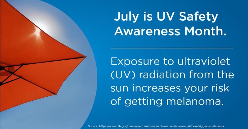
  • The major environmental risk factor:
    • Exposure to ultraviolet (UV) radiation:
      • Is reflected in geographic and ethnic patterns of melanoma rates
  • There have also been changes in the distribution and stage of melanoma at diagnosis:
    • With an overall trend toward thinner tumors:
      • Particularly among any patients with T1 / T2 melanomas:
        • While the opposite trend is seen among patients with thick, T4 lesions
  • Etiology and Epidemiology of Cutaneous Melanoma:
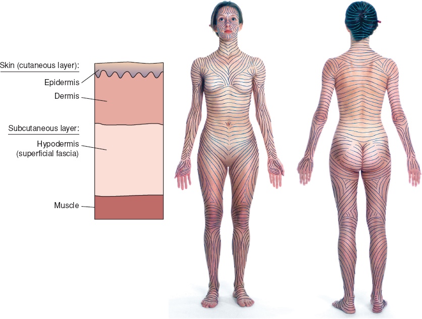
    • Cutaneous melanoma originates from melanocytes within the epidermal layer of the skin
    • Although these melanocytes represent a heterogeneous group of cells within the body:
      • They all share a common place of origin:
        • The neural crest, and the ability to produce melanin
    • In humans:
      • Melanin acts as the primary determinant of skin color and provides a layer of protection from ultraviolet (UV) radiation
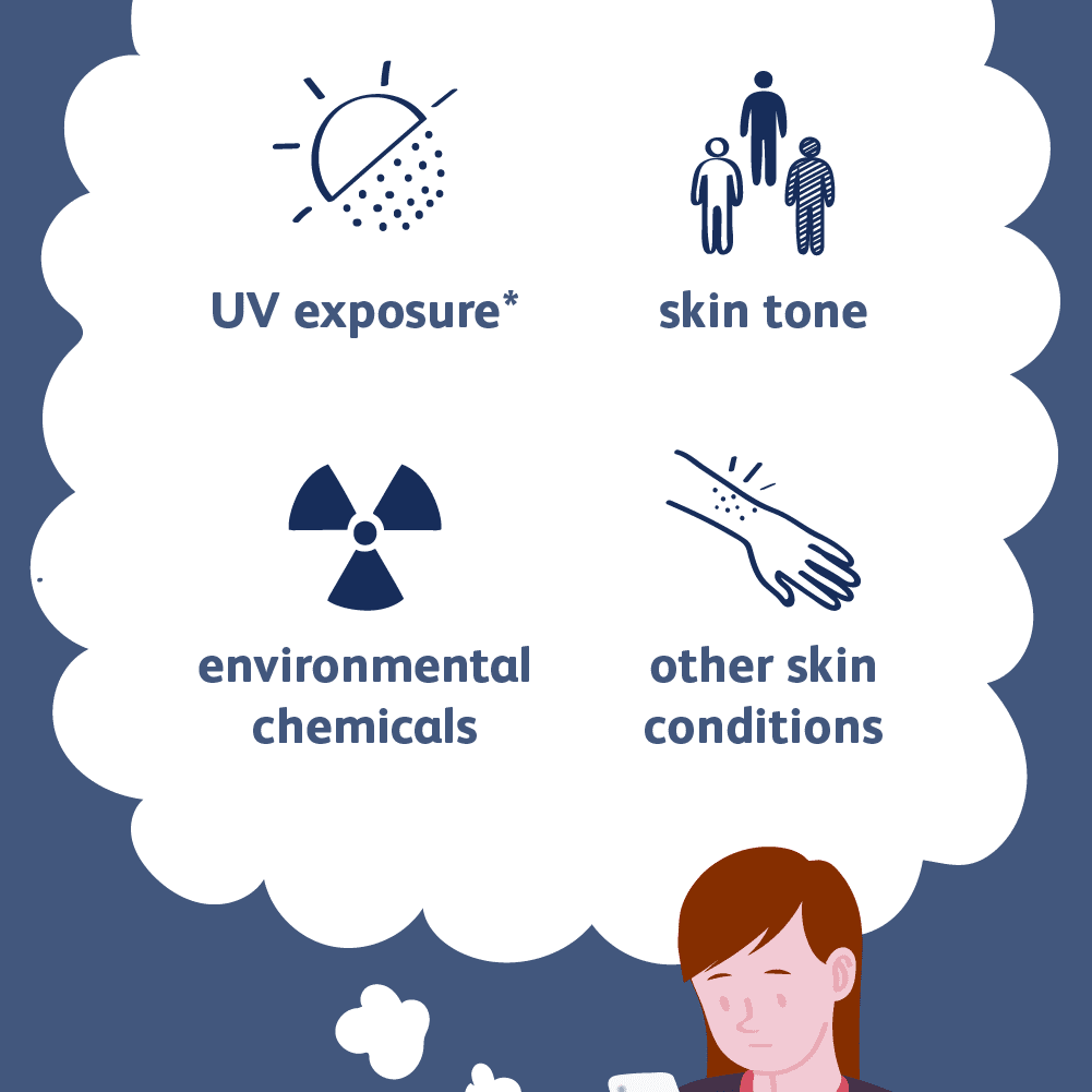
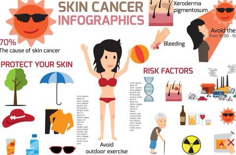
  • Risk factors for the development of cutaneous melanoma include:
    • Sun exposure
    • Blistering sunburns
    • Fair complexion
    • Family history
    • Increasing age
    • Previous melanoma
    • Dysplastic nevi
  • Although UV radiation is a critical factor in the development of most melanoma:
    • It can occur in unexposed areas of the skin:
      • Such as the perineum, palms of the hands, and soles of the feet
  • However, most melanomas start on the:
    • Trunk in men and on the legs in women
  • Cutaneous melanoma is not the most common form of skin cancer:
    • But presents a considerable health burden as the incidence of disease continues to increase rapidly for both sexes
  • In the United States:
    • There is a higher rate of melanoma in men than in women:
      • But this tends to vary by age:
        • With men more at risk after the age of 50 years
  • Despite the rising rates of melanoma:
    • The prognosis remains excellent for those diagnosed and treated early:
      • With surgery providing the best form of cure for those with early-stage disease
  • Cutaneous malignancies constitute the most commonly diagnosed cancers in the United States of America (USA):
    • More than half of all cancers diagnosed each year
    • In the USA, approximately 1.2 million to 1.4 million cases of skin cancer are diagnosed annually
    • The most common skin cancer types are:
      • Basal cell carcinoma (BCC)
      • Squamous cell carcinoma (SCC)
      • Melanoma:
        • The incidence is increasing dramatically:
          • At an overall rate of 33% for men and 23% for women from 2002 to 2006:
            • About 2.6% per year
        • These estimates for new cases may represent a substantial underestimation because many superficial and in-situ melanomas treated in the outpatient setting are not reported
        • Approximately 8000 patients will be found to have metastatic melanoma at the time of diagnosis
        • Cutaneous melanoma accounts for 4% of all skin cancer diagnosis:
          • But accounts for 75% of skin cancer deaths
        • The age-adjusted incidence of invasive melanoma in the USA:
          • Increased from approximately 4 per 100,000 to 18 per 100,000 in white males between 1973 and 1998
          • The age-adjusted incidence of invasive melanoma in the USA increased to 21.1 per 100,000 in white males between 2011 and 2015
        • The incidence of melanoma continues to increase dramatically:
          • Melanoma is increasing in men more rapidly than any other malignancy and, in women more rapidly than any other malignancy except lung cancer:
            • This disturbing increase can be ascribed to prevailing social attitudes toward sun exposure
  • Recently, The Cancer Genome Atlas (TCGA) program performed DNA-, RNA-, and protein-based analysis of 333 primary and / or metastatic melanomas to catalog the most frequently encountered somatic alterations in cutaneous melanoma:
    • Using these data, cutaneous melanoma was then divided into four genomic subtypes:
      • BRAF
      • RAS
      • NF1
      • Triple-WT
    • The BRAF subtype is the largest genomic subtype:
      • Whereas RAS and NF1 are described as the second and third major subtypes, respectively
    • Although these first three subtypes are defined by their name-specific mutations:
      • Triple-WT subtype is defined as a:
        • Heterogeneous subgroup characterized by:
          • A lack of BRAF, RAS, or NF1 mutations
    • Clinically, BRAF subtypes were:
      • Younger than patients in the other subtypes:
        • Whereas those in the NF1 subtype were older
    • BRAF, RAS, and NF1 subtypes were noted to harbor a UV signature:
      • Defined as a high fraction of cytosine to thymine transitions at dipyrimidine sites:
        • In over 90% of the samples:
          • Compared with only 30% of the Triple-WT samples
    • These distinct genomic classifications provide a framework for identification of potential therapeutic targets and predictive biomarkers
  • Risk Factors for developmenting melanoma:
    • Pigment characteristics are important determinants of melanoma susceptibility:
      • There is an inverse correlation between melanoma risk and skin color that goes from lightest skin to darkest skin:
        • Melanoma occurs infrequently in skin of color:
          • Suggesting that skin pigment plays a protective role
        • Melanoma is 10 to 20 times more common in whites, and 6 to 7 times more common in Hispanics than in African Americans (AA)
    • Fair complexion:
      • Fitzpatrick skin photo-type I and II
    • Blue or green eyes
    • Blond or red hair
    • Freckling
  • A recent meta-analysis reported, that in contrast with people with Fitzpatrick skin photo-type IV:
    • Those with Fitzpatrick skin photo-type I are at more than double (2.27 times) the risk, photo-type II at double (1.99 times) the risk, and photo-type IIIa 35% increased risk for developing malignant melanoma
  • People with red / red – blonde hair:
    • Have triple the malignant melanoma risk compared to dark-haired people
  • People with blond hair:
    • Are at double the risk
  • People with light brown hair:
    • Are at 46% increased risk
  • Individuals with freckles:
    • Have double (1.99 times) the risk of malignant melanoma, as opposed to people without freckles:
      • These individuals with freckles have increased malignant melanoma risk:
        • Irrespective of the number of moles they have
  • Individuals with blue / green-blue / green-grey eyes:
    • Are at increased risk of basal cell carcinoma (BCC):
      • The risk for melanoma is less well known

#Arrangoiz #CancerSurgeon #SurgicalOncologist #HeadandNeckSurgeon #Melanoma #SkinCancer #CASO #CenterforAdvancedSurgicalOncology

Clinical Presentation of Cutaneous Melanoma

  • Clinical features of melanoma often include:
    • Variegated color
    • Irregular raised surface
    • Irregular perimeter
    • Surface ulceration
  • A biopsy should be performed on a:
    • Pigmented lesion that changes in:
      • Size
      • Configuration or
      • Color
  • The so-called ABCDEs are a mnemonic device to help clinicians and laypersons remember potential early signs of melanoma:
    • A denotes lesion asymmetry
    • B border irregularity
    • C color variegation
    • D diameter greater than 6 mm
    • E a lesion that is elevating, evolving, or enlarging
  • When a patient presents with a lesion suggestive of melanoma:
    • In addition to biopsy:
      • A thorough physical examination must be performed, with particular emphasis on the:
        • Skin:
          • Including the scalp, interdigit webspace, and intertriginous areas
        • Nodal basins
        • Subcutaneous tissues

#Arrangoiz #CancerSurgeon #HeadandNeckSurgeon #SurgicalOncologist #Melanoma #SkinCancer #ABCDE #Teacher #CASO #CenterforAdvancedSurgicalOncology

Risk Factors for Melanoma

  • Identifying risk factors and estimating an individual’s risk of developing melanoma can be clinically useful:
    • In determining primary prevention strategies and in directing the level of screening
  • Patients identified as being at high risk for melanoma:
    • May also be recruited to prevention trials
  • Multiple factors can place a patient at risk for developing melanoma:
    • Some factors are modifiable while others are inherent to the individual
  • Skin type:
    • Caucasians have at least 20 times the melanoma incidence of African Americans and five times the melanoma incidence of American Hispanics
    • In addition, white patients with red or blond hair, fair complexion, or blue eyes are at increased risk for melanoma
  • Age and Gender:
    • The incidence of melanoma increases with age
    • The incidence of melanoma is 1.7-fold higher for women than men before 49 years of age
    • Over age 70:
      • The incidence of melanoma is 2.4-fold higher for men than women
    • In general, the incidence of melanoma is higher in men than in women
    • Specifically, a man’s lifetime risk of melanoma development:
      • Is approximately 1.5 times greater than a woman’s risk
  • Overexposure to ultraviolet radiation (UVR) from the sun:
    • Overexposure of UVR from the sun has been associated with an increased risk of melanoma
    • Genetic sequencing data also support the role of UV melanomagenesis
    • Known to be a tumor with one of the highest mutational loads:
      • A seminal report of the melanoma effort within The Cancer Genome Atlas Program:
        • Revealed that a majority of somatic mutations in melanoma indeed have a “UV signature”
    • Data support that damage from sunburns in childhood or even in adulthood are associated with increased risk:
      • A correlation has been identified between the number of severe and painful sunburn episodes and the risk of melanoma:
        • For example, patients who have a history of more than 10 severe sunburns:
          • Are more than twice as likely to develop a melanoma compared to patients who have no history of sunburns
  • Use of indoor tanning devices:
    • Multiple studies support that use of an indoor tanning device is strongly also associated with increased risk of melanoma
    • A systematic review by the International Agency for Research on Cancer (IARC) demonstrated:
      • A 15% increased relative risk of melanoma in individuals who had ever used a sunbed versus those who had never (RR 1.15; 95% CU 1.00 to 1.31)
    • The dangers of indoor tanning have been corroborated by subsequent US and Australian groups
    • Young age of onset and higher frequency of use are key risk factors that are associated with even greater risk of melanoma
    • Indeed, a well-designed Minnesota case–control study showed increased risk with number of years, hours, and sessions of indoor tanning, independent of outdoor sun exposure:
      • These researchers also found that 97% of women diagnosed with melanoma before age 30 had indoor tanned
    • An Australian population-based study showed that tanning bed use more than 10 times per year:
      • Was associated with a doubling of melanoma risk in patients at least 30 years of age, and the association was stronger with earlier exposure
    • Young patients who use indoor tanning devices more than 10 times annually have more than 7 times the melanoma risk compared to individuals who do not indoor tan
    • A recent meta-analysis estimated a 1.8% increased melanoma risk for each additional tanning bed session
    • Since 2009, the World Health Organization lists tanning beds as:
      • A Class I carcinogen
  • Previous melanoma:
    • Individuals with a personal history of melanoma have an increased risk of developing a second melanoma of:
      • Approximately 3% to 7% (life time risk)
  • Benign nevi:
    • Although a benign nevus is most likely not a precursor of melanoma, the presence of large numbers of nevi has been consistently associated with an increased risk of melanoma:
      • Persons with ≥ 50 nevi, all of which are > 2 mm in diameter:
        • Have 5 to 17 times the melanoma risk of persons with fewer nevi
    • Individuals who tend to develop freckles also have an increased risk of melanoma
  • Family history:
    • Approximately 10% of individuals diagnosed with melanoma have a family member with a history of melanoma
    • A family history of melanoma increases an individual’s risk of melanoma three- to eightfold
    • Furthermore, persons who have two or more family members with melanoma are also at a particularly high risk
  • Genetic predisposition:
    • Approximately 8% to 12% of melanomas occur in individuals with a genetic predisposition
    • Specific genetic alterations have been implicated in the pathogenesis of melanoma:
      • Cyclin-dependent kinase inhibitor 2A (CDKN2A):
        • Is the most commonly identified mutation in suspected familial melanoma:
          • A tumor suppressor gene located on chromosome 9p21:
            • CDKN2A is probably involved in familial and sporadic cutaneous melanoma
        • Data from North America, Europe, and Australia correlate germline CDKN2A mutation (45%, 57%, and 20%, respectively):
          • With multiple-case families
          • Early age of onset
          • Multiple primaries within an individual patient
      • Deletions or rearrangements of chromosomes 10 and 8p are also well documented in cutaneous melanoma
      • Also associated with an increase in melanoma incidence are copy number gains of:
        • Chromosomes 2, 6p, 7, 8, 17, 19, and 20
          • These melanomas also tend to present at an earlier age and individuals may have multiple primary lesions
  • Atypical mole and melanoma syndrome:
    • Previously known as dysplastic nevus syndrome, atypical mole and melanoma syndrome is characterized by:
      • The presence of multiple, large (> 5 mm) atypical dysplastic nevi generally in nonexposed areas of skin that represent a distinct clinicopathologic type of melanocytic lesion
    • Melanomas can originate from either normal skin or from a dysplastic nevus
    • Since the actual frequency of an atypical mole progressing to melanoma is small:
      • Resection of all dysplastic nevi is not indicated
    • However, new, changing, or symptomatic lesions:
      • That appear suspicious for melanoma on clinical and / or dermoscopic examination:
        • Should be evaluated histologically

#Arrangoiz #CancerSurgeon #SurgicalOncologist #HeadandNeckSurgeon #CASO #CenterforAdvancedSurgicalOncology #Teacher #SkinCancer #Melanoma

World Head and Neck Cancer Day

#Arrangoiz #HeadandNeckSurgeon #CancerSurgeon #SurgicalOncologist #CASO #Miami #CenterforAdvancedSurgicalOncology

Management of the Axilla in the Setting of Neoadjuvant Therapy

  • Management of the axilla continues to evolve in the setting of neoadjuvant therapy
  • Sentinel lymph node biopsy (SLNB):
    • In clinically node-negative patients after neoadjuvant chemotherapy is feasible and accurate:
      • A recent systematic review:
        • Reported a pooled identification rate of 96% and false negative rate of 6%:
          • These data do not differ from studies evaluating SLNB in early breast cancer without neoadjuvant chemotherapy
  • Neoadjuvant chemotherapy:
    • Can result in down staging of the axilla
  • Performing the SLNB after chemotherapy:
    • Decreases the rate of finding a positive sentinel lymph node and subsequent axillary dissection:
      • The Alliance Z1071 trial:
        • Involved patients with:
          • Initially node-positive
        • It sought to determine the false negative rate for sentinel lymph node surgery following neoadjuvant chemotherapy in this group of patients
        • The false negative rate:
          • For the entire cohort was 12%
        • On additional analysis:
          • Retrieval of at least two sentinel nodes and the previously biopsied node:
            • Was associated with a false negative rate of 6.8%
          • Therefore, marking the biopsied node with a clip and documenting excision at time of SLNB is recommended
  • References:
  • Geng C, Chen X, Pan X, Li J. The feasibility and accuracy of sentinel lymph node biopsy in initially clinically node-negative breast cancer after neoadjuvant chemotherapy: a systematic review and meta-analysis. PLoS One. 2016;11(9):e0162605.
  • Hunt KK, Yi M, Mittendorf EA et al. Sentinel lymph node surgery after neoadjuvant chemotherapy is accurate and reduces the need for axillary dissection in breast cancer patients. Ann Surg. 2009;250(4):558-566.
  • Boughey JC, Suman VJ, Mittendorf EA, et al. Sentinel lymph node surgery after neoadjuvant chemotherapy in patients with node-positive breast cancer: the ACOSOG Z1071 (Alliance) clinical trial. JAMA. 2013;310(14):1455-1461.
  • Boughey JC, Ballman KV, Le-Petross HT et al. identification and resection of clipped node decreases the false-negative rate of sentinel lymph node surgery in patients presenting with node-positive breast cancer (T0-T4, N1-N2) who receive neoadjuvant chemotherapy: results from ACOSOG Z1071 (Alliance). Ann Surg. 2016;263(4):802-807.

#Arrangoiz #BreastSurgeon #CancerSurgeon #SurgicalOncologist #Miami #CASO #CenterforAdvancedSurgicalOncology

Thyroid Cancer New Chemotherapy

👉Dierks C et al. 2021 The lenvatinib/pembrolizumab combination is an effective treatment option for anaplastic and poorly differentiated thyroid carcinoma. Thyroid. Epub 2021 Jan 28. PMID: 33509020.

👉Combination pembrolizumab plus lenvatinib may be a treatment option for patients with anaplastic and poorly differentiated thyroid cancer

BACKGROUND
👉Most types of thyroid cancer have an excellent prognosis and patients do well. Two significant exceptions are anaplastic thyroid cancer (ATC) and poorly differentiated thyroid cancer (PDTC), both of which are rare but very aggressive types of thyroid cancer. Overall, ATC has an extremely high death rate and a 10-year survival of less than 5%. PDTC has a more favorable prognosis than ATC, however, the 10-year survival is still lower than 10%. One of the reasons for this poor prognosis is that neither of these thyroid cancers take up radioactive iodine, which serves as a magic bullet to kill thyroid cancer cells in the more common types of papillary and follicular thyroid cancer. Thus, treatment for ATC and PDTC is limited to surgery, with chemotherapy the only option if the cancer is persistent or recurrent after surgery.

👉Despite of the significant progress and the availability of numerous combination chemotherapies to treat cancer, treatment options for ATC and PDTC are still limited. The goal of this study was to evaluate the response of a combination of two new types of chemotherapy: a multikinase inhibitor (lenvatinib) and an immune checkpoint inhibitor (pembrolizumab) in patients with metastatic ATC and PDTC who failed standard chemotherapy

SUMMARY OF THE STUDY
👉This is a study of patients with metastatic ATC (6 patients) and PDTC (2 patients), who failed other treatments and received a combination therapy of lenvatinib and pembrolizumab between March 2016 and December 2019 at a medical center in Germany.

👉The patients were started on lenvatinib 20 to 24 mg daily and then pembrolizumab was added at a fixed dose of 200 mg intravenously every 3 weeks. The lenvatinib dose was progressively decreased if side effects occurred. Lenvatinib was given for at least 1 year and then stopped in patients with complete response to treatment after a maximum of 24 months. Pembrolizumab was continued after reaching a complete response for a maximum of 40 months during the study. The individual treatment duration was 1, 4, 11, 15, 19, 25, 27, and 40 months.

👉Based on the RECIST criteria, within 16 months of treatment, 4 out of 6 (66%) ATC patients had a complete remission, 1 (16%) had stable disease, and 1 (16%) had progressive disease and died within the first month of treatment. Both patients with PDTC had partial remission. The average time without disease progression was 17.75 months for all patients and 16.5 months for the ATC patients. The average survival time was 18.5 months, with 3 ATC patients being still alive without relapse (at 40, 27, and 19 months) despite metastatic disease at the start of treatment.

👉Most side effects resolved after decreasing the lenvatinib dose. However, this medication had to be discontinued in two patients due to severe weight loss/loss of appetite, while a patient had a severe bleeding leading to death while being in complete remission.

WHAT ARE THE IMPLICATIONS OF THIS STUDY?
👉The study results suggest that the combination therapy with lenvatinib and pembrolizumab is well tolerated in general and it might be an effective treatment in patients with ATC or PDTC, resulting in complete and long-term remissions. At present this combination treatment is being evaluated in patients with ATC and PDTC in a phase two clinical trial (Anaplastic Thyroid Carcinoma Lenvatinib Pembrolizumab – ATLEP).

#Arrangoiz #ThyroidSurgeon #ThyroidExpert #HeadandNeckSurgeon #Miami #CASO #CenterforAdvancedSurgicakOncology

SCREENING, PREVENTION, AND FOLLOW-UP OF NON-MELANOMA SKIN CANCERS

  • Aggressive screening of patients at risk for skin cancer is essential to minimize the morbidity and mortality of NMSC
  • Patients at risk include those with:
    • Light skin types
    • Immunosuppression
    • A family or personal history of skin cancer
    • Exposure to UV radiation should be limited in children with regular use of broad-spectrum sunscreen from an early age:
      • Appropriate SPF level and application techniques should be emphasized for all patients, especially regarding applications to the face and neck
  • Regular examination of the skin by a dermatologist is recommended for all patients at risk for skin cancer on at least a yearly basis
  • A complete skin examination includes:
    • Examination of the entire skin surface, including the scalp, with particular attention to previous areas of skin cancer
  • For patients with a history of AKs or NMSCs:
    • Regular follow-up with a dermatologist is recommended
  • For patients with SCC:
    • Most recurrences or metastases will occur within 2 years, and almost all will occur within 5 years
    • In addition, patients with a history of SCC:
      • Should undergo a thorough examination of all regional lymph node basins to evaluate for metastases

#Arrangoiz #CancerSurgeon #SurgicalOncologist #HeadandNeckSurgeon #CASO #CenterforAdvancedSurgicalOncology #SkinCancer

Which Ultrasound Classification System is Best at Reducing Unnecessary Thyroid Nodule Biopsies?

  • Laryngoscope August 2021
  • Thyroid nodules:
    • Have a prevalence of 19% to 68% in the general population:
      • With only 7% to 15% harboring thyroid cancer
  • Ultrasound (US):
    • Is now the primary radiologic tool for evaluating thyroid nodules:
      • With several US features being predictive of malignancy-risk and, therefore, able to guide when a fine needle aspiration (FNA) biopsy may be advisable
  • Multiple medical specialty societies across the world have developed US-based risk stratification guidelines for thyroid nodules:
    • In part to curb the significant testing
    • And possible overtreatment of millions of benign thyroid nodules
  • A key design feature of the guidelines:
    • Is to identify nodules with low risk of malignancy:
      • Whose cytologic assessment with FNA can be safely deferred
  • Differences among the guidelines include the:
    • US lexicon
    • Risk stratification category
    • Quantitative versus qualitative grouping
    • Nodule size thresholds for FNA
  • Few studies, however, have compared the different guidelines to determine, which is best at identifying high-risk nodules or thyroid cancers while minimizing the number of “unnecessary” thyroid biopsies
  • These paper chose to compare studies that assessed large cohorts of thyroid nodules with known outcomes, had a blinded review of the US, and then applied various classification systems to see which performed best
  • Within these investigations, “unnecessary” thyroid biopsies are defined as biopsies, which would have been indicated by the particular classification system and ultimately turned out to be benign, either by cytology or histology
  • This review, we focused on the negative predictive value (NPV) of the guidelines:
    • Representing the probability that a nodule is benign
  • They also looked at the rate of unnecessary FNAs and the probability that a nodule not selected for FNA was malignant:
    • The false negative rate (FNR)
  • They sought to clarify, which classification performed best with respect to the NPV, unnecessary biopsy rate, and FNR
  • This would indicate that the system was superior at identifying those nodules that did not require biopsy while simultaneously avoiding misclassifying malignancies as benign
  • Since most guidelines have a size threshold of 1 cm for FNA, this review only applied to thyoid nodule > 1cms
  • BEST PRACTICE SUMMARY:
    • Although all guidelines noted have relatively high NPV:
      • The ACR-TIRADS performed best at identifying nodules ≥1 cm:
        • That could safely avoid biopsy while demonstrating the lowest number of unnecessary FNAs
      • In addition, the ACR- TIRADS is unique among the other guidelines in that it is a point-based system rather than a pattern-based systemÑ
        • Favoring a synoptic report to make the decision easier for the individual physician
        • Therefore, universal application of this system could potentially result in less variability of the US interpretation of thyroid nodules across providers and institutions
    • The limitation of these studies due to the application of the classification systems to retrospective datasets, which can introduce the possibility of selection bias:
      • However, the blinded nature of the sonographic assessment does help to minimize this issue
    • The use of such classification schemes can help introduce evidence-based decision making in the management of thyroid nodules to reduce personal bias and potential unnecessary overtreatment
    • Certainly, other issues will always be at play, including patient preference
    • Future studies will be needed to determine consistency in performance across different interpreting physicians and validate the results in a prospective manner

#Arrangoiz #ThyroidSurgeon #ThyroidExpert #ThyroidCancer #ThyroidNodules #TIRADS #HeadandNeckSurgeon #CancerSurgeon #Teacher #CASO #CenterforAdvancedSurgicalOncology

Cambios en la Incidencia del Cáncer Papilar de Tiroides

La incidencia del cáncer de tiroides ha ido aumentado durante décadas. De hecho, la incidencia del cáncer de tiroides durante este período ha …

Cambios en la Incidencia del Cáncer Papilar de Tiroides

Treatment of Non-Melanoma Skin Cancers (NMSC)

  • The treatment of NMSC requires careful evaluation of:
    • Tumor size
    • Pathologic characteristics
    • Anatomical location
    • Age
    • Overall health of the patient
    • Cost to the patient
    • Cosmesis
  • Treatment modalities can be divided into:
    • Surgical and nonsurgical therapies:
      • Although surgical intervention is often the mainstay of treatment
  • Surgical Techniques:
    • Primary surgical excision with a margin of clinically normal tissue:
      • Allows subsequent evaluation of the entire specimen for clear margins
      • Complete excision with a margin of clinically normal tissue:
        • Is associated with 5-year disease-free rates of:
          • Over 98% for BCC
          • Over 92% for SCC
    • Predetermined margins of 4 to 10 mm should be performed along Langer lines to achieve the best cosmetic result
  • For well-defined, low-risk lesions less than 2 cm:
    • Excision with a 4-mm margin around the tumor border:
      • Is expected to definitively treat the tumor in 95% of cases
  • For high-risk tumors:
    • A larger margin of at least 6 mm is indicated,
    • European guidelines suggest at least 10-mm margin should be used
  • Recurrent BCC:
    • Is associated with a poor cure rate
    • A 10-mm excision margin is recommended
  • For incompletely excised lesions:
    • Re excision is recommended if possible
  • Mohs micrographic surgery:
    • Involves removal of the clinical margins of the tumor under local anesthesia with immediate evaluation of the margins with frozen sections
    • Small incremental sections are removed until the margins are clear
    • This technique allows for the best cosmetic results by preserving normal tissue, while ensuring that larger lesions with subclinical extension are entirely removed
    • Mohs micrographic surgery of primary and recurrent NMSC of the head and neck:
      • Has a cure rate (negative histologic margin):
        • Of 97%
    • The reconstructive choices after Mohs surgery:
      • Are similar to those available after traditional excision
    • Although Mohs micrographic surgery is time consuming and requires skilled practitioners, the benefits of superior cosmesis and excellent cure rates make it the treatment of choice for many patients
    • Indications for Mohs procedure are:
      • Centrofacially located tumors
      • Large tumors
      • Poorly defined tumor margins
      • Recurrent lesions
      • Lesions with perineural or perivascular involvement
      • Tumors at a site of prior radiation therapy
      • Tumors in the setting of immunosuppression
      • Patients with high-risk histologic subtypes of BCC
  • There has been considerable interest in performing sentinel lymph node (SLN) biopsy:
    • In selected patients with high-risk SCC:
      • In hopes that detection and treatment of subclinical nodal metastases:
        • Will lead to improved outcomes for these patients
    • In a pooled evaluation of 83 high-risk SCC patients:
      • All patients with a positive SLN had lesions > 2 cm in diameter
      • Among patients with tumors < 2 cm, 2.1 to 3 cm, and > 3 cm in diameter:
        • The proportions of patients with a positive SLN were:
          • 0%, 15.8%, and 30.4%, respectively
      • While the available data suggest SLN biopsy can accurately identify micrometastatic nodal disease in patients with SCC:
        • Studies to date are limited by small sample size, limited follow-up, and lack of uniform criteria
      • Additional studies will be needed before definitive guidelines can be established with respect to SLN biopsy
  • Destructive Techniques:
    • Destructive techniques for superficial BCC and SCC include:
      • Curettage
      • Cryotherapy
      • Laser ablation
    • Curettage involves debulking the tumor under local anesthesia with a sharp curette until firm underlying dermis is reached
    • The base is hyfrecated, and the process is repeated two or three times
    • This technique is reserved for:
      • Small (less than 1 cm), primary, low risk, or superficial tumors in non–hair-bearing areas:
        • As such is not suitable for recurrent or ill-defined tumors
    • The technique is based on the ability of the sharp curette to differentiate friable tumor tissue from normal dermis
    • If subcutaneous fat is reached during this technique:
      • It is necessary to convert to surgical excision:
        • As the curette will no longer be able to distinguish soft fat tissue from tumor
  • Cryotherapy:
    • Is a destructive method primarily reserved for the treatment of precancerous lesions such as:
      • Actinic keratosis (AKs)
      • Occasionally for small, superficial, low-risk, primary BCCs or SCCs
    • Liquid nitrogen:
      • Is either sprayed with a cryogen or is directly applied to the lesion with cotton-tipped applicators for a period of time such that the visible thawing of the lesions takes at least 15 seconds (30 seconds for superficial SCC or BCC)
  • Ablation with a carbon dioxide laser:
    • May be considered for precancerous lesions or low-risk BCC:
      • However, follicular involvement may be difficult to treat and lead to recurrence

#Arrangoiz #CancerSurgeon #Miami #SurgicalOncology #HeadandNeckSurgeon #CASO #CenterforAdvancedSurgicalOncology #BCC #SCC #NonMelonomaSkinCancer