- Identifying risk factors and estimating an individual’s risk of developing melanoma can be clinically useful:
- In determining primary prevention strategies and in directing the level of screening
- Patients identified as being at high risk for melanoma:
- May also be recruited to prevention trials
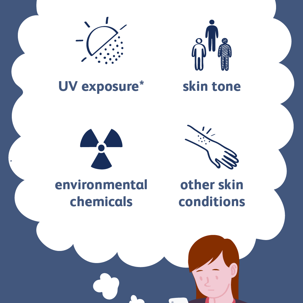
- Multiple factors can place a patient at risk for developing melanoma:
- Some factors are modifiable while others are inherent to the individual
- Skin type:
- Caucasians have at least 20 times the melanoma incidence of African Americans and five times the melanoma incidence of American Hispanics
- In addition, white patients with red or blond hair, fair complexion, or blue eyes are at increased risk for melanoma
- Age and Gender:
- The incidence of melanoma increases with age
- The incidence of melanoma is 1.7-fold higher for women than men before 49 years of age
- Over age 70:
- The incidence of melanoma is 2.4-fold higher for men than women
- In general, the incidence of melanoma is higher in men than in women
- Specifically, a man’s lifetime risk of melanoma development:
- Is approximately 1.5 times greater than a woman’s risk
- Overexposure to ultraviolet radiation (UVR) from the sun:
- Overexposure of UVR from the sun has been associated with an increased risk of melanoma
- Genetic sequencing data also support the role of UV melanomagenesis
- Known to be a tumor with one of the highest mutational loads:
- A seminal report of the melanoma effort within The Cancer Genome Atlas Program:
- Revealed that a majority of somatic mutations in melanoma indeed have a “UV signature”
- A seminal report of the melanoma effort within The Cancer Genome Atlas Program:
- Data support that damage from sunburns in childhood or even in adulthood are associated with increased risk:
- A correlation has been identified between the number of severe and painful sunburn episodes and the risk of melanoma:
- For example, patients who have a history of more than 10 severe sunburns:
- Are more than twice as likely to develop a melanoma compared to patients who have no history of sunburns
- For example, patients who have a history of more than 10 severe sunburns:
- A correlation has been identified between the number of severe and painful sunburn episodes and the risk of melanoma:
- Use of indoor tanning devices:
- Multiple studies support that use of an indoor tanning device is strongly also associated with increased risk of melanoma
- A systematic review by the International Agency for Research on Cancer (IARC) demonstrated:
- A 15% increased relative risk of melanoma in individuals who had ever used a sunbed versus those who had never (RR 1.15; 95% CU 1.00 to 1.31)
- The dangers of indoor tanning have been corroborated by subsequent US and Australian groups
- Young age of onset and higher frequency of use are key risk factors that are associated with even greater risk of melanoma
- Indeed, a well-designed Minnesota case–control study showed increased risk with number of years, hours, and sessions of indoor tanning, independent of outdoor sun exposure:
- These researchers also found that 97% of women diagnosed with melanoma before age 30 had indoor tanned
- An Australian population-based study showed that tanning bed use more than 10 times per year:
- Was associated with a doubling of melanoma risk in patients at least 30 years of age, and the association was stronger with earlier exposure
- Young patients who use indoor tanning devices more than 10 times annually have more than 7 times the melanoma risk compared to individuals who do not indoor tan
- A recent meta-analysis estimated a 1.8% increased melanoma risk for each additional tanning bed session
- Since 2009, the World Health Organization lists tanning beds as:
- A Class I carcinogen
- Previous melanoma:
- Individuals with a personal history of melanoma have an increased risk of developing a second melanoma of:
- Approximately 3% to 7% (life time risk)
- Individuals with a personal history of melanoma have an increased risk of developing a second melanoma of:
- Benign nevi:
- Although a benign nevus is most likely not a precursor of melanoma, the presence of large numbers of nevi has been consistently associated with an increased risk of melanoma:
- Persons with ≥ 50 nevi, all of which are > 2 mm in diameter:
- Have 5 to 17 times the melanoma risk of persons with fewer nevi
- Persons with ≥ 50 nevi, all of which are > 2 mm in diameter:
- Individuals who tend to develop freckles also have an increased risk of melanoma
- Although a benign nevus is most likely not a precursor of melanoma, the presence of large numbers of nevi has been consistently associated with an increased risk of melanoma:
- Family history:
- Approximately 10% of individuals diagnosed with melanoma have a family member with a history of melanoma
- A family history of melanoma increases an individual’s risk of melanoma three- to eightfold
- Furthermore, persons who have two or more family members with melanoma are also at a particularly high risk
- Genetic predisposition:
- Approximately 8% to 12% of melanomas occur in individuals with a genetic predisposition
- Specific genetic alterations have been implicated in the pathogenesis of melanoma:
- Cyclin-dependent kinase inhibitor 2A (CDKN2A):
- Is the most commonly identified mutation in suspected familial melanoma:
- A tumor suppressor gene located on chromosome 9p21:
- CDKN2A is probably involved in familial and sporadic cutaneous melanoma
- A tumor suppressor gene located on chromosome 9p21:
- Data from North America, Europe, and Australia correlate germline CDKN2A mutation (45%, 57%, and 20%, respectively):
- With multiple-case families
- Early age of onset
- Multiple primaries within an individual patient
- Is the most commonly identified mutation in suspected familial melanoma:
- Deletions or rearrangements of chromosomes 10 and 8p are also well documented in cutaneous melanoma
- Also associated with an increase in melanoma incidence are copy number gains of:
- Chromosomes 2, 6p, 7, 8, 17, 19, and 20
- These melanomas also tend to present at an earlier age and individuals may have multiple primary lesions
- Chromosomes 2, 6p, 7, 8, 17, 19, and 20
- Cyclin-dependent kinase inhibitor 2A (CDKN2A):
- Atypical mole and melanoma syndrome:
- Previously known as dysplastic nevus syndrome, atypical mole and melanoma syndrome is characterized by:
- The presence of multiple, large (> 5 mm) atypical dysplastic nevi generally in nonexposed areas of skin that represent a distinct clinicopathologic type of melanocytic lesion
- Melanomas can originate from either normal skin or from a dysplastic nevus
- Since the actual frequency of an atypical mole progressing to melanoma is small:
- Resection of all dysplastic nevi is not indicated
- However, new, changing, or symptomatic lesions:
- That appear suspicious for melanoma on clinical and / or dermoscopic examination:
- Should be evaluated histologically
- That appear suspicious for melanoma on clinical and / or dermoscopic examination:
- Previously known as dysplastic nevus syndrome, atypical mole and melanoma syndrome is characterized by:
#Arrangoiz #CancerSurgeon #SurgicalOncologist #HeadandNeckSurgeon #CASO #CenterforAdvancedSurgicalOncology #Teacher #SkinCancer #Melanoma
