Blog

Fibroadenomas of the Breast

  • Fibroadenomas of the breast are benign tumors:
    • Composed of stromal and epithelial elements:
    • That are commonly seen in young women 
    • Multiple or complex fibroadenomas:
      • May indicate a slightly increased risk for breast cancer: 
      • The relative risk of breast cancer in patients with such fibroadenomas: 
        • Is approximately twice that of patients of similar age without fibroadenomas 
    • A patient’s age determines the preferred imaging method:
      • In general, ultrasonography (US) is preferred:
      • If a palpable mass is found
      • If a patient is younger than 30 years
      • If the patient is pregnant 
      • Mammography and US are both useful if the patient has: 
      • A palpable mass
      • Is older than 30 years
      • Is not pregnant
      • In patients younger than 30 years: 
      • The most appropriate modality is ultrasound:
        • Because the patient is spared radiation exposure and because the likelihood for fibroadenoma is high
      • Mammography is not indicated as the primary imaging study in women younger than 30 years:
      • Unless high-risk factors are present
      • Computed tomography (CT) scanning: 
      • Is not initially indicated for assessing a palpable lump in a woman in women younger than 30 years:
        • Because of radiation exposure 
        • The inability of CT to demonstrate micro-calcifications
        • The lack of specificity in the findings
      • Magnetic resonance imaging (MRI):
      • Is not initially indicated for assessing a palpable lump in women younger than 30 year:
        • Mainly because of its high cost and the high likelihood of false-positive findings
      • Positron emission tomography:
      • Is expensive and is not universally available
      • On mammograms:
      • Fibroadenomas typically appear as:
        • Circumscribed oval or round masses:
          • Which occasionally have coarse calcifications
      • On ultrasonograms:
      • Fibroadenomas appear as:
        • Circumscribed, homogeneous, oval, hypoechoic masses
          • That may have gentle lobulations
          • A smooth, thin, echogenic capsule
          • Variable acoustic enhancement; and homogeneity 
      • On MRI:
      • Fibroadenomas typically appear as smooth masses with high signal intensity on T2-weighted images and enhancement with the administration of gadolinium-based contrast agent
      • Fibroadenoma:
      • Is a common benign breast lesion:
        • Results from the excess proliferation of connective tissue
      • Fibroadenomas characteristically contain both:
        • Stromal and epithelial cells 
      • Epidemiology: 
      • They usually occur in women:
        • Between the ages of 10 and 40 years
        • It is the most common breast mass:
          • In the adolescent and young adult population :
          • Their peak incidence is between:
          • 25 and 40 years
      • The incidence decreases after 40 years 
      • Clinical presentation:
      • The typical presentation is in a woman of reproductive age:
        • With a mobile palpable breast lump:
      • Due to their hormonal sensitivity:
        • Fibroadenomas commonly enlarge during pregnancy and involute at menopause:
          • Hence, they rarely present after the age of 40 years
      • The lesions are well defined and well-circumscribed clinically and the overlying skin is normal
      • The lesions are not fixed to the surrounding parenchyma and slip around under the palpating fingers:
        • Hence the colloquial term a breast “mouse”
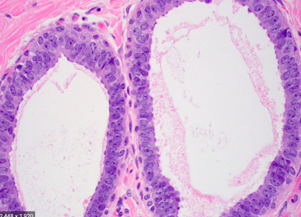
      • Pathology:
      • A fibroadenoma is a type of adenomatous breast lesion:
        • It contains epithelium:
          • Has minimal malignant potential
      • Multiple fibroadenomas occur in:
        • 10% to 15% of patients: 
          • Patients with multiple fibroadenomas:
          • Tend to have a strong family history of these tumors
        • They are assumed to be:
          • Aberrations of normal breast development (ANDI) or the product of hyperplastic processes:
          • Rather than true neoplasms
      • Fibroadenomas can be stimulated by estrogen and progesterone:
        • Some fibroadenomas also have receptors and respond to:
          • Growth hormone and epidermal growth factor
      • When found in an adolescent girl:
        • The term juvenile fibroadenoma is more appropriate 
      • Location:
      • Although they can be located anywhere in the breast:
        • There may be a predilection for the upper outer quadrant 
      • Associations:
      • Cyclosporin use o Cowden syndrome
      • Radiographic features:
      • Mammography:
        • Fibroadenomas have a spectrum of features:
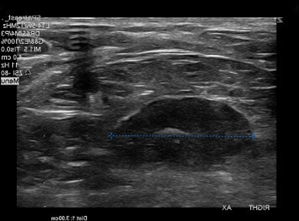
          • Well-circumscribed discrete oval mass hypodense or isodense to the breast glandular tissue
          • Mass with macro-lobulation or partially obscured margin 
          • Involuting fibroadenomas in older, typically postmenopausal patients may contain:
          • Calcification:
            • Often producing the classic, coarse popcorn calcification appearance
            • In some cases the whole lesion is calcified 
            • Calcification may also present as crushed stone-like micro-calcification:
              • Which makes differentiation from malignancy difficult 
      • Breast ultrasound: 
        • Typically seen as a well-circumscribed, round to ovoid, or macro-lobulated mass with generally uniform hypoechogenicity 
        • Intralesional sonographically detectable calcification: 
          • May be seen in approximately 10% of cases
        • Sometimes a thin echogenic rim (pseudo capsule) may be seen sonographically 
      • Breast MRI:
        • T1: typically hypo intense or isointense compared with adjacent breast tissue 
        • T2: can be hypo- or hyper intense
        • T1 C+ (Gd): can be variable but a majority will show slow initial contrast enhancement followed by a persistent delayed phase (type I enhancement curve)
          • Non-enhancing internal septations may be seen 
      • Diagnosis:
      • These lesions are easily biopsied under ultrasound guidance
      • When a lesion has the typical features of a fibroadenoma on ultrasound and there are no clinical red flags:
        • They can be safely followed clinically 
      • When lesions enlarge or have atypical imaging findings:
        • Ultrasound-guided core biopsy is a minimally invasive outpatient procedure that will give a diagnosis with virtually no complications 
        • There may be a maximum diameter above which a biopsy should be done if no previous imaging is available
          • The reason for intervention based on size is that a phyllodes tumor may be indistinguishable from a fibroadenoma on ultrasound:
          • maximum diameter of 2.5 cm may be a useful benchmark for biopsy if you have no previous imaging 
        • Interval enlargement:
          • Is an indication for biopsy 
    • Symptomatic, progressively enlarging masses or atypical presentations:
      • May warrant surgical excision
    • If a needle biopsy shows that a mass less than 2 centimeters in size is a fibroadenoma, with no other concerning features:
      • It does not have to be surgically removed
    • The patient’s core biopsy pathology demonstrating a fibroadenoma is consistent with the typical imaging findings of a smooth, round, hypoechoic mass:
      • As the biopsy is concordant, no further intervention is needed:
      • Follow-up for reassurance is acceptable
    • Treatment and prognosis:
      • They are benign lesions with minimal or no malignant potential 
      • The risk of malignant transformation is extremely low: 
      • Has been reported to range around 0.0125% to 0.3%
      • Indications for biopsy include: 
      • Enlarging lesion 
      • Atypical findings on ultrasound 
      • A lesion above 2.5 cm and there are no previous studies for comparison
      • Patient peace of mind:
        • Some patients are simply not happy with a palpable mass in the breast without a histological diagnosis:
          • This is a valid and reasonable indication for biopsy

#Arrangoiz #CancerSurgeon #BreastSurgeon #SurgicalOncologist #BreastFibroadenoma #Breast Cancer 

Fibromastosis of the Breast

  • Fibromatosis (also called desmoid fibromatosis):
    • Is characterized by a proliferation of histologically bland spindle cells arranged in long fascicles, with infiltrative architecture:
      • Similar to fibromatosis in other sites
    • Fibromatosis is a rare tumor with:
      • Locally aggressive behavior
      • High incidence of local recurrence
    • Various causes have been accused of being its etiology including:
      • Genetic factors
      • Endocrine factors
      • Surgical trauma
    • For treatment of fibromatosis:
      • Wide local excision with adequate safety margins is considered the standard of care
  • The breast is an unusual site for occurrence of desmoid type fibromatosis with few cases reported in literature:
    • It represents 0.2% of all breast tumors
    • 4% of all extra-abdominal desmoid tumors
    • Bilaterality has been reported in:
      • 4% of cases
    • Most of these cases were reported in:
      • Young fertile females:
        • With rare cases reported in males
    • The reported risk factors include:
      • Surgical trauma
      • Silicone implants
      • Association with Gardener’s syndrome
    • Clinically, it is presented as a firm mass with skin dimpling and nipple retraction in superficial and retro-areolar tumors
      • Neither nipple discharge nor axillary lymphadenopathy commonly occur with fibromatosis 
    • Mammography:
      • Presents irregular walled and highly dense lesion with no calcifications mimicking sometimes breast carcinoma
    • Microscopic examination of desmoid tumor usually reveals:
      • The characteristic irregular bundles of spindle cells with regular nuclei surrounded by abundant collagen 
      • Giant cells, macrophages and lymphocytes:
        • Are noticed mostly peripherally 
    • Although fibromatosis does not have metastatic potential:
      • It can be locally aggressive
    • Metaplastic carcinoma (‘fibromatosis like,’ low-grade spindle cell):
      • Should be ruled out with keratin or other epithelial stains in every case
    • The differential diagnosis also includes:
      • Nodular fasciitis
      • Myofibroblastoma
      • Fascicular pseudoangiomatos stromal hyperplasia (PASH)
      • Stromal overgrowth of phyllodes tumors
      • Other spindle cell sarcomas
    • Fibromatosis may be very difficult to distinguish from scar / granulation tissue pathologically:
      • Especially in patients with prior resection and concern for recurrence
    • Beta-catenin nuclear staining:
      • Is characteristic of fibromatosis:
        • But is positive in only up to 70% of cases and may be difficult to demonstrate
Spindle cell proliferation formed of bland looking cell arranged in short fascicles with dissection of breast fat lobules at the periphery 
The cells showed diffuse positive nuclear staining for B-catenin 
  • References
  • Lee A, Gobbi H. Desmoid type fibromatosis In: Lakhani SR, Ellis IO, Schnit SJ, Tan PH, van de Vijver MJ et al. eds. WHO Classification of Tumours of the Breast. Lyon: IARC Press, 2012:131-2.
  • Schnitt SJ, Collins L. Biopsy Interpretation of the Breast. 3rd edition. Philadelphia, PA: Wolters Kluwer, 2018: 412-416.
  • Kuba MG, Lester SC, Giess CS, Bertagnolli MM3, Wieczorek TJ4, Brock JE. Fibromatosis of the breast: diagnostic accuracy of core needle biopsy. Am J Clin Pathol. 2017;148(3):243-250.
  • Kim T, Jung EA, Song JY, Roh JH, Choi JS, Kwon JE. Prevalence of the CTNNB1 mutation genotype in surgically resected fibromatosis of the breast. Histopathology. 2012;60(2):347-356.

Complex Fibroadenoma

  • According to the World Health Organization definition:
    • Complex fibroadenoma is a fibroadenoma containing:
      • Cysts greater than 3 mm in size
      • Sclerosing adenosis
      • Epithelial calcifications
      • Papillary apocrine hyperplasia
  • While this is a benign diagnosis:
    • Some authors have reported a slightly increased risk of subsequent breast cancer development:
      • On par with other proliferative lesions of the breast:
        • About 2 to 3-fold
          • Thus, the terminology “complex fibroadenoma” is not always employed
  • The term “complex” does not imply any relation to radial scar or epithelial atypia in the setting of a fibroadenoma
  • References
    • Tan PH, Tse G, Lee A, et al. Fibroepithelial tumours. In: Lakhani SR, Ellis IO, Schnit SJ Tan PH, van de Vijver MJ. eds. WHO Classification of Tumours of the Breast. Lyon: IARC Press, 2012:142-143.
    • Dupont WD, Page DL, Parl FF, Vnencak-Jones CL, Plummer WD Jr, Rados MS, et al. Long-term risk of breast cancer in women with fibroadenoma. N Engl J Med. 1994;331(1):10-15.
    • Nassar A, Visscher DW, Degnim AC, Frank RD, Vierkant RA, Frost M, et al. Complex fibroadenoma and breast cancer risk: a Mayo Clinic Benign Breast Disease Cohort Study. Breast Cancer Res Treat.2015;153(2):397-405.
    • Krings G, Bean GR, Chen YY. Fibroepithelial lesions; The WHO spectrum. Semin Diagn Pathol. 2017;34(5):438-452.

#Arrangoiz #BreastSurgeon #BreastCancer #CancerSurgeon #SurgicalOncologist #ComplexFibroadenoma #Miami #Mexico

Atypical Lobular Hyperplasia

  • Atypical lobular hyperplasia (ALH):
    • Is generally an incidental finding on core needle biopsy:
      • Without specific defining characteristics on:
        • Mammography, ultrasound, or MRI
  • palpable breast mass which yields ALH at core needle biopsy is discordant:
    •  This should prompt further diagnostic work-up with a second biopsy:
      • Either core or excisional
  • ALH should only be considered for observation:
    • When there is radiologic, pathologic, and clinical concordance:
      • As the risk of upstaging to carcinoma in this scenario is:
        • Less than 5%
  • ALH alone:
    • Confers a 4 to 5-fold increased risk of future breast cancer in either breast
    • The probability of upgrade to invasive carcinoma with a diagnosis of atypical lobular hyperplasia (ALH) on core needle biopsy:
      • Is reported as less than 3%
  • Excision to achieve negative margins in ALH:
    • Is generally not required
  • Having a family history:
    • Does not appear to increase the risk associated with atypical hyperplasia
  • Relevant indications for genetic testing include:
    • A personal history of breast cancer ≤ age 45
    • Triple negative breast cancer ≤ age 60
    • A first-degree relative with breast cancer ≤ age 50
    • Two or more first- or second-degree relatives with breast cancer at any age
    • Patient or relative with bilateral breast cancer
    • Male breast cancer in a relative at any age
  • Risk-reducing mastectomy:
    • Can be considered in patients with very high lifetime breast cancer risk:
      • Usually reserved for women with high-penetrance gene mutations:
        • Such as BRCA 1 or BRCA 2
  • References
    • Morrow M, Schnitt SJ, Norton L. Current management of lesions associated with an increased risk of breast cancer. Nat Rev Clin Oncol. 2015;12(4):227-238.
    • Murray MP, Luedtke C, Liberman L, Nehhozina T, Akram M, Brogi E. Classic lobular carcinoma in situ and atypical lobular hyperplasia at percutaneous breast core biopsy: outcomes of prospective excision. Cancer. 2013;119(5):1073-1079.
    • Middleton LP, Sneige N, Coyne R, Shen Y, Dong W, Dempsey P. Most lobular carcinoma in situ and atypical lobular hyperplasia diagnosed on core needle biopsy can be managed clinically with radiologic follow-up in a multidisciplinary setting. Cancer Med. 2014;3(3):492-499.
    • Hartmann LC, Sellers TA, Frost MH, Lingle WL, Degnim AC, Ghosh K, et al. Benign breast disease and the risk of breast cancer. N Engl J Med. 2005;353(3):229-237.
    • Degnim AC, Visscher DW, Berman HK, Frost MH, Sellers TA, Vierkant RA, et al. Stratification of breast cancer risk in women with atypia: a Mayo cohort study. J Clin Oncol. 2007;25(19):2671-2677.
    • Smart CE, Furnival CM, Lakhani SR. Chapter 17. High-Risk Lesions: ALH/LCIS/ADH. In: Kuerer HM ed. Kuerer’s Breast Surgical Oncology. New York, NY: McGraw-Hill, 2010.

#Arrangoiz #BreastSurgeon #CancerSurgeon #SurgicalOncologist #AtypicalLobularHyperplasia #ALH #Miami #Mexico

Flat Epithelial Atypia

  • Flat epithelial atypia:
    • Confers a relative risk of developing breast cancer of:
      • Approximately 1.5
    • Excisional biopsy may be indicated if:
      • Calcifications are incompletely removed or 
      • If there is question of sampling error:
        • But excision to negative margins is not required
    • The probability of finding a concurrent invasive carcinoma is:
      • Approximately 1% to 5% when flat epithelial atypia is found on core needle biopsy:
        • Given the small (less than two-fold) associated increase in breast cancer risk:
          • Chemoprevention is not indicated nor is routine annual screening breast MRI
  • References:
    • Sudarshan M, Meguerditchian AN, Mesurolle B, Meterissian S. Flat epithelial atypia of the breast: characteristics and behaviors. Am J Surg. 2011;201(2)245-250.
    • Piubello Q, Parisi A, Eccher A, Barbezeni G, Franchini Z, Iannucci A. Flat epithelial atypia on core needle biopsy: which is the right management? Am J Surg Pathol. 2009;33(7):1078-1084.
    • Martel M, Barron-Rodriguez P, Tolgay Ocal I, Dotto J, Tavassoli FA. Flat DIN 1 (flat epithelial atypia) on core needle biopsy: 63 cases identified retrospectively among 1,751 core biopsies performed over an 8-year period (1992-1999). Virchow’s Arch. 2007;451(5):883-891.
    • Lamb LR, Bahl M, Gadd MA, Lehman CD. Flat epithelial atypia: upgrade rates and risk-stratification approach to support informed decision making. J Am Coll Surg. 2017;225(6):696-701.

Fat Necrosis

  • Fat necrosis of the breast:
    • Is a benign condition that most commonly occurs as the result of breast trauma or surgical intervention
  • Fat necrosis can be confused with a:
    • Malignancy on physical examination and may mimic malignancy on radiologic studies
  • It is sometimes necessary to biopsy these lesions to confirm the diagnosis:
    • Although experienced radiologists can usually determine that a lesion represents fat necrosis based on mammographic and ultrasound findings such as:
      • Oil cysts (collections of liquefied fat)
  • Once the diagnosis is established:
    • Excision is not necessary and there is no increased risk of subsequent breast cancer
  • References
    • Guaray M, Sahin AA. Benign breast diseases: classification, diagnosis and management. Oncologist. 2006;11(5):435-449.
    • Agel NM, Howard A, Collier DS. Fat necrosis of the breast: a cytological and clinical study. Breast. 2001;10(4):342-345

#Arrangoiz #BreastSurgeon #BreastCancer #FatNecrosis #CancerSurgeon #Miami #Mexico #SurgicalOncology

Periductal Mastitis

  • Periductal mastitis:
    • Is an inflammatory condition of the subareolar ducts:
      • That often presents with periareolar inflammation
  • Secondary infection of inflamed ducts can occur:
    • Leading to duct damage and subsequent rupture with abscess formation:
      • These abscesses often drain spontaneously at the edge of the areola
    • Recurrent abscesses and a draining fistula can occur.
  • Most patients with periductal mastitis are smokers:
    • It has been postulated that smoking is associated with damage of the subareolar ducts:
      • With tissue necrosis and subsequent infection
    • Therefore, smoking cessation is helpful for reducing the risk of repeat infections
  • The management of subareolar abscess consists of:
    • Antibiotic therapy and abscess drainage
    • In refractory cases;
      • Excision of the fistula track is necessary
  • References
    • Schafer P, Furrer C, Mermillod B. An association of cigarette smoking with recurrent subareolar breast abscess. Int J Epidemiol 1988;17(4):810-813.
    • Bundred NJ, Dover MS, Coley S, Morrison JM. Breast abscesses and cigarette smoking. Br J Surg. 1992;79(1):58-59.

#Arrangoiz #BreastSurgeon #CancerSurgeon #SurgicalOncologist #BreastCancer #Miami #Mexico #PerriductalMastitis

Lactational Mastitis

  • Lactational mastitis:
    • Is a condition in which a woman’s breast becomes:
      • Red, painful, and swollen:
        • Within 12 to 24 hours after breast engorgement with poor milk drainage
    • Most episodes of lactation mastitis:
      • Are caused by Staphylococcus aureus
    • The condition is characterized by:
      • Pain, redness, fever and malaise
  • References
    • Kvist LJ, Larsson BW, Hall-Lord ML, Steen A, Schalen C. The role of bacteria in lactational mastitis and some considerations of the use of antibiotic treatment. Int Breastfeed J. 2008;3:6.
    • Schoenfeld EM, McKay MP. Mastitis and methicillin-resistent staphylococcus aureus (MRSA): the calm before the storm? J Emerg Med. 2010;38(4):e31.

Nipple Discharge

  • Nipple discharge is common:
    • With more than half of women of reproductive age able to express discharge with manipulation
  • A thorough history to determine whether a discharge is:
    • Physiologic or pathologic:
      • Is important to determine the need for additional workup
  • Characteristics of pathologic nipple discharge include:
    • Spontaneous
    • Bloody or clear
    • Unilateral / single duct
    • Presence of a palpable mass associated with the discharge
  • Nipple discharge should be further evaluated if considered pathologic with:
    • Diagnostic mammography
    • Retroareolar ultrasound
    • Clinical examination
  • The differential diagnosis of focal dilation on ultrasound includes:
    • Lactational changes
    • Duct ectasia
    • Malignancy
  • Based on ultrasound findings and if the nature of the nipple discharge is concerning that it is pathologic:
    • It should be further evaluated with tissue sampling:
      • An image-guided excisional biopsy of the duct in question:
        • Is a minimally invasive simple technique to definitively rule out malignancy and establish the diagnosis:
          • This can be accomplished with placement of an intraoperative probe into the offending duct, or by placement of a preoperative wire or seed marker into the imaging abnormalities
  • Failure to identify a cause of the discharge warrants further work-up:
    • Ductogram and MRI have also been considered in the routine workup of nipple discharge:
      • Though data is inconclusive on their effectiveness
    • Ductogram can be considered operator dependent:
      • Has a reported positive predictive value of 19%
      • Negative predictive value of 63%
    • Successful localization of an offending lesion by ductogram:
      • Occurs in 50% to 60% of cases
    • MRI, on the other hand, has a reported:
      • Positive predictive value of 24% to 56%
      • Negative predictive value of 87% to 98%
        • Suggesting discussion of the technique may be reasonable in some situations but the limitations should also be reviewed with patients
  • In the absence of any palpable concerns on examination or suspicious findings on conventional mammogram and ultrasound imaging:
    • Duct excision reveals an intraductal papilloma:
      • As the cause of the discharge about 50% of the time
  • While most studies suggest that duct excision is indicated for patients with pathologic nipple discharge for both diagnosis and treatment:
    • Observation may be appropriate for selected patients with pathologic discharge who have:
      • normal examination, mammogram, and subareolar ultrasound:
        • In whom the risk of carcinoma is 3%
  • References:
    • Cabioglu N, Hunt KK, Singletary SE, et al. Surgical decision making and factors determining a diagnosis of breast carcinoma in women presenting with nipple discharge. J Am Coll Surg. 2003;196:354-364.
    • Vargas HI, Vargas MP, Eldrageely K, Gonzalez KD, Khalkhali I. Outcomes of clinical and surgical assessment of women with pathologic nipple discharge. Am Surg. 2006;72:124-128.
    • Romanoff A, Nulsen B, Mester J, Jaffer S, Weltz C. Ultrasound‐guided wire localization of focal ductal dilatation in the evaluation and treatment of pathologic nipple discharge. Breast J. 2018;24(3):356–359.
    • Morrogh M, Morris EA, Liberman L, Borgen PI, King TA. The predictive value of ductography and magnetic resonance imaging in the management of nipple discharge. Ann Surg Oncol. 2007;14(12):3369-3377.
    • Woodward S, Daly CP, Patterson SK, Joe AI, Helvie MA. Ensuring excision of intraductal lesions: marker placement at time of ductography. Acad Radiol. 2010;17(11):1444-1448.
    • Sanders LM, Daigle M. The rightful role of MRI after negative conventional imaging in the management of bloody nipple discharge. Breast J. 2016;22(2):209-212.
    • Nelson RS, Hoehn JL. Twenty-year outcome following central duct resection for bloody nipple discharge. Ann Surg. 2006;243(4):522-524.
    • Gray RJ, Pockaj BA, Karstaedt PJ. Navigating murky waters: a modern treatment algorithm for nipple discharge. Am J Surg. 2007;194(6):850-854.

#Arrangoiz #BreastSurgeon #CancerSurgeon #SurgicalOncologist #BreastCancer #NippleDischarge @PathologicNippleDischarge #Mexico #Miami