Blog

Anaplastic Thyroid Cancer

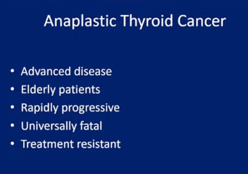
  • Anaplastic carcinoma of the thyroid (ATC):
    • Is the most aggressive thyroid gland malignancy
  • Although ATC accounts for less than 2% of all thyroid cancers:
    • It causes up to 40% of deaths from thyroid cancer
  • The aggressive nature of ATC:
    • Makes treatment studies difficult to perform
  • The overall 5-year survival rate:
    • Is reportedly less than 10%, and most patients do not live longer than a few months after diagnosis
  • Patients with ATC:
    • Typically present with a rapidly growing neck mass
  • Metastases, particularly in the lung:
    • Are likely to be present at diagnosis more than 50% of the time
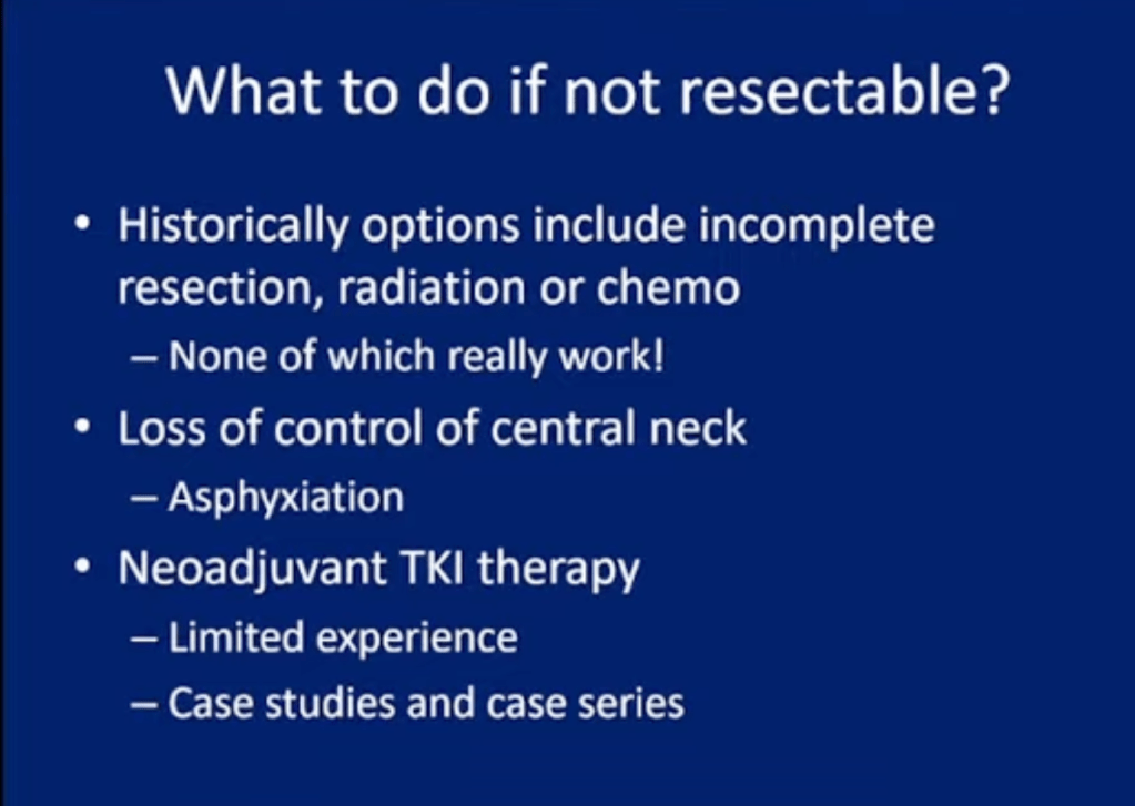
  • Treatment is mostly palliative
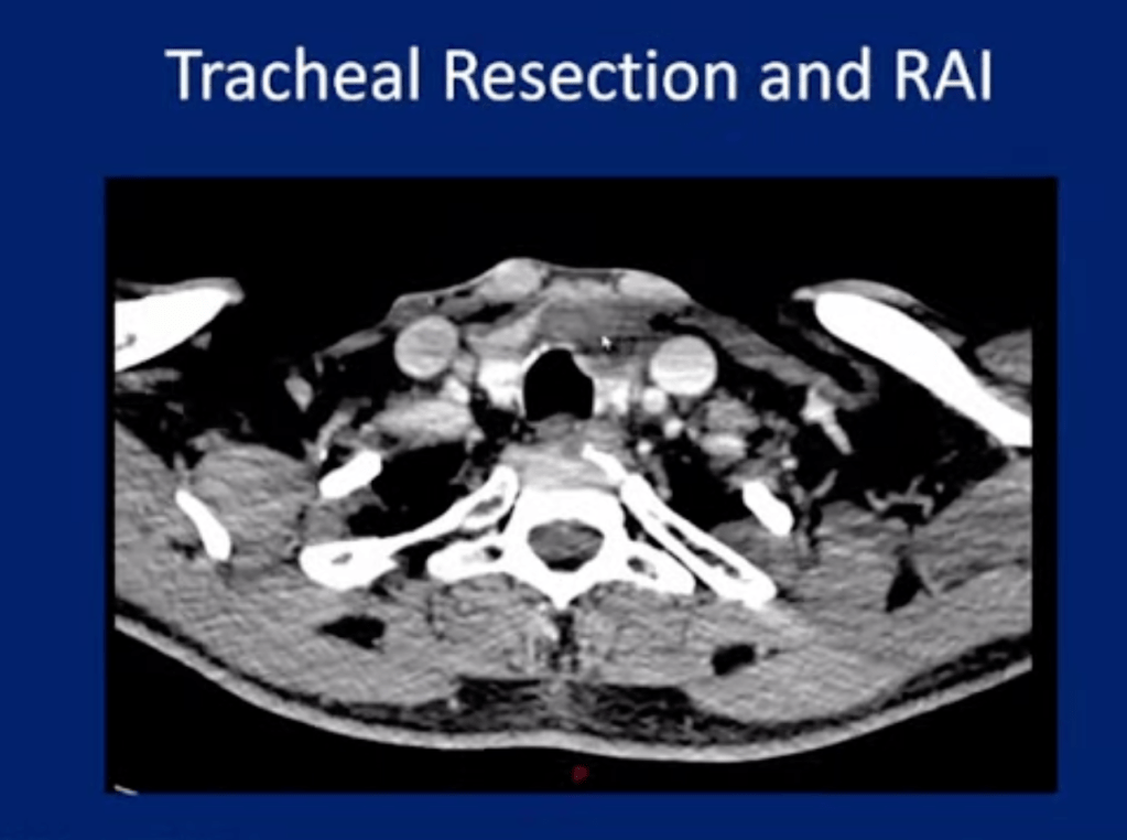
  • Surgical resection with adjuvant radiation therapy and chemotherapy:
    • May prolong survival somewhat and improve quality of life
  • Anaplastic carcinoma of the thyroid (ATC):
    • Generally occurs in people in iodine-deficient areas and in a setting of previous thyroid pathology (eg, preexisting goiter, follicular thyroid cancer, papillary thyroid cancer)
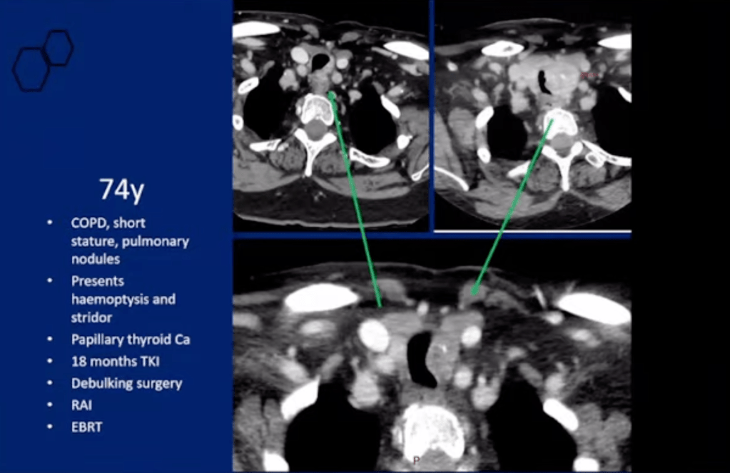
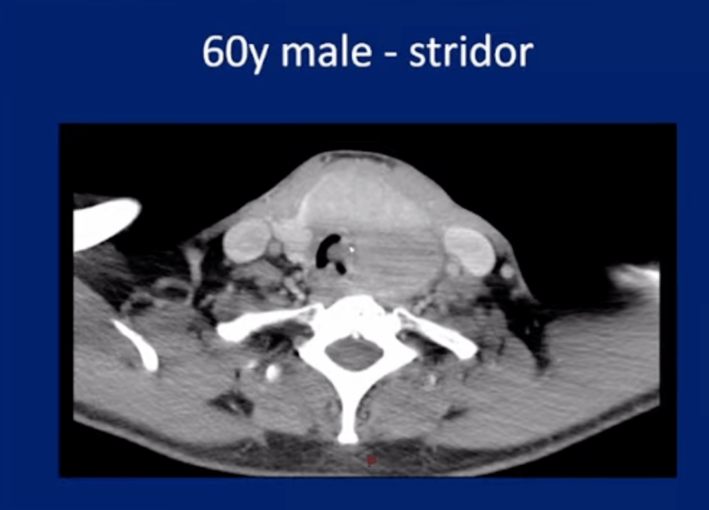
  • Local invasion of adjacent structures (eg, trachea, esophagus):
    • Commonly occurs
  • ATC is believed to occur from a:
    • Terminal dedifferentiation of previously undetected long-standing thyroid carcinoma (eg, papillary, follicular)
  • ATC has a genetic association with:
    • Oncogenes C-myc, H-ras, and Nm23
  • Mutations in genes that code for BRAF, RAS, catenin (cadherin-associated protein), beta 1, PIK3CA, TP53, AXIN1, PTEN, and APC:
    • Have been found in ATC, and chromosomal abnormalities are common 
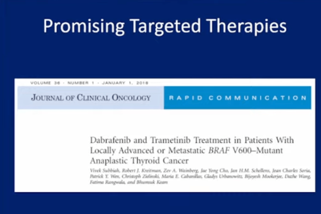
  • Jonker and collegues performed functional genomic RNA profiling on 25 anaplastic thyroid carcinoma and 80 normal thyroid samples and identified 301 significantly upregulated genes, of which the following were seen as potential therapeutic targets:
    • MTOR
    • MET 
    • WEE1
    • PSMD1
    • MERTK
    • FGFR3
    • RARG
    • ESR2
  • Anaplastic carcinoma of the thyroid (ATC):
    • Constitutes less than 2% of all thyroid malignancies in the United States:
      • Which equates to slightly more than 1000 new cases annually
    • Fortunately, the incidence appears to be declining
    • Worldwide frequency likely approximates that in the United States
  • The female-to-male ratio:
    • Is approximately 3:1
  • Peak incidence occurs during the sixth to seventh decades of life
  • The age range of affected patients reportedly is 15-90 years

#Arrangoiz #ThyroidSurgeon #CancerSurgeon #SurgicalOncologist #HeadandNeckSurgeon #EndocrineSurgery #ThyroidCancer #Miami #Mexico #MountSinaiMedicalCenter #Surgeon #Teacher #ThyroidNodules #RadioactiveIodine #RAI #PTC #MSMC

Parathyroid Glands

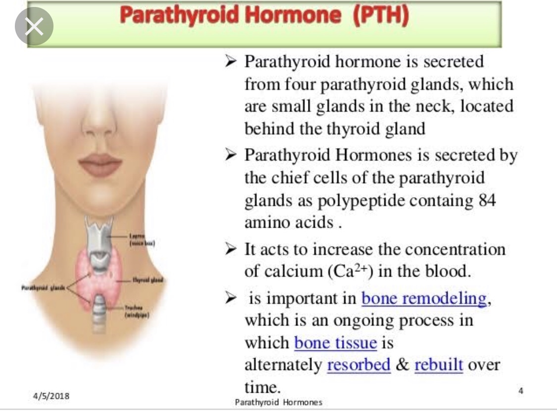
  • Through their secretion of parathyroid hormone (PTH):
    • The parathyroid glands are primarily responsible for:
      • Maintaining extracellular calcium concentrations
  • Hyperparathyroidism:
    • Is a disease characterized by excessive secretion of parathyroid hormone:
      • An 84–amino acid polypeptide hormone
  • The secretion of parathyroid hormone:
    • Is regulated directly by the plasma concentration of ionized calcium
  • The main effects of parathyroid hormone (PTH):
    • Are to increase the concentration of plasma calcium:
      • By increasing the release of calcium and phosphate:
        • From bone matrix
      • Increasing calcium reabsorption:
        • By the kidney
      • Increasing renal production of:
        • 1,25-dihydroxyvitamin D-3 (calcitriol):
          • Which increases intestinal absorption of calcium
    • Thus, overproduction of parathyroid hormone results in:
      • Elevated levels of plasma calcium
  • Parathyroid hormone also causes phosphaturia:
    • Thereby decreasing serum phosphate levels
  • Hyperparathyroidism is usually subdivided into:
    • Primary, secondary, and tertiary hyperparathyroidism
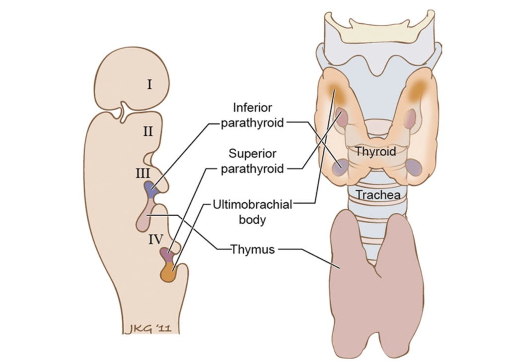
  • Usually, four parathyroid glands are situated posterior to the thyroid gland:
    • A small number of patients have 3, 5, or, occasionally, more glands:
      • The glands are identified based on their location as right or left and superior or inferior.
  • The inferior glands are derived from:
    • The third pharyngeal pouch:
      • This structure is also the embryologic origin of the thymus:
        • Therefore, the inferior glands originate more cephalad than the superior glands:
          • But they migrate along with the thymus to finally become situated more inferiorly than the superior glands
        • Because of their embryologic association with the thymus:
          • The inferior glands are often found adjacent to or within the thymus
          • They are usually located near the inferior pole of the thyroid
  • The superior glands are more consistent in location:
    • Usually found just superior to the intersection of the inferior thyroid artery and the recurrent laryngeal nerve
    • The superior glands are derived embryologically from the fourth pharyngeal pouch:
      • This structure also gives rise to the C cells of the thyroid gland
    • Because of their embryologic origin, the superior glands are occasionally found within the substance of the thyroid gland

#Arrangoiz #ParathyroidSurgeon #ParathyroidSurgeon #HeadandNeckSurgeon #EndocrineSurgery #Hyperparathyroidism #MSMC #MountSinaiMedicalCenter #Mexico #Miami

Identification of the Recurrent Laryngeal Nerve

  • In order to limit any bleeding or tissue distortion that may compromise the identification of the parathyroid glands:
    • The RLN is identified ONLY after all parathyroid glands have been exposed AND a decision made as to which one to remove
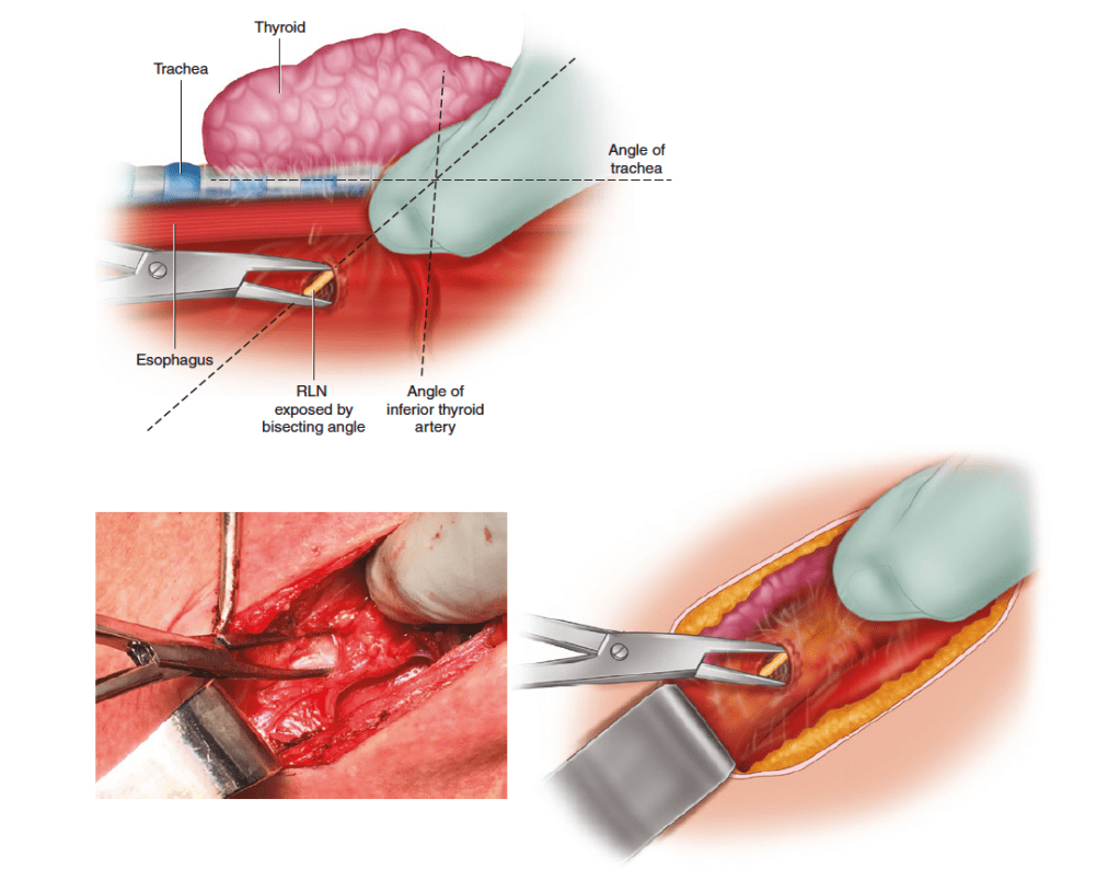
  • The RLN is identified in the inferior portion of the neck caudal to the ITA:
    • The angle between the trachea and the ITA is visually bisected, and careful blunt dissection is performed along this line to identify the nerve (Figure)
  • Further dissection on the perineurium of the nerve toward the ITA can be performed to approximate the course of the RLN
Identification of the recurrent laryngeal nerve allows for safe and confident dissection and ligation of the vascular pedicle supplying the parathyroid gland. The angle between the inferior thyroid artery and the trachea is visually bisected, and areola tissue along this angle is spread with a fine hemostat to identify the nerve

#Arrangoiz #ParathyroidSurgeon #HeadandNeckSurgeon #Surgeon #EndocrineSurgery #MSMC #Teacher #ParathyroidExpert #Hyperparathyroidism #MountSinaiMedicalCenter #Miami #Mexico

Thyroid Cancer

Hurtle Cell Carcinoma

#Arrangoiz #ThyroidSurgeon #CancerSurgeon #SurgicalOncologist #HeadandNeckSurgeon #EndocrineSurgery #ThyroidCancer #Miami #Mexico #MountSinaiMedicalCenter #Surgeon #Teacher #ThyroidNodules #RadioactiveIodine #RAI #PTC #MSMC
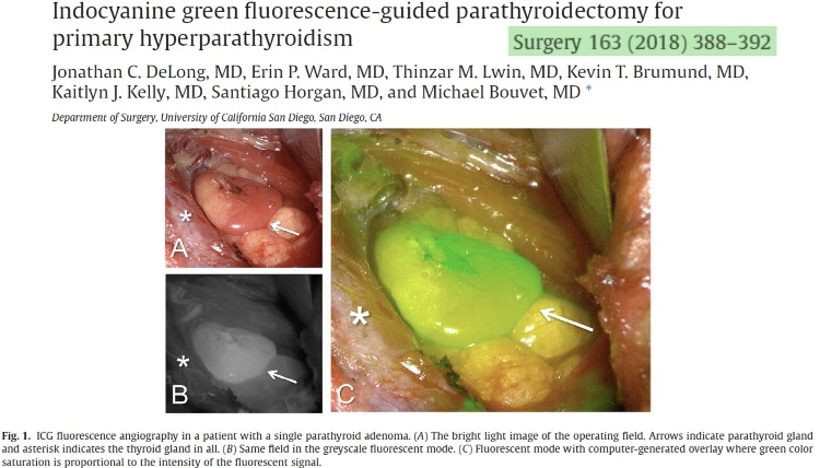
Indocyanine Green Flourescence Guided Parathyroidectomy

  • Indocyanine green fluorescence angiography may be a useful adjunct during parathyroidectomy and thyroid surgery.
  • https://doi.org/10.1016 j.surg.2017.08.018

Thyroid Cancer

#Arrangoiz #ThyroidSurgeon #CancerSurgeon #SurgicalOncologist #HeadandNeckSurgeon #EndocrineSurgery #ThyroidCancer #Miami #Mexico #MountSinaiMedicalCenter #Surgeon #Teacher #ThyroidNodules #RadioactiveIodine #RAI #PTC #MSMC
What is Parathyroid Hormone (PTH)?

  • Did you know that the first report for using intraoperative parathyroid hormone (IOPTH) measurement as an adjunct to guide removal of parathyroid tissue in a patient with hyperparathyroidism:
    • Was published greater than 25 years ago:
      • By Dr. G.L. Irvin the 3rd and colleagues? – Rodrigo Arrangoiz MS, MD, FACS, FSSO

👉CheckYourCalcium (realízate un calcio total en sangre).

#Arrangoiz #ParathyroidSurgeon #ParathyroidExpert #Hyperparathyroidism #PrimaryHyperparathyroidism #CancerSurgeon #EndocrineSurgery #Teacher #Surgeon #HeadandNeckSurgeon #SurgicalOncologist #ParathyroidAdenoma #Hypercalcemia #ElevatedCalciumLevels #Miami #MountSinaiMedicalCenter #MSMC #Mexico #Hialeah

https://www.ncbi.nlm.nih.gov/pubmed/8256205

Skeletal Manifestations of Primary Hyperparathyroidism (PHPT)

  • Skeletal manifestations including osteopenia, osteoporosis, and osteitis fibrosa cystica:
    • Are found in approximately 15% of patients with PHPT
  • PHPT is linked with a reduction in bone mineral density (BMD):
    • Particularly in the cortical bone:
      • Such as in the distal third of the radius
    • In the lumbar region, composed all most exclusively by trabecular bone, and in the femoral region, composed by cortical and trabecular bone:
      • The decrease in BMD is less severe
  • Osteitis fibrosa cystica:
    • A skeletal manifestation that is rarely seen today:
      • Seen in less than five percent of patients with PHPT
    • Is caused by an increase in bone turnover:
      • Can be determined by finding an:
        • Elevated serum alkaline phosphatase level
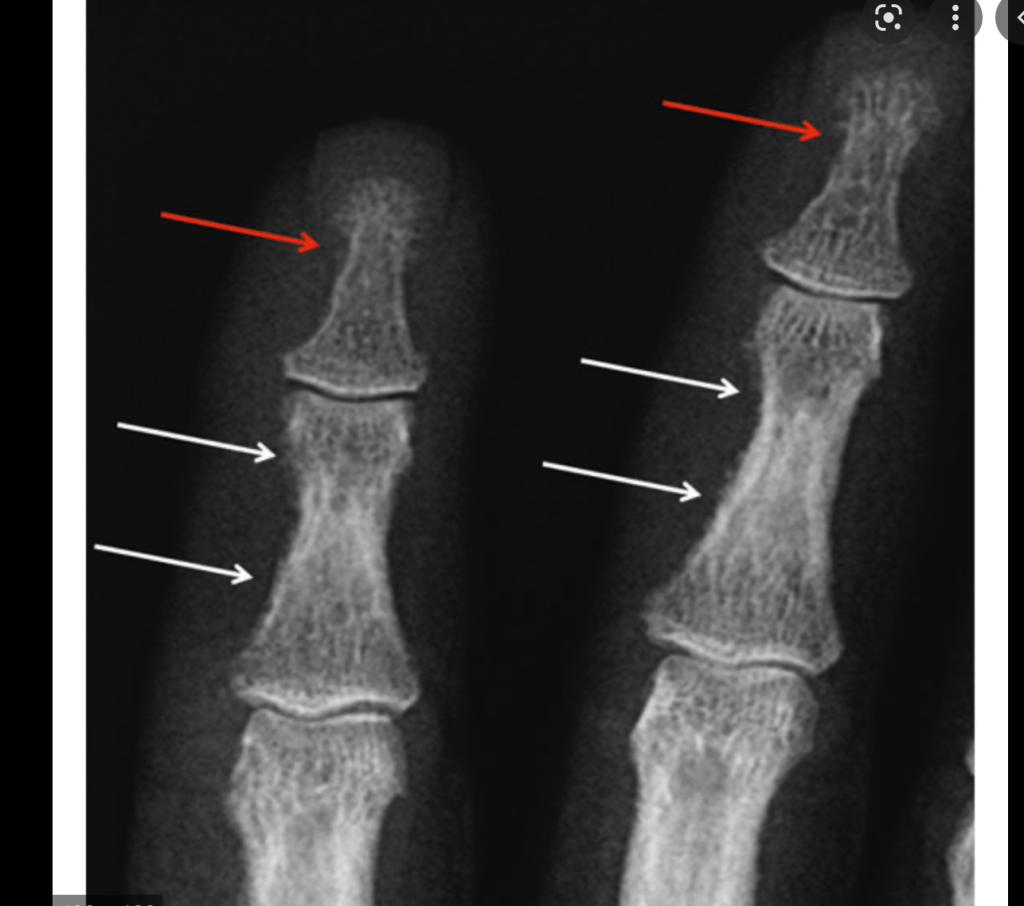
    • The radiologic findings seen in patients with PHPT with bone disease are characterized by:
      • Subperiosteal resorption:
        • Most obvious on the radial aspect of the middle phalanx of the second and third fingers
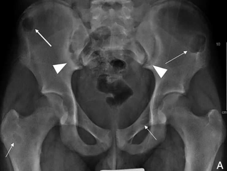
      • Bone cysts
      • Tufting of the distal phalanges:
        • Which are best evaluated on plain x-rays of the hands
      • Brown or osteoclastic tumors:
        • Caused by the accumulations of osteoclasts and fibrous tissue:
        • Brown tumors have a slightly greater incidence in PHPT than in secondary HPT:
          • 3% versus 2%
        • In patients with chronic kidney disease:
          • Persistent and excessive urinary calcium elimination:
            • Can lower serum calcium level:
              • And lead to an increase in PTH secretion
        • This results in mobilization of calcium from the bones:
          • Through rapid osteoclastic turnover of bone to maintain normal serum calcium levels
        • In regions where bone loss is exceptionally fast:
          • Hemorrhage, and reparative granulation tissue, with active, vascular, proliferating fibrous tissue may replace the normal marrow contents:
            • Resulting in a brown tumor
        • Hemosiderin imparts the brown color (hence the name of the lesions
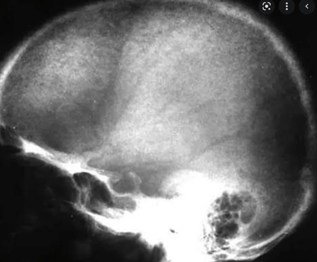
      • The skull also may be affected:
        • Appears mottled with a loss of definition of the inner and outer cortices
  • Patients with normal serum alkaline phosphatase levels:
    • Almost never have clinically apparent osteitis fibrosa cystica
  • Bone disease correlates with serum PTH and vitamin D levels
Generalized osteopenia, erosion of the terminal phalangeal tufts and subperiosteal resorption of bone particularly the radial aspects of the 2nd and 3rd middle phalanges.
Brown Tumors
Salt and Pepper Skull

#Arrangoiz #ParathyroidSurgeon #ParathyroidExpert #HeadandNeckSurgeon #EndocrineSurgery #MountSinaiMedicalCenter #Miami #Mexico #Teacher #Surgeon #Parathyroidectomy #Hypercalcemia #ElevatedCalcium #MSMC

Thyroid Cancer

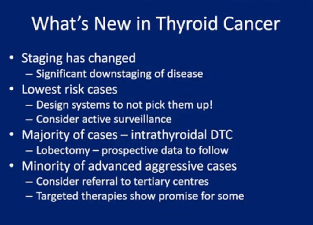
  • Clinical and pathologic staging of patients with cancer is valuable for many reasons, including the following:
    • To estimate risk of recurrence and disease-specific mortality for an individual patient
    • To tailor decisions regarding postoperative adjunctive therapy (such as the need for radioiodine [RAI] ablation and degree of thyroid-stimulating hormone [TSH] suppression):
      • To the patient’s risk for disease recurrence and mortality
    • To make decisions regarding the frequency, modality, and intensity of follow-up based upon an individual patient’s risk of recurrence and mortality
    • To enable accurate communication regarding a patient among health care professionals
    • To allow evaluation of differing therapeutic strategies applied to comparable groups of patients in clinical studies
    • To provide “a method of conveying clinical experience to others without ambiguity
  • Initial postoperative staging:
    • You can use the several staging systems to estimate disease-specific mortality:
      • Tumor, node, metastasis (TNM) staging systems
      • MSKCC:
        • Grade, Age, Metastases, Extent, Size (GAMES) 
      • Mayo Clinic:
        • Metastases, Age, Complete Resection, Invasion, Size  (MACIS)
      • Karolinska Hospital and Institute:
        • DNA ploidy, Age, Metastases, Extent, Size (DAMES)
      • Age, Grade, tumor Extent, Size (AGES)
      • Lahey Clinic:
        • Age, distant Metastasis, Extent, Size (AMES)
    • Clinical and pathologic staging system:
      • Such as the American Thyroid Association (ATA) system:
        • To estimate the risk of recurrence  
  • Re-staging during follow-up:
    • These initial risk assessments are then:
      • Actively modified based upon data obtained during follow-up:
        • That reflect the individualized patient’s response to therapy
    • This dynamic risk assessment approach:
      • Allows for more accurate risk assessments:
        • Than either a static estimate of risk based on the:
          • TNM staging system or the ATA risk of recurrence classification system 
  • The ATA recommends the TNM staging system:
    • For all patients with differentiated thyroid cancer and any of the:
      • Postoperative clinicopathologic staging systems:
        • To achieve more accurate risk factor stratification 
  • It should be recognized, however:
    • No staging system reliably predicts outcome in individual patients:
      • Clinicians should use individual clinicopathologic characteristics:
        • To determine the specific treatment for a given patient
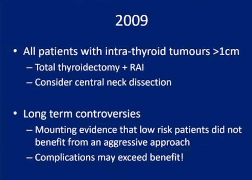
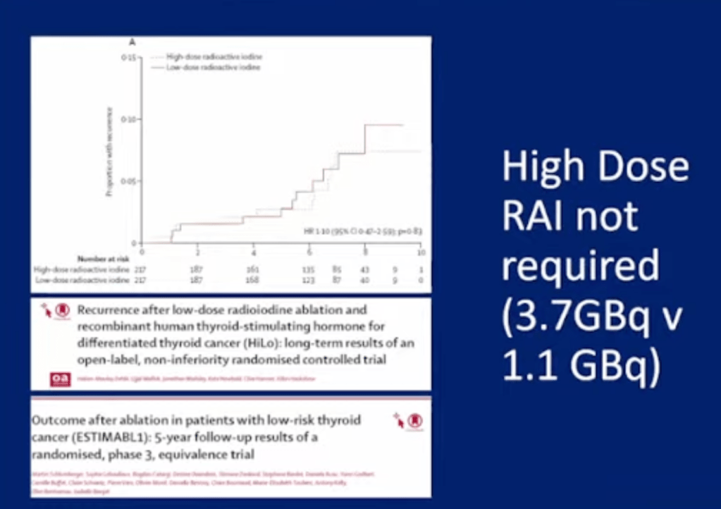
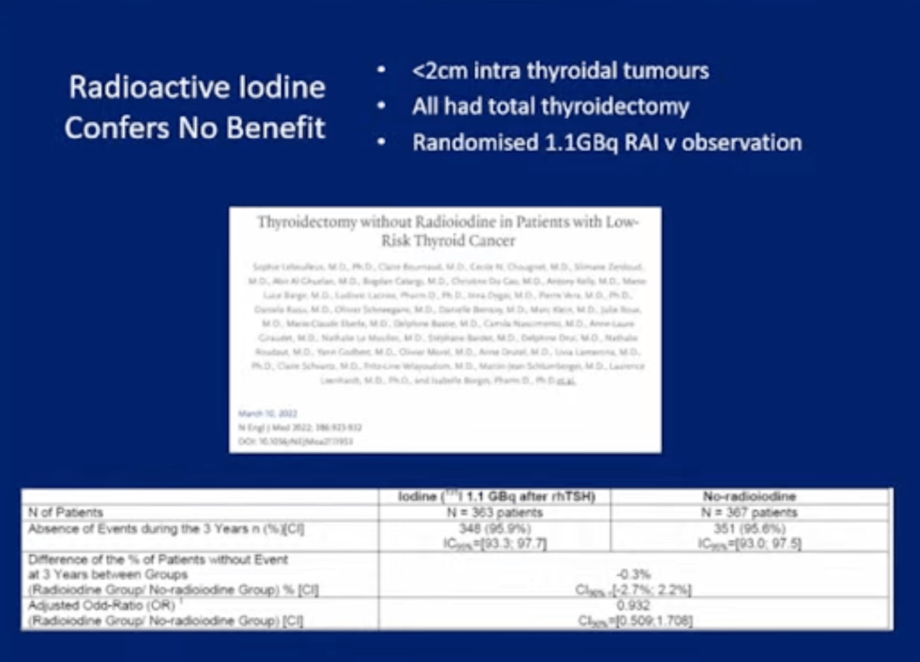
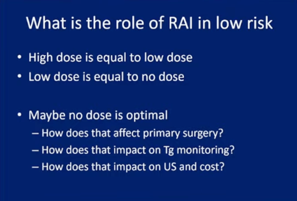
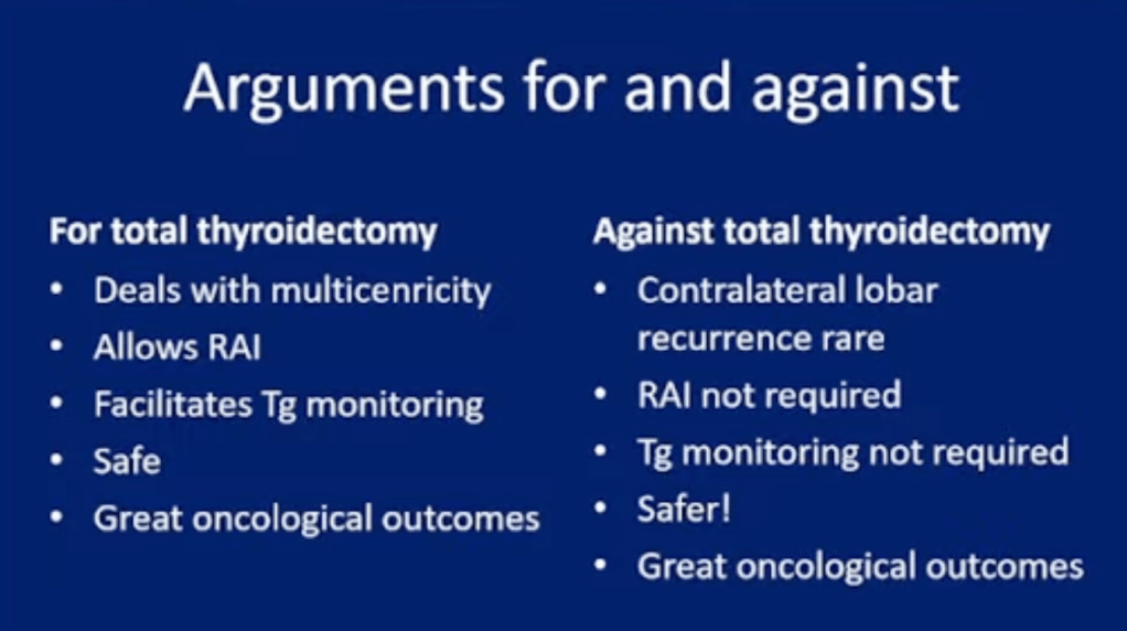
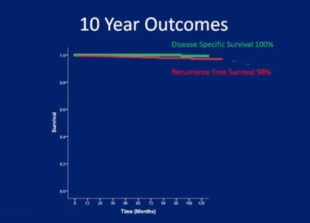
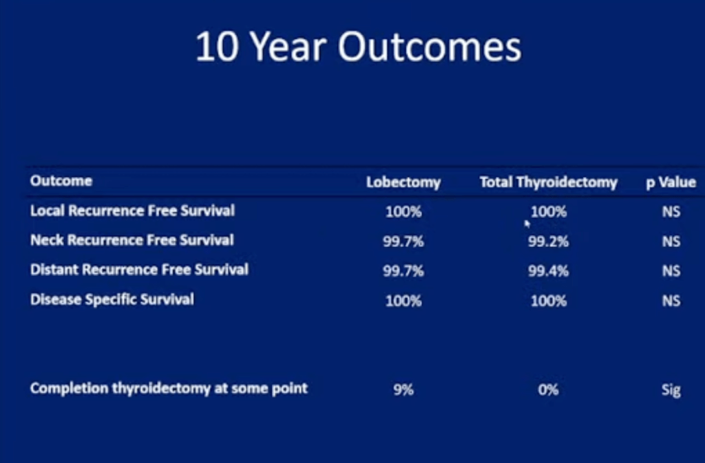
No Difference in Outcome in High Dose vs Low Dose Radioactive iodine

#Arrangoiz #ThyroidSurgeon #CancerSurgeon #SurgicalOncologist #HeadandNeckSurgeon #EndocrineSurgery #ThyroidCancer #Miami #Mexico #MountSinaiMedicalCenter #Surgeon #Teacher #ThyroidNodules #RadioactiveIodine #RAI #PTC #MSMC
Elderly patients with Primary Hyperparathyroidism

  • Elderly patients with pHPT are at increased risk of bone-related disease and fractures and are more likely to have neuropsychiatric complaints; however, referral for parathyroidectomy in the elderly is often limited due to the perceived risk of surgery.
  • Parathyroidectomy is the only curative treatment.
  • This study found that parathyroidectomy in the elderly was not associated with higher morbidity.
  • Elderly patients with hypercalcemia should be evaluated for pHPT and if confirmed should be considered for parathyroidectomy.
  • Am J Surg. 2021 Sep;222(3):549-553. doi: 10.1016/j.amjsurg.2021.01.027.