
#Arrangoiz #ThyroidSurgeon #ThyroidExpert #HeadandNeckSurgeon #ThyroidCancer #Goiter #MountSinaiMedicalCenter #Miami #Mexico #ThyroidCancer #ThyroidNodule #SurgicalOncology
#Arrangoiz #ThyroidSurgeon #ThyroidExpert #HeadandNeckSurgeon #ThyroidCancer #Goiter #MountSinaiMedicalCenter #Miami #Mexico #ThyroidCancer #ThyroidNodule #SurgicalOncology
• Fibroadenomas of the breast are benign tumors:
• Composed of stromal and epithelial elements:
• That are commonly seen in young women
• It is a common benign breast lesion:
• Results from the excess proliferation of connective tissue:
• Fibroadenomas characteristically contain both:
• Stromal and epithelial cells
• Epidemiology:
• They usually occur in women:
• Between the ages of 10 and 40 years
• It is the most common breast mass:
• In the adolescent and young adult population:
• Their peak incidence is between:
• 25 and 40 years:
• The incidence decreases after 40 years
• Clinical presentation:
• The typical presentation is in a woman of reproductive age:
• With a mobile palpable breast lump
• Due to their hormonal sensitivity:
• Fibroadenomas commonly:
• Enlarge during pregnancy and involute at menopause:
• Hence, they rarely present after the age of 40 years
• The lesions are well defined and well-circumscribed clinically and the overlying skin is normal:
• The lesions are not fixed to the surrounding parenchyma and slip around under the palpating fingers:
• Hence the colloquial term a breast “mouse”
• Pathology:
• A fibroadenoma is a type of adenomatous breast lesion:
• It contains epithelium (stromal and epithelial elements):
• Has minimal malignant potential
• Multiple fibroadenomas occur in:
• 10% to 15% of patients
• Patients with multiple fibroadenomas:
• Tend to have a strong family history of these tumors
• Fibroadenomas are assumed to be:
• Aberrations of normal breast development (ANDI) or the product of hyperplastic processes:
• Rather than true neoplasms
• Fibroadenomas can be stimulated by:
• Estrogen and progesterone:
• Some fibroadenomas also have receptors and respond to:
• Growth hormone and epidermal growth factor
• When found in an adolescent girl:
• The term juvenile fibroadenoma is more appropriate
• Location:
• Although they can be located anywhere in the breast:
• There may be a predilection for the upper outer quadrant
• Associations:
• Cyclosporin use
• Cowden syndrome
• Multiple or complex fibroadenomas:
• May indicate a slightly increased risk for breast cancer:
• The relative risk of breast cancer in patients with such fibroadenomas:
• Is approximately twice that of patients of similar age without fibroadenomas
• A patient’s age:
• Determines the preferred imaging method
• In general, ultrasonography (US) is preferred:
• If a palpable mass is found
• If a patient is younger than 30 years
• If the patient is pregnant
• Mammography and US are both useful if the patient has:
• A palpable mass
• Is older than 30 years
• Is not pregnant
• In patients younger than 30 years:
• The most appropriate modality is ultrasound:
• Because the patient is spared radiation exposure and because the likelihood for fibroadenoma is high
• Mammography is not indicated as the primary imaging study in women younger than 30 years:
• Unless high-risk factors are present
• Computed tomography (CT) scanning:
• Is not initially indicated for assessing a palpable lump in a woman in women younger than 30 years:
• Because of radiation exposure
• The inability of CT to demonstrate micro-calcifications
• The lack of specificity in the findings
• Magnetic resonance imaging (MRI):
• Is not initially indicated for assessing a palpable lump in women younger than 30 year:
• Mainly because of its high cost and the high likelihood of false-positive findings
• Positron emission tomography:
• Is expensive and is not universally available
• On mammograms:
• Fibroadenomas have a spectrum of features:
• Well-circumscribed discrete oval to round mass:
• Hypodense or isodense to the breast glandular tissue:
• Mass with macro-lobulation or partially obscured margin
• Involuting fibroadenomas in older, typically postmenopausal patients may contain:
• Calcification:
• Often producing the classic:
• Coarse popcorn calcification appearance
• In some cases the whole lesion is calcified
• Calcification may also present as crushed stone-like micro-calcification:
• Which makes differentiation from malignancy difficult
• On ultrasound:
• Fibroadenomas appear as:
• Circumscribed, homogeneous (generally uniform hypoechogenicity), round to oval, hypoechoic masses:
• That may have gentle lobulations:
• Macro-lobulated lesion
• A smooth, thin, echogenic capsule:
• Sometimes a thin echogenic rim (pseudocapsule) may be seen sonographically
• Variable acoustic enhancement; and homogeneity
• Intralesional sonographically detectable calcification:
• May be seen in approximately 10% of cases
• On MRI:
• Fibroadenomas typically appear as smooth masses:
• With high signal intensity on T2-weighted images
• Enhancement with the administration of gadolinium-based contrast
• T1:
• Typically hypointense or isointense compared with adjacent breast tissue
• T2:
• Can be hypointense or hyperintense compared with adjacent breast tissue
• T1 C+ (Gd):
• Can be variable:
• But a majority will show slow initial contrast enhancement:
• Followed by a persistent delayed phase:
• Type I enhancement curve:
• Non-enhancing internal septations may be seen
• Diagnosis:
• These lesions are easily biopsied under ultrasound guidance
• When a lesion has the typical features of a fibroadenoma on ultrasound and there are no clinical red flags:
• They can be safely followed clinically
• When lesions enlarge or have atypical imaging findings:
• Ultrasound-guided core biopsy is a minimally invasive outpatient procedure that will give a diagnosis with virtually no complications
• There may be a maximum diameter above which a biopsy should be done if no previous imaging is available:
• The reason for intervention based on size is that a phyllodes tumor may be indistinguishable from a fibroadenoma on ultrasound:
• A maximum diameter of 2.5 cm may be a useful benchmark for biopsy if you have no previous imaging
• Interval enlargement:
• Is an indication for biopsy
• Symptomatic, progressively enlarging masses or atypical presentations:
• May warrant surgical excision
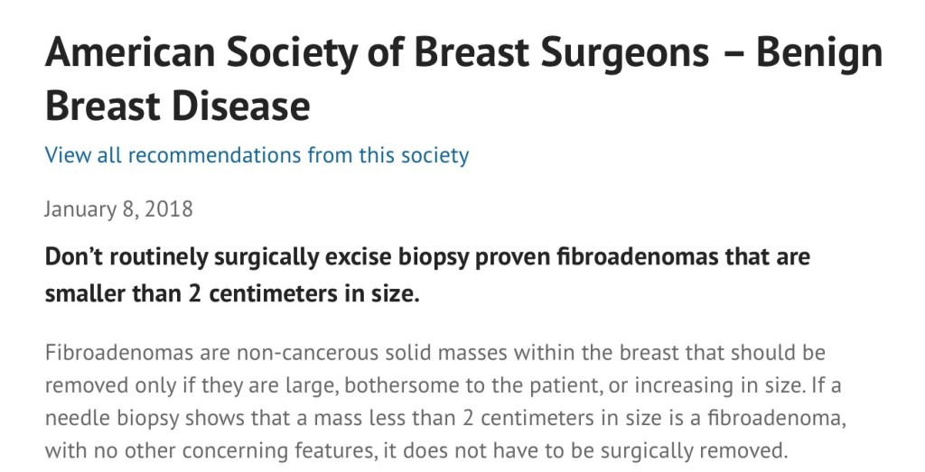
• If a needle biopsy shows that a mass less than 2 centimeters in size is a fibroadenoma, with no other concerning features:
• It does not have to be surgically removed
• The patient’s core biopsy pathology demonstrating a fibroadenoma is consistent with the typical imaging findings of a smooth, round, hypoechoic mass:
• As the biopsy is concordant:
• No further intervention is needed:
• Follow-up for reassurance is acceptable
• Treatment and prognosis:
• They are benign lesions with minimal or no malignant potential
• The risk of malignant transformation is extremely low:
• Has been reported to range around 0.0125% to 0.3%
• Indications for biopsy include:
• Enlarging lesion
• Atypical findings on ultrasound
• A lesion above 2.5 cm and there are no previous studies for comparison
• Patient peace of mind:
• Some patients are simply not happy with a palpable mass in the breast without a histological diagnosis:
• This is a valid and reasonable indication for biopsy
• References:
• Tan BY, Tan PH A Diagnostic Approach to Fibroepithelial Breast Lesions. Surg Pathol Clin. 2018 Mar;11(1):17-42.
• American Society of Breast Surgeons – benign breast disease. January 8, 2018. Choosing Wisely website. http://www.choosingwisely.org/clinician-lists/asbrs-benign-breast-disease-biopsy-proven-fibroadenomas-smaller-than-2-cm/. Accessed October 17, 2019.

#Arrangoiz #BreastSurgeon #BreastFibroadenoma #Fibroadenoma #SurgicalOncology #MSMC #MountSinaiMedicalCenter #Miami #Mexico
• The decision to administer radioiodine after thyroidectomy in patients with differentiated thyroid cancer is based upon the clinicopathologic features of each case
• The efficacy of radioiodine depends upon:
• Tumor-specific characteristics
• Sites of disease
• Patient preparation
• Dose
• Because of the careful risk stratification used in some studies, it is possible to identify specific patient and tumor characteristics which suggest that radioiodine may be beneficial:
• My approach outlined below is in agreement with the American Thyroid Association (ATA) guidelines on the role of postoperative radioiodine ablation:
• Low risk:
• The ATA guidelines do not routinely recommend administration radioiodine after lobectomy or total thyroidectomy to low-risk patients with differentiated thyroid cancer:
• This includes patients with:
• Unifocal cancer less than 1 cm without other high-risk features (eg, without distant metastases, vascular invasion, gross extrathyroidal extension, worrisome histologic subtypes):
• Even in the presence of small-volume regional lymph node metastases (less than five lymph nodes measuring less than 2 mm)
• Multifocal cancer when all foci are less than 1 cm and there are no other high-risk features:
• Without distant metastases
• Vascular invasion
• Gross extrathyroidal extension
• Worrisome histologic subtypes
• Intrathyroidal cancer in the 1 to 4 cm range without other high-risk features:
• Without distant metastases, vascular invasion, gross extrathyroidal extension, worrisome histologic subtypes
• Individual tumor- and patient-specific features may warrant radioiodine ablation in selected low-risk patients:
• As an example, low-dose radioiodine ablation (30 mCi) in low-risk patients with intrathyroidal tumors greater than 4 cm may be considered
• In a randomized trial evaluating post-thyroidectomy radioiodine (1.1 GBq [30 mCi] after recombinant human TSH [rhTSH]) or no radioiodine therapy in 730 patients with low-risk differentiated thyroid cancer (multifocal pT1a with the sum of the longest diameters less than 2 cm, or pT1b, both with N0 or Nx, and without aggressive pathological subtypes or extrathyroidal extension, outcomes at three years were similar in the two groups, including the following:
• Primary disease-related events (identification of residual or recurrent disease or an elevated level of thyroglobulin or thyroglobulin antibodies) at three years occurred in:
• 4.1% and 4.4% of patients, respectively
• The frequency of BRAF mutation did not differ between cases with or without disease-related events:
• 61.5% versus 53.1% respectively
• An ATA excellent response at 10 months and 3 years occurred in:
• 86.8% and 86.3%, respectively, after 10 months
• 73% and 74.1%, respectively, after 3 years
• Quality-of-life scores related to:
• Anxiety, distress, and fear of recurrence
• In systematic reviews and meta-analyses of observational data:
• There was no benefit of radioiodine in low-risk patients with regard to either:
• Overall recurrence rate or disease-specific mortality
• As an example, in a retrospective analysis of 1129 patients who underwent total thyroidectomy for differentiated thyroid cancer and received radioiodine based upon risk assessment:
• The majority of patients with low-risk local disease (age less than 45 years, papillary cancer less than 4 cm without extrathyroidal extension, and without distant metastases):
• Had low rates of recurrence and high rates of survival when managed without radioiodine;
• Five-year recurrence-free survival greater than 97%
• Another retrospective study similarly showed no benefit of radioiodine ablation on disease recurrence in patients with papillary thyroid microcarcinoma (unifocal or multifocal):
• In this study, there was no disease-related mortality
• Intermediate risk:
• The administration of radioiodine after total thyroidectomy is considered in selected intermediate-risk patients depending upon specific tumor characteristics, including:
• Microscopic invasion into the perithyroidal soft tissue
• Clinically significant lymph node metastases outside of the thyroid bed
• Other higher-risk features:
• Vascular invasion
• More aggressive histologic subtypes such as:
• Tall cell histology
• Columnar cell histology
• Insular histology
• Poorly differentiated histology
• When the combination of age, tumor size or multifocality, lymph node status, and individual histology:
• Predicts an intermediate to high risk of recurrence or death from thyroid cancer
• In the absence of evidence supporting survival benefit for all of the factors listed:
• Clinical judgment and an individualized approach to care are important
• Postoperative serum thyroglobulin is a critical factor that should be routinely integrated into clinical decision-making:
• For example, in a retrospective cohort study, there was no difference in the five-year recurrence-free survival among intermediate-risk patients who did or did not receive radioiodine therapy:
• When postoperative unstimulated serum thyroglobulin levels were less than 1 ng/mL
• There are limited data showing a benefit of radioiodine in intermediate-risk patients:
• In a study using the National Cancer Database registry, which included 21,870 patients with intermediate-risk papillary thyroid cancer who had total thyroidectomy with or without radioiodine:
• Patients who received radioiodine had improved overall survival (hazard ratio [HR] 0.71, 95% CI 0.62-0.82)
• In the National Thyroid Cancer Treatment Cooperative Study Group (NTCTCSG), a multicenter thyroid cancer registry that has analyzed the outcomes of nearly 5000 patients with differentiated thyroid cancer:
• Multivariable analysis showed that radioiodine ablation was associated with:
• Improvement in overall survival in stage II patients:
• But this did not reach statistical significance (relative risk [RR] 0.67, 95% CI 0.36-1.28)
• High risk:
• Based on the ATA guidelines high risk patients are routinely treated with radioiodine after total thyroidectomy, including patients with:
• Distant metastases
• Macroscopic tumor invasion,
• Incomplete tumor resection with gross residual disease
• In the National Thyroid Cancer Treatment Cooperative Study Group:
• Radioiodine was associated with improved overall survival in stage III patients (RR 0.66, 95% CI 0.46-0.98), with similar but nonsignificant improvement in stage IV (RR 0.70, 95% CI 0.46-1.10)
• In addition, data prospectively collected from the Surveillance, Epidemiology, and End Results (SEER) database showed:
• Benefit from radioiodine in patients older than 45 years with primary tumors greater than 2 cm, with disease in the lymph nodes at initial diagnosis, and with distant metastatic disease
• The benefits and dose limits of iodine-131 (131-I) therapy for metastatic disease were evaluated in a retrospective analysis of 444 patients treated between 1953 and 1994 (analysis of whole-body iodine scans and conventional radiographs):
• 43% of the 295 patients with radioiodine uptake achieved resolution of radioiodine-avid metastases on iodine scan and negative conventional radiographs
• Additional features of patients in this group included the following:
• They were more likely to be younger:
• With differentiated tumors
• 96% of these patients were given cumulative doses of 100 to 600 mCi (3700 to 22,000 MBq)
• 7% had a recurrence
• 10-year survival was 92% in this group:
• Compared with only 19% in patients who did not achieve resolution of the radioiodine-avid metastatic lesions
#Arrangoiz #ThyroidSurgeon #CancerSurgeon #SurgicalOncologist #HeadandNeckSurgeon #EndocrineSurgery #ThyroidCancer #Miami #Mexico #MountSinaiMedicalCenter
👉Common labs ordered in the workup of patients with Primary hyperparathyroidism include a serum calcium (+/- ionized calcium), albumin (for correction of total serum calcium level), PTH, phosphate, creatinine, 25-hydroxy vitamin D, and a 24-hour urine calcium & creatinine.
👉Hypercalcemia with with an elevated PTH with normal kidney function and without hypocalciuria indicates primary hyperparathyroidism.
👉However, some patients with this disease will have hypercalcemia with an “inappropriately normal” PTH – this can be a common cause of delayed diagnosis.

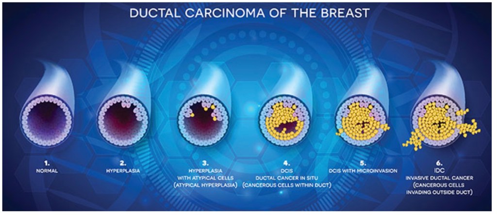
#Arrangoiz #CancerSurgeon #BreastSurgeon #SurgicalOncologist #BreastCancer #LCIS #DCIS #DuctalCarcinomaInsitu #LobularNeoplasia #LobularCarcinomaInsitu #Surgeon #Teacher #Miami #Mexico #MSMC #MountSinaiMedicalCenter
#Arrangoiz #ThyroidSurgeon #CancerSurgeon #SurgicalOncologist #HeadandNeckSurgeon #EndocrineSurgery #ThyroidCancer #TallCellVariantofPapillaryThyroidCancer #Miami #Mexico #MountSinaiMedicalCenter

