- Skeletal manifestations including osteopenia, osteoporosis, and osteitis fibrosa cystica:
- Are found in approximately 15% of patients with PHPT
- PHPT is linked with a reduction in bone mineral density (BMD):
- Particularly in the cortical bone:
- Such as in the distal third of the radius
- In the lumbar region, composed all most exclusively by trabecular bone, and in the femoral region, composed by cortical and trabecular bone:
- The decrease in BMD is less severe
- Particularly in the cortical bone:
- Osteitis fibrosa cystica:
- A skeletal manifestation that is rarely seen today:
- Seen in less than five percent of patients with PHPT
- Is caused by an increase in bone turnover:
- Can be determined by finding an:
- Elevated serum alkaline phosphatase level
- Can be determined by finding an:
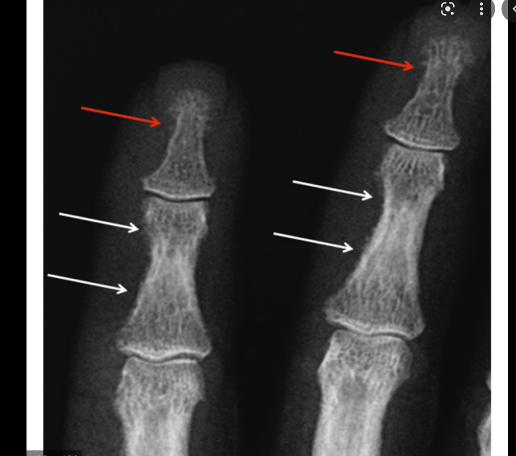
- The radiologic findings seen in patients with PHPT with bone disease are characterized by:
- Subperiosteal resorption:
- Most obvious on the radial aspect of the middle phalanx of the second and third fingers
- Bone cysts
- Tufting of the distal phalanges:
- Which are best evaluated on plain x-rays of the hands
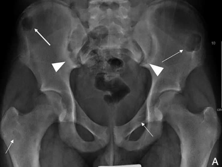
- Brown or osteoclastic tumors:
- Caused by the accumulations of osteoclasts and fibrous tissue:
- Brown tumors have a slightly greater incidence in PHPT than in secondary HPT:
- 3% versus 2%
- In patients with chronic kidney disease:
- Persistent and excessive urinary calcium elimination:
- Can lower serum calcium level:
- And lead to an increase in PTH secretion
- Can lower serum calcium level:
- Persistent and excessive urinary calcium elimination:
- This results in mobilization of calcium from the bones:
- Through rapid osteoclastic turnover of bone to maintain normal serum calcium levels
- In regions where bone loss is exceptionally fast:
- Hemorrhage, and reparative granulation tissue, with active, vascular, proliferating fibrous tissue may replace the normal marrow contents:
- Resulting in a brown tumor
- Hemorrhage, and reparative granulation tissue, with active, vascular, proliferating fibrous tissue may replace the normal marrow contents:
- Hemosiderin imparts the brown color (hence the name of the lesions
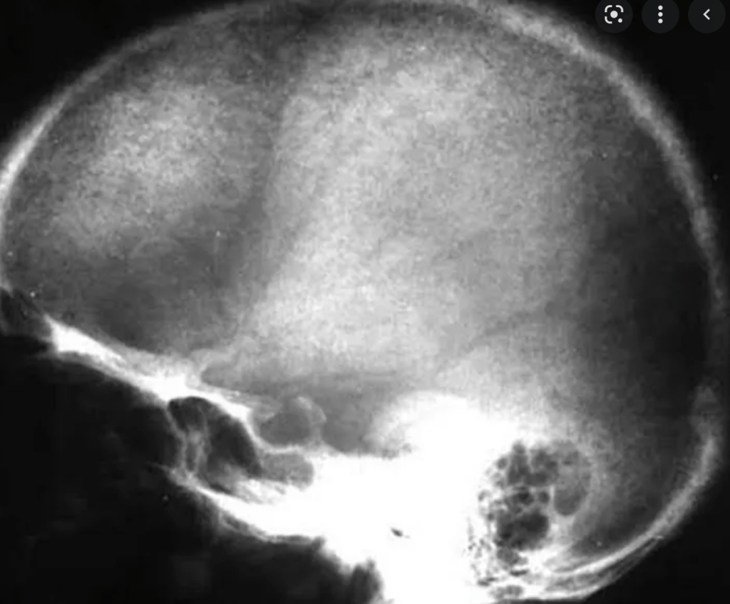
- The skull also may be affected:
- Appears mottled with a loss of definition of the inner and outer cortices
- Subperiosteal resorption:
- A skeletal manifestation that is rarely seen today:
- Patients with normal serum alkaline phosphatase levels:
- Almost never have clinically apparent osteitis fibrosa cystica
- Bone disease correlates with serum PTH and vitamin D levels
#Arrangoiz #ParathyroidSurgeon #ParathyroidExpert #HeadandNeckSurgeon #EndocrineSurgery #MountSinaiMedicalCenter #Miami #Mexico #Teacher #Surgeon #Parathyroidectomy #Hypercalcemia #ElevatedCalcium #MSMC
