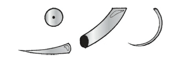
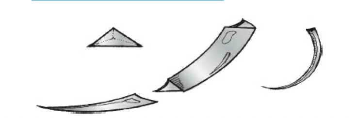
Triangular body with the sharp edge toward the inner circumference:
Leaves a triangular hole in tissue
What are its uses?
Suturing of skin
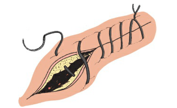
What is a simple interrupted stitch?
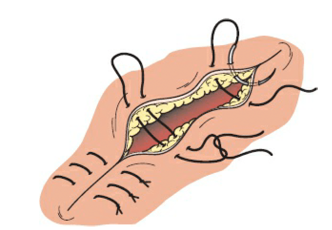
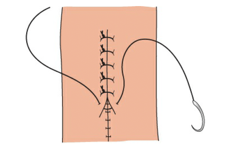
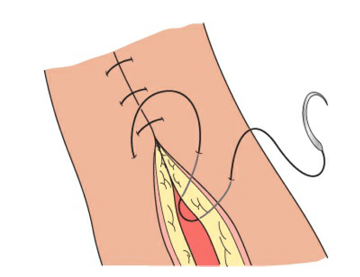
What is a vertical mattress stitch?
Simple stitch is made;
The needle is reversed, and a small bite is taken from each wound edge:
The knot ends up on one side of the wound
What is the vertical mattress stitch also known as?
Far–far, near–near stitch:
Oriented perpendicular to wound
What is it used for?
Difficult-to-approximate skin edges:
Everts tissue well
What is a simple running (continuous) stitch?
Stitches made in succession without knotting each stitch
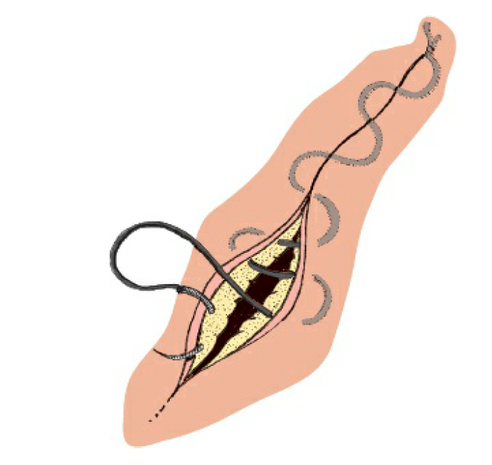
What is a subcuticular stitch?
Stitch (usually running) placed just underneath the epidermis:
Can be either absorbable or non-absorbable:
Pull-out stitch if non-absorbable
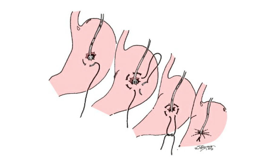
What is a purse-string suture?
Stitch that encircles a tube perforating a hollow viscus:
For example:
Jejunostomy tube
Gastrostomy tube:
Allowing the hole to be drawn tight and thus preventing leakage
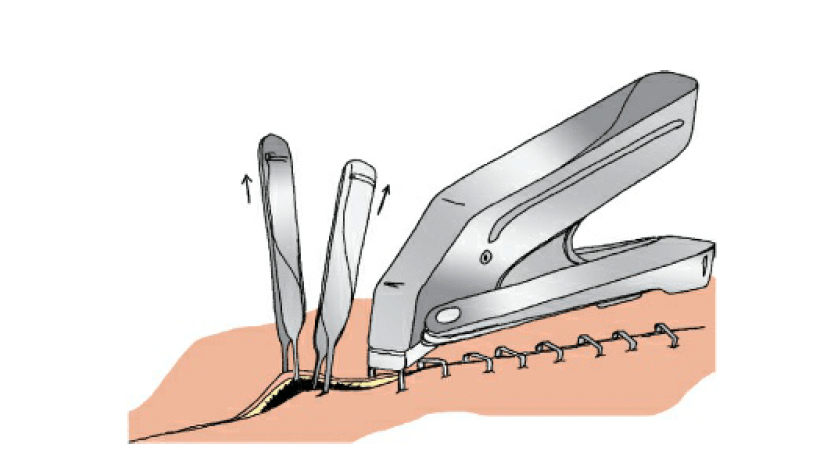
What are metallic skin staples?
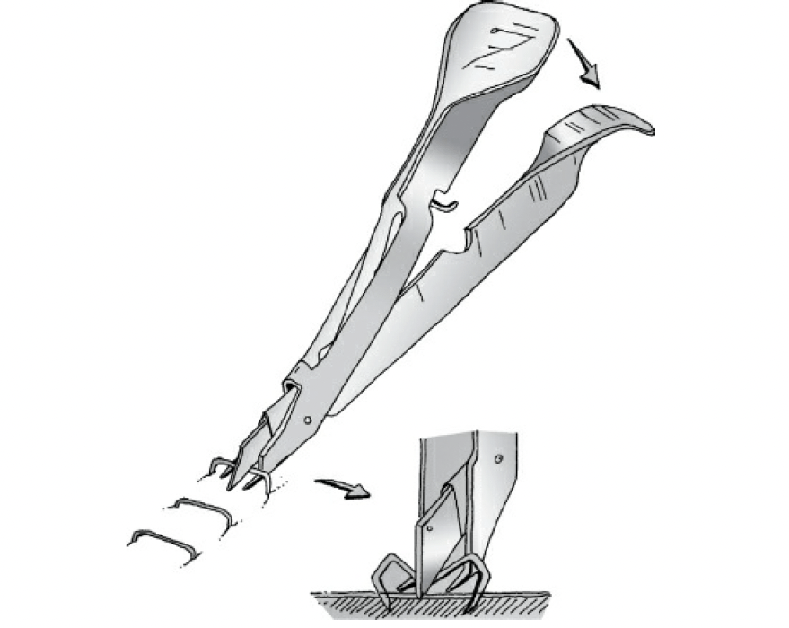
What is a staple removal device?
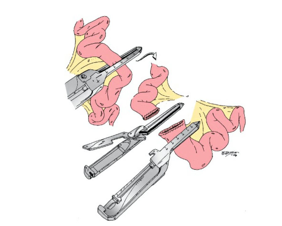
What is a gastrointestinal anastomosis (GIA) device?
Stapling device that lays two rows of small staples in a hemostatic row:
And automatically cuts in between them
What is a Lembert stitch?
It is a second layer in bowel anastomoses
Technique:
The needle is inserted perpendicular to the epidermis, approximately 8 mm distant to the wound edge.
With a fluid motion of the wrist, the needle is rotated superficially through the dermis, and the needle tip exits the skin 2 mm distant from the wound edge on the ipsilateral side.
The needle body is grasped with surgical forceps in the left hand and reloaded onto the needle driver.
The needle is then inserted perpendicular to the skin on the contralateral side of the wound edge, 2 mm distant from the wound edge.
The needle is again rotated superficially through its arc, exiting 8 mm from the incised wound edge.
The suture material is then tied off gently, with care being taken to minimize tension across the epidermis and avoid overly constricting the wound edges
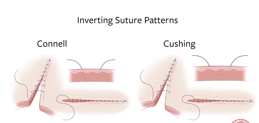
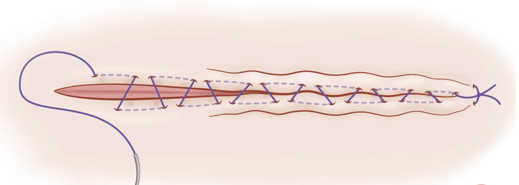
What is a Connell’s stitch?
The first mucosa-to-mucosa layer in an anastomosis:
Basically, a running U stitch
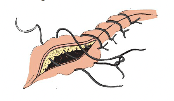
The Cushing and Connell suture technique:
Is often used to close the incisions in hollow organs such as the stomach, urinary bladder, and uterus
In the Cushing suture technique:
The suture penetrates into the submucosa without penetrating the organ lumen
The suture runs from both sides of the incision:
Parallel to each other
The Connell suture technique is almost identical to the Cushing suture technique:
These two suture techniques are separated according to the tissue they penetrate during suture passage
While the Cushing suture technique is also passed through the submucosa, Connell suture technique is used to pass through the lumen
While applying these techniques, the following steps are followed:
A directionally opposed suture passage is made parallel to the incision.
Suture passage is made from the other side of the incision in the same direction as the incision, parallel to the first passage
The beginning of the suture line is fixed with a knot.
Starting from the back of the knot, a suture passage is made in the direction of the incision
A passage is made from the other side of the incision parallel to the first pass and in the same direction
When the suture is pulled, the tissue becomes inverted and the knot is buried under the skin
A suture passage is made in the direction of the incision
A passage is made from the other side of the incision parallel to the first pass and in the same direction
The last two steps are repeated throughout the incision
After the incision line is crossed, End of suture line is fixed by repeating first three steps
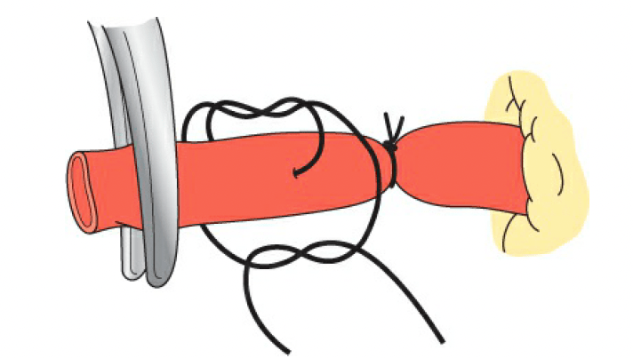
What is a suture ligature (a.k.a. “stick tie”)?
Suture is anchored by passing it through the vessel on a needle before wrapping it around and occluding the vessel:
May be associated with a 3.5% to 5% occurrence of occult carcinoma:
Depending on the indication for the operation
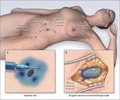
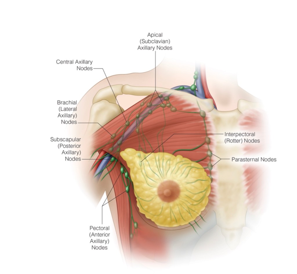
SLNB at the time of prophylactic mastectomy:
May eliminate the need for ALND if occult disease is identified
A recent meta-analysis reviewed 14 studies where SLNB was routinely performed for prophylactic mastectomy in patients undergoing bilateral mastectomy for unilateral cancer:
This study found metastatic disease in the SLNB of the prophylactic mastectomy:
In 0 to 4% of patients with contralateral cancer
The majority of metastatic disease was associated with:
Contralateral axillary tumor spread from the primary tumor:
Not an occult primary tumor
In patients who were found to have occult malignancy in the prophylactic mastectomy breast:
Less than 1% of sentinel lymph nodes were positive for metastatic disease
Given the low rates of occult malignancy and axillary metastasis:
SLNB is not indicated for patients undergoing prophylactic mastectomy
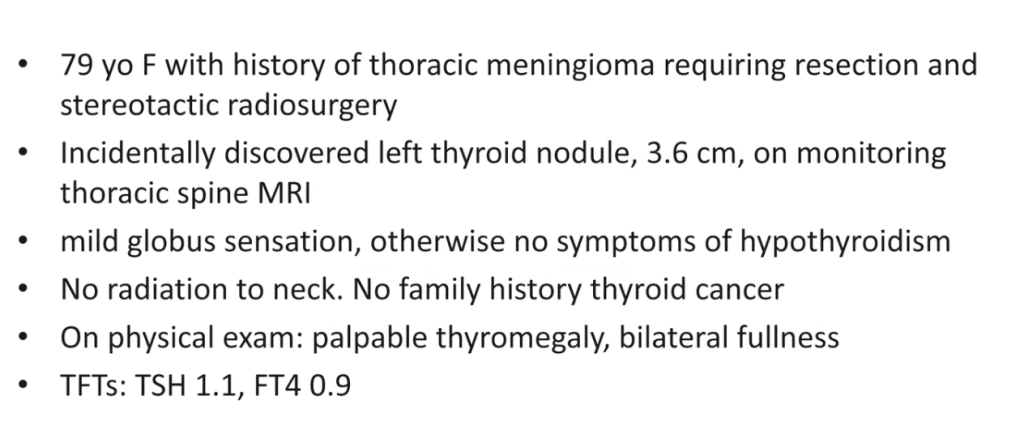
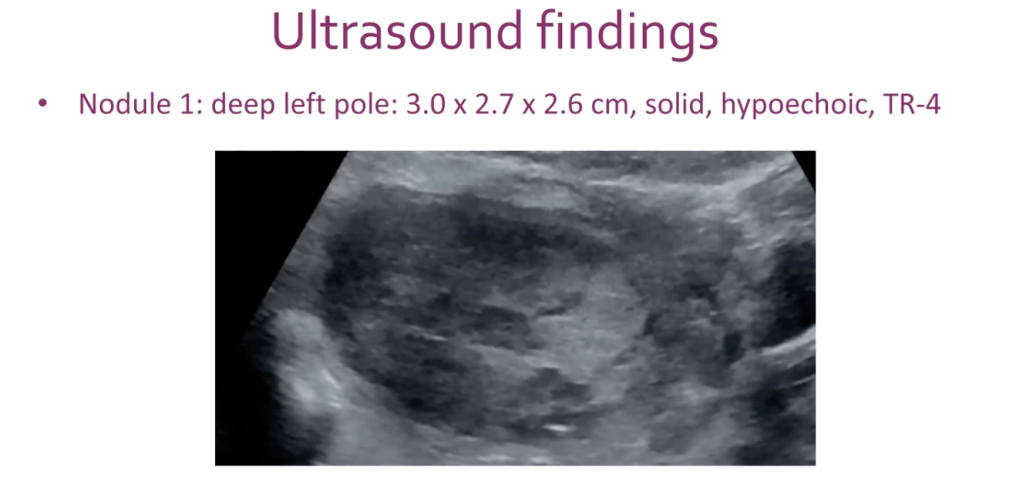
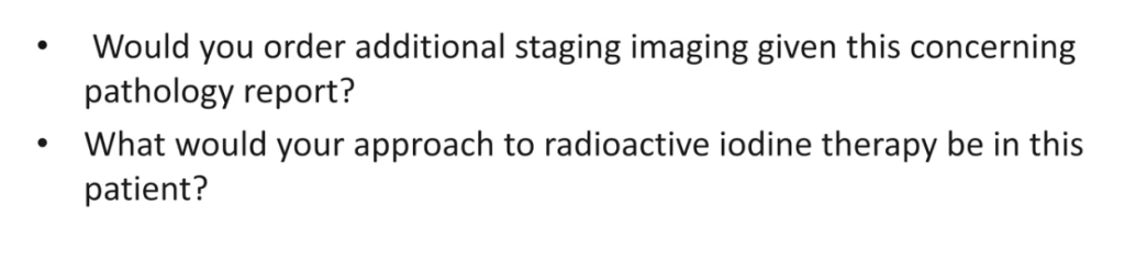
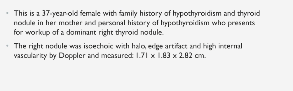
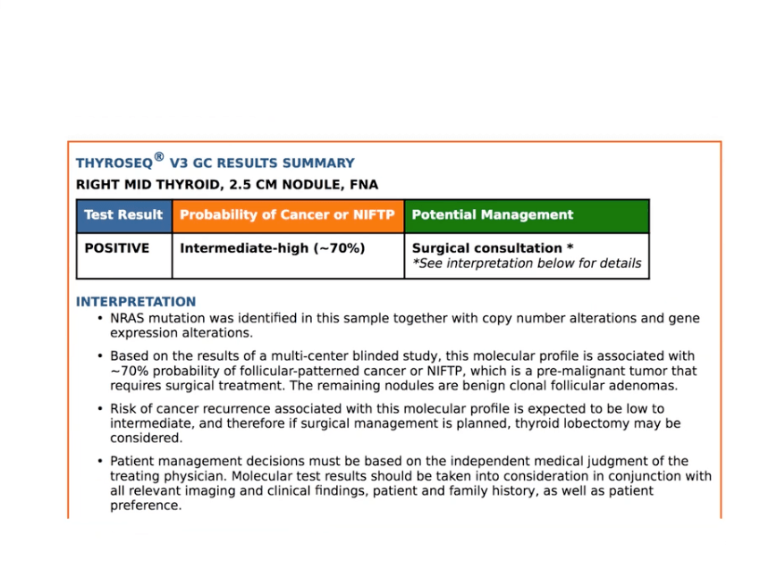
US Guided FNA Performed. Very close look at the central and lateral compartment lymph nodes on ultrasound, may be CT scan of the neck with IV contrast.
CT scan of the neck and chest, in the future may be PET/CT scan. Postoperative radiation iodine management.