Blog

RAS Gene Mutations in Thyroid Cancer

The RAS genes (HRAS, KRAS and NRAS) encode highly related G-proteins that play a central role in the intracellular transduction of signals arising from cell membrane receptors.

In its inactive state, RAS protein is bound to GDP. Upon activation, it releases GDP and binds GTP, thereby activating the MAPK and other signaling pathways, such as PI3K / AKT. Normally, the activated RAS-GTP protein becomes quickly inactive due to its intrinsic GTPase activity and the action of cytoplasmic GTPase-activating proteins.

Point mutations in the discrete domains of the RAS gene either increase its affinity for GTP (mutations in codons 12 and 13) or inactivate its autocatalytic GTPase function (mutation in codon 61). As a result, the mutant protein becomes permanently switched in the active position and continuously activates its downstream targets.

Point mutations involving several specific sites (codons 12, 13 and 61) of the NRAS, HRAS or KRAS genes are found in 10% to 20% of papillary thyroid carcinomas. Papillary thyroid carcinomas harboring RAS mutation almost always have follicular variant histology; this mutation also correlates with significantly less prominent nuclear features of papillary thyroid carcinoma, more frequent encapsulation and low rate of lymph node metastases.

Some studies have reported the association between RAS mutations and more aggressive behavior of papillary thyroid carcinoma, such as a higher frequency of distant metastases. Mutations of the RAS gene are not restricted to papillary thyroid carcinoma and also found in other benign and malignant thyroid neoplasms, as well as in tumors from other tissues.

The molecular pathogenesis of thyroid cancer involves dysregulation of the mitogen-activated protein kinase (MAPK) and phosphatidylinositol-3 kinase (PI3K)/AKT pathways. Common activating mutations in the MAPK pathway include RET / PTC and NTRK rearrangements, and RAS and BRAF mutations. Common genetic alterations in the PI3K pathway include RAS mutations, PTEN mutations or deletions, PIK3K mutations or amplifications, and AKT1 mutations. PAX8 / PPARG fusions are common in FTC. Activation of Wnt/b-catenin pathway, inactivating mutations in TP53, and activating mutations in TERT promoter are frequent in undifferentiated thyroid cancer.
#Arrangoiz #ThyroidSurgeon
  • The BI-RADS lexicon was developed by the American College of Radiology (ACR) and was mandated by the Mammographic Quality and Standard Act of 1992. This act pertains only to mammography.
  • Since this lexicon was successful, ACR has now developed a similar lexicon for breast ultrasound and breast MRI.
  • The 5th edition of the BI-RADS system for mammography is the most current edition.
  • Mammographic findings are grouped into seven categories including masses, calcifications, architectural distortion, asymmetries, intramammary lymph node, skin lesions, and solitary dilated duct (see the table).
  • A group of calcifications is defined as 5 calcifications within a 1-cm area of tissue but less than 2 cm.
  • BI-RADS is reported on a scale from 0 to 6, with BI-RADS 0 indicating further work-up is needed and BI-RADS 6 indicating biopsy-proven malignancy.
  • BI-RADS 3 indicates that a short-term follow-up is needed.
  • The table shows the term associated with each of the six BI-RADS categories, the recommended action, and the likelihood of malignancy.
  • References:
  • D’Orsi CJSE, Mendelson EB, Morris EA, et al. ACR BI RADS Atlas, Breast Imaging Reporting and Data System. Reston, VA: American College of Radiology. 2013.
  • Sedgwick EL, Ebuoma L, Hamame A, et al. BI-RADS update for breast cancer caregivers. Breast Cancer Res Treat. 2015;150:243-254.
  • Tomosynthesis is a newer imaging modality:
    • That allows for reconstruction of planes from breast tissue volume:
      • Which overcomes the limitations of 2-dimensional imaging caused by overlapping normal and pathologic breast tissue
  • Tomosynthesis has been shown to increase the detection of invasive cancer:
    • By more than a third compared with 2D mammography alone and reduces false-positive results by 15%
  • Screening mammography does reduce mortality:
    • Pooled estimates from multiple trials demonstrate a reduction of 20%
  • MRI is recommended for screening in high-risk women:
    • High-risk is defined as:
      • A lifetime risk of 20%
  • There is a national trend toward adopting legislation:
    • That requires the reporting of breast density to women undergoing mammography:
      • As well, there is a need for supplemental screening for this group of women with dense breasts
    • Automated ultrasound may be a good alternative for these patients:
      • In one study of patients with 50% breast density, automated ultrasound detected 12.3 cancers per 1,000 breast cancers compared with 4.6 per 1,000 by mammography alone
  • Imaging with all three modalities has not been validated
  • References:
    • Friedewald SM, Rafferty EA, Rose SL, et al. Breast cancer screening using tomosynthesis in combination with digital mammography. JAMA.
    • Giuliano V, Giuliano C. Improved breast cancer detection using automated whole breast ultrasound and mammography in radiographically dense breasts. Clin Imaging. 2013;20:480-
    • Smith RA, Duffy SW, Gabe R, Tabar L, Yen AM, Chen TH. The randomized trials of breast cancer screening: what have we learned? Radiol Clin North Am. 2004;42:793-806.486.2014;311:2499-2507.
    • Smith RA, Duffy SW, Gabe R, Tabar L, Yen AM, Chen TH. The randomized trials of breast cancer screening: what have we learned? Radiol Clin North Am. 2004;42:793-806.

#Arrangoiz #BreastSurgeon #CancerSurgeon #SurgicalOncologist #BreastCancer #Mammography #MountSinaiMedicalCenter #MSMC

Hyperparathyroidism during Pregnancy

  • Hyperparathyroidism during pregnancy:
    • Is often unrecognized:
      • Is associated with a 3.5-fold increase in miscarriage
  • Loss of the pregnancy:
    • Most often occurs during the late second trimester
  • The incidence of hyperparathyroidism in pregnancy is:
    • 0.7%
  • Maternal complications include:
    • Hyperemesis
    • Nephrolithiasis
    • Pancreatitis
  • Fetal complications include:
    • Spontaneous abortion
    • Growth retardation
  • In those who reach delivery, neonatal complications include:
    • Hypocalcemic crisis:
      • Within the first few days of life
  • Calcimimetic medications:
    • Have not been used in the setting of hyperparathyroidism in pregnancy
  • Calcitonin:
    • Has no role in the management of hyperparathyroidism

Low vitamin D levels increase chances of developing low parathyroid hormone levels after thyroid surgery

BACKGROUND
Surgery to remove all, or part, the thyroid gland is commonly needed to treat some types of thyroid disease. One problem that can happen after thyroid surgery is development of low calcium levels due to damage to the parathyroid glands. The parathyroid glands are four very small structures (each about the size of a grain of rice) that live in the neck on the surface of the thyroid gland. Their job is to make a single hormone (parathyroid hormone), which works with vitamin D in the body to control a person’s calcium levels. When calcium levels are low, parathyroid hormone levels are increased to help increase calcium levels.

It is critical that the parathyroid glands work normally after thyroid surgery. If they do not, which can happen if the parathyroid glands are damaged, removed or irritated during surgery, calcium levels will be too low. This can cause serious side effects, including severe muscle cramps and seizures. For this reason, understanding how best to preserve parathyroid gland function after thyroid surgery is very important.

The research described here studied people who had surgery to remove all, or part, of their thyroid to learn if low vitamin D levels before thyroid surgery increases the risk of low parathyroid hormone levels after surgery.

THE FULL ARTICLE TITLE
Vaitsi K et al 2021 Pre-operative vitamin D deficiency is a risk factor for post-thyroidectomy hypoparathyroidism: A systematic review and meta-analysis of observational studies. J Clin Endocrinol Metab. Epub 2021 Jan 23:dgab039. PMID: 33484571.

SUMMARY OF THE STUDY
In order to better understand the possible effect of low vitamin D levels on parathyroid gland function after thyroid surgery, the authors of this study collected information published between 2009 and 2020 on the relationship between parathyroid gland activity, thyroid surgery and vitamin D levels. They then used mathematical testing (statistics) to see if low vitamin D levels before thyroid surgery increased the risk that the parathyroid glands would not work well after surgery (hypoparathyroidism, or low parathyroid hormone levels).

Overall, the authors included 755,585 people who underwent thyroid surgery. They found that mild or moderately low vitamin D levels before thyroid surgery increased the risk of temporary (lasting less than 6 months) low parathyroid hormone levels after thyroid removal. If very low vitamin D levels were present before thyroid surgery, patients also had an increased risk of having permanent low parathyroid hormone levels after surgery.

WHAT ARE THE IMPLICATIONS OF THIS STUDY?
The study authors found that low vitamin D levels before thyroid surgery increased the risk that the parathyroid glands would not work well after surgery, which could cause significant health problems. For this reason, the authors suggest that people who are planning to undergo thyroid surgery should have their vitamin D levels checked and, if these levels are low, should take a vitamin D supplement before surgery. This might decrease the risk of having a low parathyroid hormone level, and associated side effects, after thyroid surgery.

Localization of Parathyroid Glands Preoperatively

  • Routine preoperative localization in patients with PHPT includes:
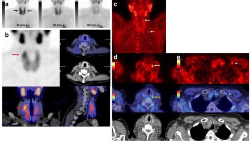
    • Neck ultrasound and technetium-99m– labeled sestamibi scan
  • Sestamibi scan:
    • Has a reported sensitivity as high as:
      • 90%
  • Ultrasound:
    • Is slightly less sensitive:
      • 75%
    • But the ease of in-office use:
      • Makes it a useful tool for the head and neck surgeon
  • SPECT:
    • When used with planar sestamibi:
      • Is very good at locating potential ectopic glands such as those in the mediastinum
  • 4D-CT:
    • Incorporates contrast perfusion:
      • In hyperfunctioning parathyroid tissue over time:
        • This additional layer provides functional information in addition to the anatomic information provided by a standard CT scan
    • In a recent study, 4D-CT has shown improved sensitivity over all other modalities:
      • Especially when used in combination with ultrasound

#Arrangoiz #ParathyroidSurgeon #ParathyroidExpert #Hypercalcemia #PHPT #Hyperparathyroidism #PrimaryHyperparathyroidism #EndocrineSurgery #HeadandNeckSurgeon #CancerSurgeon #Surgeon #Teacher #Miami #Mexico #MountSinaiMedicalCenter #MSMC

Classification of Thyroid Tumors

The conventional classification based on morphology and clinical features is largely supported by molecular data currently available. Genetic profiles of four main categories appear distinctly different from each other with a few areas of overlap.

The classification of thyroid tumors modified from WHO classification (2004) is as follows:

Tumors of Follicular Epithelium:
■„Follicular adenoma (including Hurthle cell adenoma)
■„Hyalinizing trabecular adenoma ■„Follicular carcinoma (including Hurthle cell carcinoma)
– Minimally invasive – – Widely invasive
■„Papillary carcinomas
■„Poorly differentiated carcinoma ■„Anaplastic carcinoma
■„Squamous cell carcinoma ■„Mucoepidermoid carcinoma ■„Sclerosing mucoepidermoid carcinoma with eosinophilia ■„Mucinous carcinoma

Tumors with C Cell Differentiation
Medullary carcinoma

Tumors with Mixed Differentiation
■„Collision tumor—follicular/papillary or follicular/medullary
■„Mixed differentiated carcinoma intermediate type

Tumors Showing Thymic or Related Branchial Pouch Differentiation
■„Ectopic thymoma
■„Spindle epithelial tumor with thymus-like
element (SETTLE)
■„Carcinoma showing thymus-like element (CASTLE).

Tumors of Lymphoid Cells
■„Malignant lymphoma ■„Plasmacytoma


Mesenchymal Tumors
■„Smooth muscle tumors ■„Peripheral nerve sheath tumors ■„Paragangliomas
■„Solitary fibrous tumors ■„Follicular dendritic cell tumors ■„LCH
■„Angiosarcoma.

Teratomas

Secondaries

Thyroid Tumors

Thyroid tumors account to 1% of all malignancies in developed countries and 0.2% of cancer deaths. They are the most common malignancies of the endocrine system and pose a significant challenge to pathologists, surgeons, and oncologists. Most of the carcinomas affect young and middle-aged adults and are indolent malignancies with a 10-year survival that exceeds 90%. There has been an increase in the incidence rate of these tumors worldwide which can be largely attributed to more sophisticated diagnostic methods and a change in diagnostic practices with an increasing number of smaller tumors being detected of late. Thyroid tumor pathology is an area replete with diagnostic challenges. Though there are typical morphological patterns described, overlaps with non-neoplastic entities pose diagnostic difficulties. Updates in this field include ancillary and research aiming at techniques that can further narrow down our diagnosis from the different “indeterminate/gray zone” lesions detected on screening.

Axillary Management in Patients with Isolated Tumor Cells (ITCs) and Micrometastatic Disease

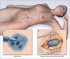
  • It is clear that patients with pathologically negative sentinel lymph nodes:
    • Do not require completion ALND:
      • But the management of patients with ITCs and micrometastatic disease in sentinel lymph nodes:
        • Has extensively been debated
  • Micrometastatic disease is defined as:
    • Tumor deposits spanning:
      • 0.2 mm to 2.0 mm:
        • Within lymph nodes
  • ITCs are:
    • Groupings of cells not greater than 0.2 mm or 200 cells:
      • In a single lymph node cross section
  • According to the American Joint Committee on Cancer (AJCC) staging guidelines:
    • Patients with ITCs are N0(i+)
    • Patients with micrometastases in one to three axillary lymph nodes are N1mi
  • ACOSOG Z0010:
    • Is one of the largest trials to prospectively evaluate the significance of small metastases in sentinel nodes
    • Hematoxylin and eosin (H&E) tumor-free sentinel nodes:
      • From patients with early breast cancer were evaluated in a central laboratory with:
        • Immunohistochemistry (IHC)
    • Micrometastatic or ITC disease:
      • Was found in 11% of 3,326 sentinel lymph nodes
    • With a median follow-up of 6.3 years:
      • Occult sentinel lymph node metastases:
        • Were not associated with differences in overall survival, disease-free survival, or recurrence:
          • When compared with patients with IHC-negative lymph nodes
  • A subset analysis of NSABP-32:
    • Looked retrospectively at patients with occult metastatic disease:
      • Including patients with micrometastatic or isolated tumor cells
    • In patients who were sentinel node negative:
      • 16% had occult metastases detected on further evaluation:
        • 11% of occult metastases were isolated tumor-cell clusters, 4% were micrometastases, and less than 1% of patients had macrometastatic deposits seen on additional sectioning of the lymph node
    • Log-rank tests indicated that patients with occult metastasis:
      • Had worse overall survival (95% versus 96%), disease-free survival (87% versus 89%), and distant disease-free interval (90% versus 93%):
        • When compared with patients without occult metastases
      • Although statistically significant:
        • These differences were not felt to be clinically relevant
    • There was no improvement in overall or disease-free survival:
      • When patients with occult metastasis underwent completion ALND

#Arrangoiz #CancerSurgeon #BreastSurgeon #SurgicalOncologist #BreastExpert #BreastCancer #SLNM #SLNB #SentinelLymphNodeBiopsy #Surgeon #Teacher #MSMC #MountSinaiMedicalCenter #Miami #Mexico

Management of Patients with Tumor-Free Sentinel Lymph Nodes in Breast Cancer

  • Randomized clinical trials support the use of SLNB without ALND:
    • For node-negative patients
  • These studies demonstrated very low axillary recurrence rates:
    • In patients with negative sentinel lymph nodes:
      • Regardless of receptor status
      • Type of breast operation
      • Patient age
  • The ASCO guidelines state:
    • That women with early-stage breast cancer without sentinel nodal metastases:
      • Should not undergo ALND
  • The first use of SLNB without ALND was a prospective observational study in 1994:
    • Which evaluated 125 patients with SLNB alone
  • NSABP B-32:
    • A phase III trial involving 80 centers in Canada and the United States
    • Prospectively randomized 3,989 sentinel lymph node–negative patients to SLNB plus ALND or SLNB alone
    • Patients were stratified based on age, tumor size, and surgical approach
    • Regional recurrence was rare in both patients who underwent SLNB alone and those who had a completion ALND and was not statistically significantly different
    • Of the 22 regional events in both groups:
      • 10 breast cancer recurrences were in the axilla:
        • For a rate of less than 1%
      • Two of these recurrences occurred in patients who were treated with ALND and eight in patients who underwent SLNB alone
      • There was no difference in overall or disease-free survival between groups:
        • And there were fewer complications in the SLNB-alone group
    • The results of NSABP B-32:
      • Have also been seen in other studies
  • Guided by this strong evidence:
    • SLNB alone has replaced ALND for sentinel node–negative patients

#Arrangoiz #CancerSurgeon #BreastSurgeon #SurgicalOncologist #BreastExpert #BreastCancer #SLNM #SLNB #SentinelLymphNodeBiopsy #Surgeon #Teacher #MSMC #MountSinaiMedicalCenter #Miami #Mexico