HER2-targeted agents are highly effective in improving survival in HER2-positive patients.
Therefore, trastuzumab (+/- pertuzumab), both of which target HER2, are part of every regimen endorsed by NCCN for HER2-positive disease.
These are often combined with standard chemotherapy, composed of anthracyclines (such as doxorubicin) and/or taxanes (such as docetaxel and paclitaxel).
National Comprehensive Cancer Network. NCCN Clinical Practice Guidelines in Oncology. Breast Cancer. Available at http://www.nccn.org.
Slamon D, Eiermann W, Robert N, et al. Adjuvant trastuzumab in her2 positive breast cancer. N Engl J Med. 2011;365:1273-1283.
We have two trials out there looking at anti-HER2 therapy plus/minus endocrine therapy in the setting. One trial is the ADAPT Trial looking at hormone receptor-positive, HER2-positive disease at 12 weeks of T-DM1 (ado-trastuzumab) plus/minus endocrine therapy, where there was no difference in the pCR (pathologic complete response) rate but a substantial pCR of 41% with 12 weeks of TDM1 alone.
The second trial is the NSABP B-52 trial, a phase III trial that looked at TCHP (docetaxel, carboplatin, trastuzumab, and pertuzumab) plus/minus endocrine therapy.
Both trials involved pre- and postmenopausal patients. And again, the NSABP B-52 trial did not show an improvement of the pCR in hormone receptor-positive HER2-positive disease by adding endocrine therapy to anti-HER2 therapy.
What does that mean for clinical practice?
It means, first of all, the endocrine therapy did not negatively impact the pCR rate. So it’s safe to use endocrine therapy plus anti-HER2 therapy in the postoperative setting, but there is no need, so far, to combine it with the anti-HER2 therapy with a chemotherapy backbone in the neoadjuvant setting. We don’t know why that is; it may be that the times used in the studies were not long enough and endocrine therapy added to anti-HER2 therapy needs a longer time. But I think for the time being, we should not combine endocrine therapy plus chemotherapy and anti-HER2 therapy in the neoadjuvant setting.
What is the utility and prognostic value of Ki-67 as a biomarker to guide treatment decisions regarding adjuvant abemaciclib in combination with endocrine therapy for patients with hormone receptor (HR)-positive / HER2-negative early breast cancer?
The FDA approval of abemaciclib for patients with high-risk HR-positive / HER2-negative early breast cancer represents a significant advancement in the field, and it is notably the first regulatory approval in the adjuvant HR-positive/HER2-negative setting after more than 15 years. Based on the results of the phase III monarchE trial, the FDA approved the use of abemaciclib in combination with endocrine therapy (tamoxifen or an aromatase inhibitor) for the adjuvant treatment of adult patients with node-positive, HR-positive/HER2-negative early breast cancer and a high risk of recurrence based on clinicopathologic features (either ≥ 4 axillary lymph nodes or 1-3 positive axillary lymph nodes with tumor grade 3 and / or tumor size ≥ 50 mm) and a Ki-67 score ≥ 20%, as determined by an FDA-approved test. The details of the approval, specifically the inclusion of a Ki-67 score of ≥ 20% as a requirement, has created some controversy in the oncology community.
The monarchE trial results demonstrated that patients with the highest risk of recurrence—specifically those with both high-risk clinical features and a high Ki-67 score—derived the greatest absolute benefit in terms of invasive disease-free survival with the addition of abemaciclib to endocrine therapy. Not surprisingly, this subgroup was the basis for the FDA approval. However, the study also demonstrated that although a high Ki-67 score is prognostic for recurrence, it is not a predictive marker for benefit from abemaciclib, as patients with a low Ki-67 score also demonstrated improved invasive disease-free survival with the addition of abemaciclib to endocrine therapy. Some in the community feel that a longer follow-up period is needed for the population of patients with a low Ki-67 score to better understand the role of adjuvant abemaciclib in this patient group. Historical data suggest that this group of patients with a low Ki-67 score is more likely to experience later recurrence compared with the group of patients with a high Ki-67 score. In view of this, both the National Comprehensive Cancer Network and American Society of Clinical Oncology guidelines currently recommend the consideration of treatment with adjuvant abemaciclib in otherwise eligible patients regardless of the Ki-67 score. This underscores the notion that the most significant impact of novel agents such as CDK4/6 inhibitors is likely to be in the localized, curable breast cancer setting, where they could reduce the risk of progression to metastatic disease.
The use of the Ki-67 score in the monarchE trial is a natural choice given that it is a marker of cellular proliferation and therefore mechanistically linked to CDK4/6 inhibitors. Also, the nuclear staining of the Ki-67 protein as a prognostic biomarker for HR-positive/HER2-negative breast cancer is well established, widely used, and inexpensive. However, the scoring methodology for Ki-67 stains remains controversial. Data from the International Ki67 in Breast Cancer Working Group (IKWG) have shown dramatic heterogeneity in Ki-67 assessment across laboratories, and the IKWG has expressed serious concerns regarding its routine use. The IKWG has systematically analyzed the causes of Ki-67 variability across laboratories and developed guidelines for its assessment using immunohistochemistry (IHC).The IKWG is of the opinion that consistency in Ki-67 scoring is difficult to achieve in the 5% to 30% range. Of note, the FDA approval of a Ki-67 score using a ≥20% cutoff as a companion diagnostic has not been generally embraced in the breast oncology community.
Ki-67 is a pan-cell cycle marker that is expressed in all tissues, albeit at low levels. This has led to issues regarding antibody titration for IHC analysis. A consequence of this is that antibodies are used at different dilutions, with resultant differences in nuclear staining intensities. The FDA approval of the Ki-67 IHC MIB-1 assay now requires the use of a ready-to-use standard concentration of an antibody. Similarly, it provides standardized criteria and analytical methods for defining Ki-67 positivity. Regarding the monarchE trial and the FDA-approved criteria, a nucleus is considered positive for Ki-67 if: (1) the signal is unequivocally brown; (2) the staining corresponds to a nucleus; (3) the staining covers the whole chromatin distribution within the nucleus; and (4) the nuclear staining is observed in viable, nonapoptotic cells. This definition differs from the one proposed by the IKWG in that the IKWG defines a nucleus as positive if it is not blue; therefore, grey nuclei are considered positive. In addition, focal nuclear positivity is considered sufficient. Both the IKWG- and FDA-approved methods recommend the analysis of the entire slide, although the IKWG method is more involved in requiring counting of larger areas and a higher number of cells. Both methods exclude necrotic tumor areas, foci of carcinoma in situ, edge effects, and fixation- and processing-related artifacts.
Overall, CDK4/6 inhibitors have revolutionized the treatment for patients with HR-positive/HER2-negative advanced breast cancer, and the FDA approval of adjuvant abemaciclib plus endocrine therapy in the early breast cancer setting is a significant advancement in the field.
Poorly differentiated carcinoma is a rare thyroid tumor that arises from follicular cells and is characterized by a partial loss of thyroid differentiation and less favorable prognosis in comparison with well-differentiated papillary or follicular carcinomas.
Anaplastic (undifferentiated) carcinoma represents the most undifferentiated type of thyroid tumors. In thyroidectomy samples of poorly differentiated and anaplastic thyroid carcinomas, it is not infrequent to find areas of well-differentiated papillary, conventional follicular or oncocytic carcinoma. This suggests that these tumors may represent distinct steps in the step-wise progression: well-differentiated carcinoma derived from follicular cells → poorly differentiated carcinoma → anaplastic carcinoma.
In agreement with such a progression, some molecular alterations, considered to be early events in thyroid carcinogenesis (i.e., mutations of RAS and BRAF), are found in tumors with all levels of dedifferentiation, whereas other, late events (i.e., TP53 mutations) occur with increasing frequency in tumors that progressively loose thyroid differentiation.
Inactivating point mutations of the TP53 tumor suppressor gene are among the most common mutations found in human cancer.
In thyroid tumors, point mutations of TP53 are a late event, reported in 60% to 80% of anaplastic thyroid carcinomas and 15% to 30% of poorly differentiated carcinomas, but only in single cases of follicular and papillary carcinomas.
Most of them involve exons 5 to 8 of the gene and alter its DNA binding properties.
p53 inactivation in thyroid cells is not only responsible for accelerated tumor growth, but is also associated with the progressive loss of differentiated markers.
Indeed, the recovery of wild-type p53 expression in cultured thyroid anaplastic carcinoma cells leads to the reduction in proliferation rate, re-expression of thyroid-specific genes (e.g., TPO, PAX-8), and re-acquisition the ability to respond to thyroid-stimulating hormone stimulation. This suggests that the progressive loss of differentiation in poorly differentiated and anaplastic carcinomas is mediated, at least in part, by inactivation of the p53 gene.
It also points to the restoration of TP53 function as a possible therapeutic approach for these highly aggressive tumors. Indeed, viral TP53 gene therapy has been tested in preclinical and clinical trials for various cancer types, and is under evaluation for anaplastic thyroid carcinoma.
β-catenin is a cytoplasmic protein, which is encoded by the CTNNB1 gene and is an important intermediate in the wingless (Wnt) signaling pathway. In thyroid tumors, point mutations in exon 3 of CTNNB1 have been found in 25% of poorly differentiated carcinomas and 66% of anaplastic thyroid carcinomas, but not in well-differentiated carcinomas.
Most of the tumors carrying the mutation also demonstrated an aberrant nuclear expression of the protein determined by immunohistochemical analysis, although there was no full correlation between these findings.
Point mutations of the RAS genes have been reported in 18% to 27% of poorly differentiated carcinomas and in 50% to 60% of anaplastic thyroid carcinomas. It is likely that mutant RAS stimulates genomic instability in the affected cells and predisposes them to accumulation of additional genetic abnormalities, such as mutations of the TP53gene. This can be illustrated by a case report of an anaplastic carcinoma developed in a well-differentiated follicular carcinoma, where RAS mutation was found in both tumor components, whereas TP53 was only found in the anaplastic carcinoma.
BRAF mutations occur in approximately 15% of poorly differentiated carcinomas and approximately 20% of anaplastic carcinomas, typically in those tumors that also contain areas of well-differentiated papillary thyroid carcinoma. In these tumors, BRAF mutation is detectable in both well-differentiated and poorly differentiated or anaplastic tumor areas, providing evidence that it occurs early in tumorigenesis.
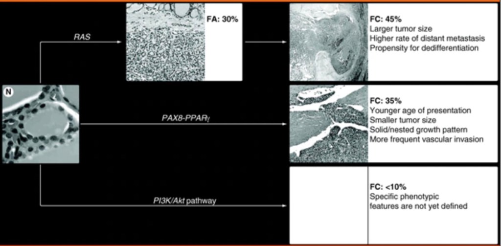
The most frequent genetic alterations in follicular thyroid carcinomas (FTC) include point mutations of the RAS genes and PAX8 / PPARγ rearrangement (Figure).
Molecular pathways in thyroid FC and typical microscopic presentation and clinical-pathological features of tumors associated with specific mutations. FA: Follicular adenoma; FC: Follicular cancer; N: Normal thyroid.
In addition, mutations and other alterations of the genes coding for the effectors of the PI3K/AKT signaling pathway can be found in these tumors, although the frequency of those mutations is low and their biological and diagnostic value remains to be elucidated.
As RAS point mutations are also frequently found in follicular adenomas (FA), it is likely that follicular thyroid carcinomas may arise as a result of malignant transformation of follicular adenomas
RAS mutations are found in 40% to 50% of conventional follicular thyroid carcinomas and in 20% to 40% of adenomas.
In adenomas, the mutations are more common in tumors with a microfollicular growth pattern. Most frequently affected hotspots are NRAS codon 61 and HRAS codon 61.
A lower incidence of RAS mutation has been reported in oncocytic (Hurthle cell) tumors, where only 0% to 4% of adenomas and 15% to 25% of carcinomas appeared to be affected.
The presence of RAS mutation in follicular thyroid carcinomas has been found to correlate with tumor dedifferentiation and a less favorable prognosis.
Several studies have found a significant correlation between RAS mutation and metastatic behavior of follicular thyroid carcinomas, especially with respect to bone metastases.
The more aggressive biological properties of these tumors may be due to the effect of the mutant RAS protein on promoting chromosomal instability, which has been demonstrated al least in the in vitro setting.
The increasing chromosomal instability may predispose the tumor cells for acquiring additional mutations which would result in more malignant phenotype.
The diagnostic use of RAS mutation detection is controversial. On the one hand, it is not specific for malignancy since RAS mutations also occur with significant prevalence in benign follicular adenomas. On the other hand, RAS mutations frequently occur in follicular thyroid carcinomas and the follicular variant papillary carcinomas, both of which are difficult to diagnose cytologically in thyroid FNA samples. Moreover, since mutant RAS is likely to predispose to progression from follicular adenoma to follicular thyroid carcinoma and to further tumor dedifferentiation, it may be justifiable to surgically remove the RAS-positive adenomas to prevent such a progression.
In a prospective study aimed to assess the role of detection of different mutations in improving the preoperative FNA diagnosis of thyroid nodules, the detection of RAS mutations was found to improve the diagnostic accuracy and allowed to diagnose malignant tumors in several samples with negative or insufficient cytology.
PAX8 / PPARγ rearrangement is a result of the translocation t(2;3)(q13;p25). It leads to the fusion between the PAX8 gene, which encodes a thyroid-specific paired domain transcription factor, and the PPARγ gene.
PAX8 / PPARγ occurs in approximately 35% of conventional follicular thyroid carcinomas, and with lower prevalence in oncocytic (Hurthle cell) carcinomas.
Tumors harboring PAX8 / PPARγ tend to present at a younger age, be smaller in size, have a solid / nested growth pattern and more frequently reveal vascular invasion.
The rearrangement results in overexpression of the PPARγ protein that can be detected by immunohistochemistry. However, only strong diffuse nuclear staining correlates with the presence of rearrangement.
PAX8 / PPARγ can also be found in a small fraction (2% to 10%) of follicular adenomas. It has been suggested that follicular adenomas positive for this rearrangement may in fact be pre-invasive (in situ) follicular carcinomas or tumors where invasion was overlooked during histological examination.
The mechanisms of cell transformation induced by PAX8 / PPARγ are not fully understood. Some evidence has been presented for inhibition of normal PPARγ function via a dominant negative effect of the PAX8 / PPARγ protein on wild-type PPARγ. Other studies have found the activation of known PPAR target genes in tumors harboring PAX8 / PPARγ, arguing against the dominant negative effect.
Other possible mechanisms include deregulation of PAX8 function, known to be critical for thyroid cell differentiation, and activation of a set of genes related to neither wild-type PPARγ nor wild-type PAX8 pathways. The lack of understanding of the definitive mechanism of PAX8 / PPARγ action hampers its use as a therapeutic target for follicular thyroid carcinomas, despite the fact that modulators of the PPAR pathway (retinoic acid derivatives as well as thiazolidenediones) are available and have already been used for other diseases.
The detection of PAX8 / PPARγ rearrangement may be of diagnostic value since it occurs almost exclusively in follicular thyroid carcinomas.
It can be achieved by reverse transcriptase-PCR, FISH, or immunohistochemistry with PPARγ antibody. Immunohistochemical detection might be challenging to set up and validate since not all commercially available PPARγ antibodies give a reliable result. The identification of PAX8 / PPARγ rearrangement by reverse-transcriptase PCR or FISH, or finding strong diffuse PPARγ inmmunoreactivity in tumor cells during pathological evaluation should justify the submission of additional sections of the tumor capsule and obtaining deeper levels of all suspicious areas in search for capsular or vascular invasion.
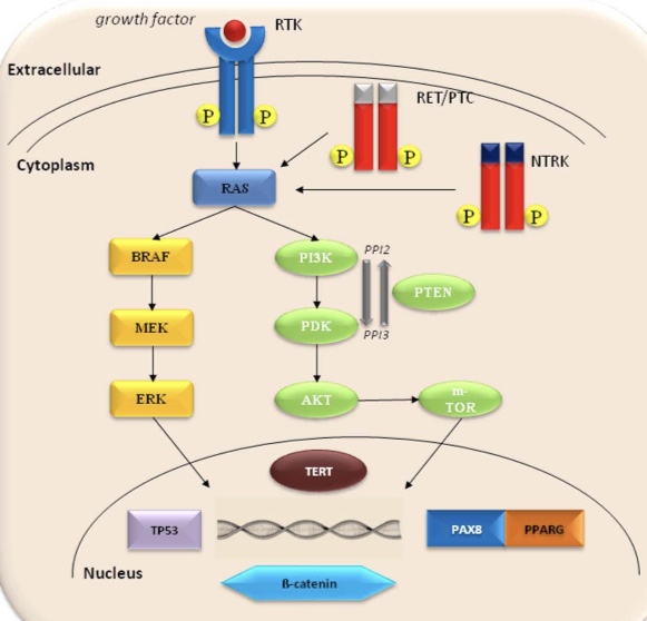
The PI3K / Akt signaling pathway plays an important role in the regulation of cell growth, proliferation and survival. This pathway can be activated by the upstream stimulatory molecules (i.e., RAS, RET /PTC), through the loss of function of PTEN protein that normally inhibits PI3K signaling or as a result of activating mutations or amplification of genes coding for the effectors of this pathway.
The PIK3CA gene, coding for a catalytic subunit of PI3Ks, has been shown to harbor mutations in thyroid tumors, although at low frequency. Specifically, it has been found in 6% to 13% of follicular thyroid carcinomas and in 0% to 6% of follicular adenomas. Mutations typically involve various nucleotides in exons 20 and 9 of the PIK3CA gene.
Mutations of the PTEN gene have been reported in a small proportion of follicular thyroid carcinomas (~7%), but not in follicular adenomas.
Loss of heterozygosity (LOH) of chromosomal regions harboring different tumor suppressor genes is another genetic alteration found in follicular carcinomas and adenomas. The most frequently deleted chromosomal regions in these tumors are on chromosomes 2p, 3p, 9q, 9p, 10q, 11p, 15q and 17p. The average rate of allelic loss of the target regions is significantly higher in follicular carcinomas (30% to 50%) than in follicular adenomas (6% to 15%).
Some studies have found correlation between the frequency of LOH and tumor aggressiveness and outcome in patients with follicular carcinomas. The minimally invasive tumors had overall lower frequency of allelic loss (~30%) compared with the widely invasive follicular carcinomas (~50%). In addition, higher frequency of LOH was associated with disease recurrence.
Allelic loss of the VHLgene on 3p26 was highly specific for malignancy and associated with death from disease in one study of a small series of follicular carcinomas, suggesting that it may serve as an important diagnostic and prognostic marker for follicular carcinomas. However, its clinical utility has to be validated in a larger series of tumors.
Oncocytic (Hurthle cell) neoplasms usually reveal a comparable or even higher rate of LOH than conventional follicular tumors. In oncocytic carcinomas, the most frequently lost regions were on chromosomes 3q and 18q in one study, and on chromosomes 1q, 2p, 8q and 14q in another observation. In the latter report, chromosomal loci at 1q and 2p showed a significantly higher rate of LOH in oncocytic carcinomas than in adenomas, with a 100% sensitivity and 65% specificity in the detection of malignant tumors.
Somatic point mutations and large deletions in mitochondrial DNA have been found in a significant proportion of oncocytic neoplasms. These mutations occur in oncocytic adenomas and carcinomas, and with lower frequency in other types of thyroid neoplasms, including papillary thyroid carcinoma and conventional follicular thyroid carcinoma. More recently, somatic mutations of the GRIM-19 gene have been identified in sporadic oncocytic tumors. GRIM-19 encodes a protein linked to retinoid-interferon-induced pathway of cell death and also involved in mitochondrial metabolism. Mutations in GRIM-19 were found in 15% of oncocytic carcinomas, but were not detected in other types of thyroid cancer, suggesting that alteration of GRIM-19 may serve as a specific marker of oncocytic tumors.
The oncology community shouldn’t be rushing to prescribe the CDK 4/6 inhibitor abemaciclib (Verzenio) for early breast cancer. There are still too many unknowns, given the lack of overall survival benefit observed so far, and too much risk of harm, given the reported side effects and high cost of treatment.
Considering the dubious or even non-existent benefit in a small percentage of patients, it may be premature to recommend adjuvant abemaciclib to patients with early breast cancer without waiting for mature data.
Abemaciclib and its rival CDK 4/6 inhibitors palbociclib and ribociclib are currently standard of care for advanced/metastatic hormone receptor (HR)– positive, HER2-negative breast cancer.
For now, it’s safe to say that CDK 4/6 inhibitors can be reserved for later lines of therapy.
Last October, the US Food and Drug Administration approved the use of abemaciclib with endocrine therapy in the adjuvant setting for women with early HR-positive, HER2-negative breast cancer who are at high risk for relapse and who have a Ki-67 score of 20% or higher.
At the time, drugmaker Eli Lilly noted in a press release that the approval made abemaciclib the first add-on that was approved for adjuvant endocrine therapy in nearly 20 years. A researcher on the company’s approval study called the new indication “practice-changing” and said this combination could become a new standard of care.
A month later, the American Society of Clinical Oncology updated its guidelines to give a strong recommendation for the use of abemaciclib with adjuvant endocrine therapy for patients who meet the indication requirements.
As for uptake by the oncology community, Eli Lilly reported earning $384.3 million for abemaciclib in the US in the second quarter of 2022 — up 83% from the second quarter of 2021.
The approval was based on a significant improvement in invasive disease-free survival at 36 months — 79% with endocrine therapy alone, vs 86% with the addition of abemaciclib; however, invasive disease-free survival has not been validated as a surrogate for overall survival. And in the trial, an overall survival benefit had not emerged at 27 months.
And with a price tag of more than $300,000 over 2 years, the authors worry about “substantial financial toxicity” for a drug with “uncertain benefit” and “known toxicities.”
In addition, abemaciclib’s side effects are notable. Almost 50% of patients experienced grade 3 or higher adverse events with abemaciclib, vs 16% with placebo.
Another potential downside: Giving the drug too early might generate treatment-resistant clones, potentially wiping out a benefit from abemaciclib in the advanced/metastatic setting.
In fact, the possibility of a worse overall survival also exists, if early CDK 4/6 exposure leads to the development of an aggressive, treatment resistant clone.
The team also probed why another CDK 4/6 inhibitor, palbociclib, which has a similar mechanism of action to abemaciclib and is similar in efficacy for the treatment of metastatic disease, struck out twice in trials for early hormone- positive breast cancer — wondering, for instance, whether there is a “genuine difference” between the two that can explain the outcomes in the adjuvant setting.
Overall, the editorialists “urge caution” before accepting abemaciclib as standard of care in early breast cancer.
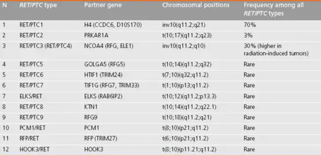
RET / PTC rearrangement is another genetic alteration that is frequently found in papillary thyroid carcinomas (PTC), and is a result of the fusion between the 3´-portion of the RET receptor tyrosine kinase gene and the 5´-portion of various genes.
Two most common rearrangement types, RET / PTC1 and RET / PTC3, are paracentric inversions, because both RET and its respective fusion partner, H4 or NCOA4 (ELE1), reside on the long arm of chromosome 10.
RET / PTC2 and nine more recently identified types of RET / PTC are all interchromosomal translocations. Most of these rare RET / PTC types have been found in papillary thyroid carcinomas from patients with a history of either environmental or therapeutic exposure to ionizing radiation, with the exception of the ELKS-RET and HOOK3-RET fusions, which have been identified in papillary thyroid carcinoma with no apparent history of radiation exposure.
All fusions leave the tyrosine kinase domain of the RET receptor intact and enable the RET / PTC oncoprotein to bind SHC and activate the RAS-RAF-MAPK cascade.
RET / PTC is found on average in approximately 20% of adult sporadic papillary thyroid carcinomas, although its prevalence is highly variable between different observations due to either geographic variability or different sensitivity of the detection.
RET / PTC fusion is typically more common in tumors from patients with a history of radiation exposure (50% to 80%) and in papillary thyroid carcinomas from children and young adults (40% to 70%).
The distribution of RET / PTC rearrangement within the tumor may be quite heterogeneous and vary from involving almost all neoplastic cells (clonal RET / PTC) to being detected only in a small fraction of tumor cells (nonclonal RET / PTC).
Although RET / PTC has been found in several studies in adenomas and other benign thyroid lesions, it can be assumed that clonal RET / PTC (i.e., rearrangement that is found in most cells within the tumor) is reasonably specific for papillary thyroid carcinomas.
RET / PTC rearrangements can be detected in thyroid FNA samples. Several studied have shown that RET / PTC detection can refine the preoperative diagnosis of thyroid nodules, particularly in samples that are indeterminate by cytology or have insufficient amount of cells for cytological evaluation. Although these studies have provided important evidence for potential diagnostic utility in RET / PTC detection in thyroid FNA samples, the performance characteristics of this test must be defined in a large prospective study before considering the implementation of this test into clinical practice. One potential problem with such a test lies in the requirement for isolation of acceptable quality RNA, which is difficult to achieve in the fixed samples. This limitation can be resolved by collecting a small portion of the aspirated FNA material directly into nucleic acid preservative solution. This approach typically yields sufficient quality and quantity of RNA that can be used for successful RET / PTC testing.
Papillary thyroid carcinomas with RET / PTC rearrangements typically present at a younger age and have a high rate of lymph node metastases, classic papillary histology and lower stage at presentation, particularly those harboring RET / PTC1
In papillary thyroid carcinomas associated with exposure to ionizing radiation (i.e., post-Chernobyl tumors), RET / PTC1 was found to be associated with classic papillary histology, whereas RET / PTC3 type was more common in the solid variant.
Schematic representation of RET / PTC1 rearrangement: a centromeric inversion of chromosome 10 (A) generates a chimeric gene through the fusion of the RET tyrosine-kinase domain with the 5′ terminal region of a new gene denominated CCD6, formerly called H4 (B).
With more than 2 million breast cancer survivors in the United States, breast cancer has a higher cost than other prevalent cancers, with the greatest proportion of the cost related to follow-up care after active treatment.
For patients with breast cancer who have been treated with curative intent, there is no evidence-based data showing that routine measurement of serum tumor markers, CT, or bone scans provide earlier detection of recurrence, or survival benefit, in asymptomatic patients.
Similarly, there are no data to support routine PET scans to assess for metastatic disease in an asymptomatic patient.
The Choosing Wisely campaign has specifically advocated that routine blood tests and imaging with CT, bone or PET scans not be performed routinely in patients who present with early-stage disease and who are treated with curative intent.
Both the American Society of Clinical Oncology and the National Comprehensive Cancer Network recommend annual mammography for all women treated with breast-conservation therapy.
Referecnes:
Khatcheressian JL, Wolff AC, Smith TJ, et al. American Society of Clinical Oncology 2006 update of the breast cancer follow-up and management guidelines in the adjuvant setting.
J Clin Oncol. 2006;24:5091-5097. Lewis JL, Tartter PI. The value of mammography within 1 year of conservative surgery for breast cancer. Ann Surg Oncol. 2012;19:3218-3222.
Locker GY, Hamilton S, Harris J, et al. ASCO 2006 update of recommendations for the use of tumor markers in gastrointestinal cancer. J Clin Oncol. 2006;24:5313-5327.
Mariotto AB, Yabroff KR, Shao Y, Feuer EJ, Brown ML. Projections of the cost of cancer care in the United States: 2010-2020. J Natl Cancer Inst. 2011;103:117-128.
National Comprehensive Cancer Network NCCN Clinical Practice Guidelines in Oncology. Breast Cancer. Available at http://www.nccn.org.
American Society of Clinical Oncology. Ten Things Physicians and Patients Should Question. Released April 4, 2012 (1-5) and October 29, 2013 (6-10).
Taxanes bind free tubulin and stabilize microtubules.
This inhibits the spindle apparatus needed for mitosis.
Cyclophosphamide is an alkylating agent, which forms DNA cross-linkages
Doxorubicin interferes with topoisomerase II.
Fluorouracil acts as a nucleotide analog
Trastuzumab targets the epidermal growth factor receptor.
References Abal M, Andreu JM, Barasoain I. Taxanes: microtubule and centrosome targets, and cell cycle dependent mechanisms of action. Curr Cancer Drug Targets. 2003;3:193-203.
Priestman T. The theoretical basis of cancer chemotherapy. In Cancer Chemotherapy in Clinical Practice. 2nd ed. London, UK: Springer-Verlag London; 2012:1-43.