Soft Palate

👉The soft palate is located in the oropharynx posteriorly.

👉It is mobile, and comprised of muscle fibres covered by a mucous membrane (stratified squamous epithelium).

👉The soft palate is a musculoaponeurotic curtain that hangs down from the posterior border of the hard palate and maintains velopharyngeal competence during speech and swallowing.

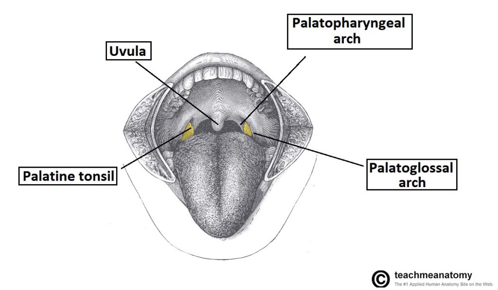
👉The midline uvula is located on the free inferior border and the sides blend into the faucial arches that attach to the lateral pharyngeal walls.

👉The mucosa of the soft palate is of the stratified squamous epithelium.

👉The submucosa of the soft palate contains minor salivary glands that mainly secret mucus.

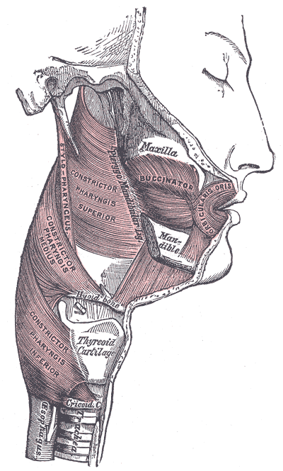
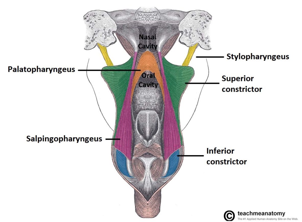
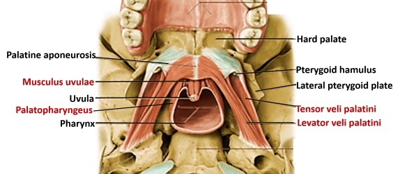
👉The muscles of the soft palate include the palatopharyngeus, palatoglossus, tensor veli palatini, levator veli palatini, and musculus uvulae.

👉Anteriorly, the soft palate is continuous with the hard palate and with the palatine aponeurosis.

👉The posterior border of the soft palate is free (i.e. not connected to any structure), and has a central process that hangs from the midline – the uvula.

👉The soft palate also forms the roof of the fauces; an area connecting the oral cavity and the pharynx.

👉Two arches bind the palate to the tongue and pharynx:

👉The palatoglossal arches anteriorly and the palatopharyngeal arches posteriorly.

👉Between these two arches lie the palatine tonsils, which reside in the tonsillar fossae of the oropharynx.

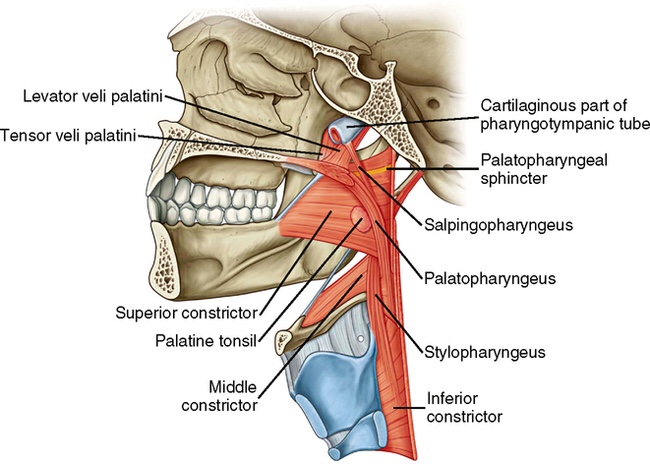
Muscles of the Soft Palate

👉There are five muscles which give the actions of the soft palate.

👉They are all innervated by the pharyngeal branch of the vagus nerve (CN X).

👉The Tensor veli palatini is the only muscle of the soft palate that is not innervated by the pharyngeal plexus derived from the vagus nerve.

👉The Tensor Veli palatini is innervated by the medial pterygoid nerve (madibular division of the trigeminal nerve / CN V).

Tensor Veli Palatini

  • Attachments: Originates from the medial pterygoid plate of the sphenoid bone and inserts into the palatine aponeurosis.
  • Function: Tenses the soft palate.

Levator Veli Palatini

  • Attachments: Arises from the petrous portion of the temporal bone and the eustachian tube, before inserting into the palatine aponeurosis.
  • Function: Elevation of the soft palate.

Palatoglossus

  • Attachments: Originates from the palatine aponeurosis, and travels anteriorly, laterally and inferiorly to insert into the side of the tongue.
  • Function: Pulls the soft palate towards the tongue.
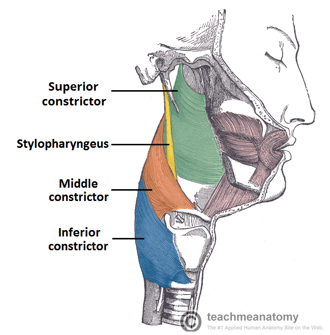
Palatopharyngeus

  • Attachments: Arises from the palatine aponeurosis and the hard palate, and inserts into the upper border of the thyroid cartilage.
  • Function: Tenses soft palate and draws the pharynx anteriorly on swallowing.

Musculus Uvulae

  • Attachments: Arises from the posterior nasal spine and the palatine aponeurosis, and inserts into the mucous membrane of the uvula.
  • Function: Shortens the uvula.

Vasculature of the Soft Palate

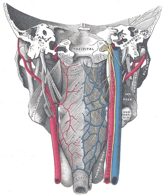
👉The palate receives its arterial supply primarily from the greater palatine arteries, which run anteriorly from the greater palatine foramen.

👉In addition, the anastomosis between the lesser palatine artery and ascending palatine artery provide collateral supply to the palate.

👉Venous drainage is into the pterygoid venous plexus.

Innervation of the Soft Palate

👉Sensory innervation of the palate is derived from the maxillary branch of the trigeminal nerve (CN V).

👉The greater palatine nerve innervates most of the glandular structures of the hard palate.

👉The nasopalatine nerve innervates the mucous membrane of the anterior hard palate and the lesser palatine nerves innervate the soft palate.

#Arrangoiz #HeadandNeckSurgeon #CancerSurgeon #SurgicalOncologist

Managing Breast and Ovarian Cancer in Mutation Carriers

👉BRCA1-associated breast cancer is frequently high-grade basal-type breast cancer.

👉One recent meta-analysis found that BRCA1 mutation was associated with worse survival, but a second recent meta-analysis failed to confirm this.

👉It is becoming apparent that BRCA1/BRCA2-associated breast cancer is more sensitive to chemotherapy of all types than sporadic breast cancer.

👉The outcome for BRCA1/BRCA2-associated breast cancer is likely no worse than that for sporadic breast cancer in the era of multimodal treatment.

👉Breast conservation is an option for nearly any mutation carrier provided that they are willing to accept the risk of second primary breast cancer.

👉The risk of ipsilateral breast tumor events ranges from 1.7% to 2.7% per year, but could be as high as 4% per year for very early onset breast cancer (e.g., age 42 or younger).

👉This risk is modified downward by hormonal therapy for estrogen receptor–positive breast cancer and by chemotherapy.

👉Breast conservation should be avoided in homozygous ATM mutation carriers (i.e., patients with ataxia-telangiectasia) in whom radiation is contraindicated and possibly TP53 mutation carriers, who may be at increased risk for radiation-induced malignancies.

👉Lifetime contralateral breast cancer risk is as high as 62% to 83% for BRCA1/BRCA2 mutation carriers.

👉There is some evidence that bilateral mastectomy improves survival in this population.

👉Although follow-up is limited, available data show acceptable local recurrence risk for BRCA 1 / 2 mutation–associated breast cancer treated by nipple-sparing mastectomy.

👉It is reasonable to encourage oophorectomy in BRCA1 and BRCA2 mutation carriers who are treated for breast cancer as this may reduce mortality by 43% to 70%, an effect that seems greatest for estrogen receptor–negative breast cancer.

👉Bilateral salpingo-oophorectomy was associated with improved all-cause and breast cancer–specific mortality in BRCA1 mutation carriers with stage 1 to 3 breast cancer.

👉There is evidence that outcome for BRCA 1 / BRCA 2 mutation–associated ovarian cancer is better than for sporadic ovarian cancer.

👉This is particularly true for BRCA2-associated ovarian cancer, which shows enhanced sensitivity to chemotherapy.

👉Nevertheless, ovarian cancer is the main mortality driver for BRCA 1 / BRCA 2 mutation carriers, and clinicians will sometimes be called upon to manage breast cancer risk in patients who have completed ovarian cancer treatment.

👉One recent study that included 364 patients with BRCA 1 / BRCA 2-associated ovarian cancer reported a 10-year survival of 11%, with 10% of patients developing breast cancer.

👉There is growing evidence that BRCA 1 / BRCA 2-associated breast cancer is less sensitive to taxanes than sporadic breast cancer and more sensitive to DNA-damaging agents, such as mitomycin C and platins.

👉BRCA 1 / BRCA 2 mutations also predict sensitivity to poly (ADP-ribose) polymerase (PARP) inhibition.

👉Trials of platin agents and PARP inhibitors consistently identify BRCA 1 / BRCA 2 mutation as the strongest predictor of response.

👉There is a growing list of platinum and PARP inhibitor trials open and accruing for BRCA1 and BRCA2 mutation carriers.

👉Whether this approach will be equally efficacious for individuals with mutations in other DNA repair genes, such as PALB2, RAD51C, BRIP, ATM, and CHEK2, is not certain, but it is hoped that the development and validation of predictive homologous recombination assays will make it possible to prospectively identify the right patients for these agents.

#Arrangoiz #BreastSurgeon #CancerSurgeon #SurgicalOncologist #BreastCancer #HereditaryBreastCancer

Differentiating Detectable Findings from Actionable Findings in Thyroid Cancer Management

👉As it is clear that not all detectable findings require immediate diagnostic or therapeutic intervention, it is imperative that we develop a risk-stratification decision-making framework to differentiate actionable findings from non-actionable findings.

👉Whether we are considering a highly suspicious subcentimeter thyroid nodule without cytologic confirmation of disease, a biopsy-proven thyroid nodule with low-risk thyroid cancer, or persistent/recurrent disease in the neck or elsewhere, we find it useful to consider five key factors that when taken together, allow us to predict the likelihood that a specific tumor focus represents clinically
important disease
that may require additional evaluations, ongoing observation, or therapeutic intervention.

👉Both tumor size and tumor location are the
major factors that determine whether a tumor focus is
likely to cause clinically substantial invasion into local structures, such as the recurrent laryngeal nerve, airway, gastrointestinal tract, major vessels, or other important structures.

👉A third important factor is the tumor growth rate (measured as tumor volume doubling time), with an observational management approach being much more appropriate for tumors either anticipated
to have a slow tumor growth rate
or with actual documented slow growth rates over time.

👉Obviously, tumors that are either symptomatic or likely to have symptomatic progression would be considered actionable.

👉Finally, patient preference plays a key role when
deciding whether a particular lesion is actionable or non-actionable, as it is important to integrate the patients understanding of the risks and benefits of intervention vs observation with their value system and goals.

👉In addition to providing initial guidance as to whether the detectable lesion is actionable at the time of detection, ongoing re-evaluation of these same factors, using the basic concepts of dynamic-risk stratification, can also assist the clinician in the determination of when it is time to transition from an observational management approach to active therapeutic intervention.

👉Thus, risk stratification has moved from a single
postoperative static assessment
of the risk of disease-
specific mortality to an all-encompassing evaluation of the patient that is continually modified over time, beginning from the first detection of a suspicious thyroid nodule and continuing throughout the life of the patient.

#Arrangoiz #ThyroidSurgeon #ThyroidExpert #HeadandNeckSurgeon #CancerSurgeon #ThyroidCancer #ThyroidNodules #SurgicalOncologist

Risk Reducing Surgery in High Risk Breast Cancer Patients

Bilateral Prophylactic Mastectomy

👉A recent meta-analysis that included 2,635 BRCA1/BRCA2 mutation carriers reported a 93% reduction in breast cancer incidence for the 627 women who underwent bilateral risk-reducing mastectomy.

👉Based on the four studies included in this meta-analysis, it can be estimated that baseline breast cancer risk was 1.7% to 2.8% per year for BRCA1/BRCA2 gene mutation carriers (34% to 56% after 20 years) and 0% to 0.8% per year after bilateral risk-reducing mastectomy (0% to 16% after 20 years).

👉Median follow-up was only 3 to 6 years for these studies, so the durability of these estimates is not certain.

👉After mastectomy, terminal duct-lobular units (TDLUs) can be identified in peripheral skin, in the inframammary crease, and in the nipple-areola complex.

👉Although removing the nipple-areola complex reduces the number of residual TDLUs, it is not clear that this is required to achieve an acceptably low breast cancer risk.

👉To date, outcomes for bilateral risk-reducing nipple-sparing mastectomy have been reported for over 300 BRCA1/BRCA2 gene mutation carriers.

👉Incidental breast cancers were identified in 3% to 6%, and with a median follow-up of 33 months, subsequent breast cancers have developed in three patients.

👉Modern nipple-sparing mastectomy should not intentionally leave any breast tissue behind.

👉Bilateral risk-reducing mastectomy has not yet been shown to improve breast cancer–specific or overall survival in BRCA1/BRCA2 mutation carriers, but a 2010 Cochrane review concluded that risk-reducing mastectomy is likely to confer a survival advantage in the highest-risk women.

#Arrangoiz #BreastSurgeon #BreastCancer #CancerSurgeon #SurgicalOncologist

Pharyngeal Muscles

👉The pharyngeal muscles are a group of muscles that form the pharynx, which is posterior to the oral cavity, determining the shape of its lumen, and affecting its sound properties as the primary resonating cavity.

👉The pharyngeal musculature (involuntary skeletal muscles) function is to push the food bolus into the esophagus.

👉There are two muscular layers of the pharynx: the outer circular layer and the inner longitudinal layer.

👉The outer circular layer includes:

  • Inferior pharyngeal constrictor muscle:
    • Located in the laryngopharynx
    • It has two components:
      • Superior component (thyropharyngeus):
        • Has oblique fibres that attach to the thyroid cartilage.
      • Inferior component (cricopharyngeus):
        • Has horizontal fibres that attach to the cricoid cartilage.
    • Inserts posteriorly into the midline fibrous pharyngeal raphe
  • Middle pharyngeal constrictor muscle:
    • Located in the laryngopharynx
    • Originates from the stylohyoid ligament and the horns of the hyoid bone.
    • Inserts posteriorly into the midline fibrous pharyngeal raphe
  • Superior constrictor muscle:
    • The uppermost pharyngeal constrictor
    • It is located in the oropharynx
    • Originates from the pterygomandibular ligament, alveolar process of mandible and medial pterygoid plate and pterygoid hamulus of the sphenoid bone
    • Inserts posteriorly into to the pharyngeal tubercle of the occiput and the medial fibrous pharyngeal raphe

👉During swallowing, these muscles constrict to propel food bolus downwards (an involuntary process).

👉The inner longitudinal layer includes:

  • Stylopharyngeus muscle:
    • Arises from the styloid process of the temporal bone, inserts into the pharynx
    • Unlike the other pharyngeal muscles:
      • It is innervated by the glossopharyngeal nerve (CN IX)
  • Salpingopharyngeus muscle:
    • Arises from the Eustachian tube, inserts into the pharynx
    • Innervated by the vagus nerve (CN X)
    • In addition to contributing to swallowing:
      • It also opens the Eustachian tube to equalize the pressure in the middle ear
  • Palatopharyngeus muscle:
    • Arises from hard palate of the oral cavity, inserts into the pharynx
    • Innervated by the vagus nerve (CN X)

👉During swallowing, these muscles act to shorten and widen the pharynx.

👉They are innervated by the pharyngeal branch of the Vagus nerve (CN X) with the exception of the stylopharyngeus muscle which is innervated by the glossopharyngeal nerve (CN IX).

#Arrangoiz #HeadandNeckSurgeon #CancerSurgeon #SurgicalOncologist

RISK-REDUCING SURGERY

Bilateral Salpingo-oophorectomy

👉The lifetime ovarian cancer risk is 39% to 59% for BRCA1 mutation carriers and 11% to 18% for BRCA2 carriers.

👉Other genes associated with increased ovarian cancer risk include BRIP1, RAD51C, RAD51D, MSH2, MLH1, PMS2, MSH6, PALB2, and BARD1.

👉BRCA 1 / 2 associated ovarian cancers are high-grade serous or undifferentiated carcinomas that are typically metastatic at presentation.

👉Most of these tumors arise in one of the fallopian tubes.

👉Median age at ovarian cancer diagnosis for BRCA 1 /2 mutation carriers is 51 years.

👉Annual incidence peaks between ages 50 and 59 for BRCA1 carriers and 60 and 69 for BRCA2 carriers.

👉Risk-reducing bilateral salpingo-oophorectomy (RRSO) reduces ovarian cancer risk by 72% to 96% (79% in a recent meta-analysis) in BRCA1/BRCA2 gene mutation carriers and is also associated with a 60% to 77% reduction in all-cause mortality.

👉Specialized protocols for performing the surgery and processing the specimens have been recommended as clinically occult cancer can be identified in 4% to 5%.

👉Case-control and cohort data suggest that bilateral salpingo-oophorectomy also reduces breast cancer risk by 50%.

👉There is some evidence that risk reduction is greater when the procedure is performed at a younger age.

👉The effect may be larger for BRCA2 carriers than for BRCA1 carriers.

👉Some have argued that the breast cancer risk reduction observed with RRSO is a statistical aberration resulting from unmanaged bias.

👉Others have refuted this.

👉For the time being, it seems prudent to recommend RRSO for the ovarian cancer and mortality benefits without relying too heavily on the procedure for breast cancer risk reduction.

👉Because it is increasingly recognized that hereditary ovarian cancers most commonly arise in the tubes and not the ovary proper, there is increasing interest in performing salpingectomy alone and leaving the ovaries intact.

👉There are currently no data quantifying the risk reduction afforded by this approach, and it is unlikely that breast cancer risk would be reduced.

👉However, there is likely merit to this approach; several studies and registries are currently accruing, so more data will be forthcoming.

👉There is also discussion about whether the uterus should be removed at the time of RRSO.

👉Current guidelines do not mandate that it should, but some advocate it to improve the safety of future tamoxifen use.

👉Of note, one recent study reported five serous or serouslike endometrial cancers among 1,083 BRCA1/2 mutation carriers undergoing RRSO and followed for a median of 5 years.

👉Less than one cancer of this type was expected.

👉The current standard of care is to consider bilateral salpingo-oophorectomy after childbearing is complete or about age 35 to 40.

👉NCCN guidelines recommend consideration of risk-reducing salpingo-oophorectomy for women found to carry pathogenic mutations in BRCA1, BRCA2, Lynch syndrome genes, BRIP1, RAD51C, or RAD51D.

👉Abrupt surgical menopause can adversely affect quality of life due to severe hot flashes, vaginal dryness, sexual dysfunction, sleep disturbances, and cognitive changes.

👉HRT should not be withheld for symptom control as short-term HRT does not seem to negate the risk-reducing effect of RRSO.

#Arrangoiz #BreastSurgeon #CancerSurgeon

Next Generation Sequencing Multigene Panel Test

👉Since the 2013 Supreme Court decision revoking patent protection for raw DNA sequences, BRCA1 and BRCA2 have been included in most multigene panel tests.

👉Next-generation sequencing tests are accurate, with analytic sensitivity and specificity measured at greater than 99.9% for detection of known pathogenic variants.

👉The 2013 Supreme Court decision also introduced competition into the genetic testing marketplace, leading to several low-cost multigene panel testing options.

👉Panel tests that include dozens of genes still identify BRCA1 and BRCA2 as the genes most frequently affected by pathologic mutation.

👉Mutations in PALB2, CHEK2, and ATM are the next most common.

👉There is a 20% to 40% chance that a multigene panel test will return a variant of uncertain clinical significance compared with about 3% for standard BRCA1 and BRCA2 tests.

#Arrangoiz #BreastSurgeon #BreastCancer #CancerSurgeon #HereditaryBreastCancer #SurgicalOncologist

Lumpectomy and radiation therapy for the treatment of intraductal breast cancer: findings from National Surgical Adjuvant Breast and Bowel Project B-17.

, , , , , Show Morehttps://doi.org/10.1200/JCO.1998.16.2.441


N Engl J Med.
1993 Jun 3;328(22):1581-6.

Fisher, Costantino J, Redmond C, Fisher E, Margolese R, Dimitrov N, Wolmark N, Wickerham DL, Deutsch M, Ore L, et al.

Abstract

BACKGROUND AND METHODS: 

👉Women with ductal carcinoma in situ have been treated both by lumpectomy and by lumpectomy followed by radiation therapy, but the benefit of combined therapy is uncertain.

👉A group of 818 women with ductal carcinoma in situ were randomly assigned to undergo lumpectomy or lumpectomy followed by breast irradiation (50 Gy).

👉Sufficient tissue was removed that the margins of the resected specimens were histologically tumor-free.

👉The mean duration of follow-up was 43 months (range, 11 to 86).

👉The principal end point of the study was event-free survival, as defined by the presence of no new ipsilateral or contralateral breast cancers, regional or distant metastases, or other cancers and by no deaths from causes other than cancer.

RESULTS:

👉Five-year event-free survival was better in the women who received breast irradiation (84.4%, vs. 73.8% for the women treated by lumpectomy alone; P = 0.001).

👉The improvement was due to a reduction in the occurrence of second ipsilateral breast cancers; the incidence of each of the other events was similar in the two groups.

👉Of 391 women treated by lumpectomy alone, ipsilateral breast cancer developed in 64 (16.4%); it was noninvasive in 32 and invasive in the remaining 32.

👉Of 399 women treated with lumpectomy and breast irradiation, ipsilateral breast cancer developed in 28 (7.0%) (noninvasive in 20 and invasive in 8).

👉The five-year cumulative incidence of second cancers in the ipsilateral breast was reduced by irradiation from 10.4% to 7.5% for noninvasive cancers and from 10.5% to 2.9% for invasive cancers (P = 0.055 and P < 0.001, respectively).

CONCLUSIONS:

👉Breast irradiation after lumpectomy is more appropriate than lumpectomy alone for women with localized ductal carcinoma in situ.

Abstract

👉In 1993, findings from a National Surgical Adjuvant Breast and Bowel Project (NSABP) trial to evaluate the worth of radiation therapy after lumpectomy concluded that the combination was more beneficial than lumpectomy alone for localized intraductal carcinoma-in-situ (DCIS). This report extends those findings.

PATIENTS AND METHODS

👉Women (N = 818) with localized DCIS were randomly assigned to lumpectomy or lumpectomy plus radiation (50 Gy).

👉Tissue was removed so that resected specimen margins were histologically tumor-free.

👉Mean follow-up time was 90 months (range, 67 to 130).

👉Size and method of tumor detection were determined by central clinical, mammographic, and pathologic assessment.

👉Life-table estimates of event-free survival and survival, average annual rates of occurrence for specific events, relative risks for event-specific end points, and cumulative probability of specific events comprising event-free survival are presented.

RESULTS

👉The benefit of lumpectomy plus radiation was virtually unchanged between 5 years and 8 years of follow-up and was due to a reduction in invasive and noninvasive ipsilateral breast tumors (IBTs).

👉Incidence of locoregional and distant events remained similar in both treatment groups; deaths were only infrequently related to breast cancer.

👉Incidence of noninvasive IBT was reduced from 13.4% to 8.2% (P = .007), and of invasive IBT, from 13.4% to 3.9% (P < .0001).

👉All cohorts benefited from radiation regardless of clinical or mammographic tumor characteristics.

CONCLUSION

👉Through 8 years of follow-up, our findings continue to indicate that lumpectomy plus radiation is more beneficial than lumpectomy alone for women with localized, mammographically detected DCIS. When evaluated according to the mammographic characteristics of their DCIS, all groups benefited from radiation.

#Arrangoiz #BreastSurgeon #BreastCancer #CancerSurgeon #SurgicalOncologist

Enhanced Surveillance for Breast Cancer in Patients at High Risk

👉Women with genetic predisposition to breast cancer tend to develop the disease at a younger age, when breast tissue is dense and the sensitivity of mammography is low.

👉The sensitivity of MRI ranges from 77% to 100% in high-risk women (compared with 12.5% to 40.0% for mammography) and the specificity from 81.0% to 98.9% (compared with 93% to 100% for mammography).

👉The false positive rate is higher for magnetic resonance imaging (MRI) than mammography but not excessively so.

👉Screening MRI increases the diagnosis of smaller, lymph node–negative breast cancers but has not yet been shown to improve survival.

👉Per NCCN guidelines, women with a genetic predisposition to breast cancer should be familiar with their breasts and report any changes to their health care provider.

👉This breast awareness can include periodic, consistent breast self-examination and should begin at age 18.

👉Clinical breast examinations by a health care provider should be done every 6 to 12 months starting at age 20 to 25 years or 5 to 10 years before the earliest known breast cancer in the family (whichever comes first).

👉Enhanced surveillance includes breast imaging starting between ages 20 and 29 years in Li-Fraumeni syndrome, between 25 and 29 years in hereditary breast and ovarian cancer syndrome, and between 30 and 35 years in Cowden syndrome, or 5 to 10 years before the earliest known breast cancer in the family (whichever comes first).

👉Mammography and breast MRI are usually obtained yearly at 6-month intervals.

👉Annual mammography is generally not recommended before age 30 because of evidence that exposure to diagnostic radiation before age 30 is associated with an increased breast cancer risk.

👉The American Cancer Society supports screening MRI for anyone with lifetime breast cancer risk greater than 20%.

👉This would include most anyone with a pathogenic mutation in any of the established breast cancer predisposition genes included in the modern genetic testing panels.

👉The NCCN guidelines have specifically endorsed enhanced surveillance with MRI for BRCA1, BRCA2, ATM, CDH1,CHEK2, NBN, NF1, PALB2, PTEN, STK11, and TP53.

👉There are no established guidelines for ovarian cancer screening.

👉Transvaginal sonography and cancer antigen 125 measurements can be performed every 6 months at the physician’s discretion, but this approach will not diagnose ovarian cancer at an earlier, more treatable stage and will not improve survival.

👉It is not a substitute for bilateral salpingo-oophorectomy.

👉Men with a BRCA1 or BRCA2 mutation should practice breast self-examination starting at age 35.

👉Routine screening mammography is not recommended.

👉Prostate cancer screening with digital rectal examination and prostate-specific antigen measurements is recommended for BRCA2 mutation carriers beginning at age 40.

#Arrangoiz #BreastSurgeon #CancerSurgeon #SurgicalOncologist

Chemoprevention in High Risk Women

👉Tamoxifen has been shown to reduce breast cancer risk by nearly 50% even for women with up to three first-degree relatives with breast cancer but has not yet been shown to improve survival.

👉Because tamoxifen is associated with an increased risk of endometrial cancer and thromboembolic events, especially in postmenopausal women, its safety profile is better in premenopausal women.

👉Chemoprevention options for postmenopausal women also include raloxifene, exemestane, and anastrozole.

👉These estrogen and estrogen response manipulators preferentially reduce the risk of hormone-sensitive breast cancer, so they may be most appropriate for women with gene mutations associated with a predominance of hormone-sensitive breast cancer, such as BRCA2, PALB2, CHEK2, and TP53.

👉However, there are data showing a 42% to 50% reduction in the risk of contralateral breast cancer in BRCA1 mutation carriers, a group that is at greatest risk for estrogen receptor–negative breast cancer.

👉It should be noted, however, that data from the National Surgical Adjuvant Breast and Bowel Project (NSABP) P1 Breast Cancer Prevention Trial suggested that tamoxifen reduced risk in BRCA2 but not BRCA1 carriers.

👉The number of mutation carriers included in this trial was quite small, so this conclusion may be suspect.

👉Chemoprevention can reasonably be considered for any high-risk woman, but the age distribution of breast cancer risk, the Food and Drug Administration statements approving tamoxifen for women over 35 years of age, and a trend toward later age at childbearing make chemoprevention an uncommon choice among genetically high–risk patients.

👉Oral contraceptive use is convincingly associated with a 50% reduction in ovarian cancer risk among BRCA1 and BRCA2 gene mutation carriers.

👉The effects of modern, low-dose oral contraceptives on breast cancer risk are uncertain as available data are conflicting.

👉NCCN guidelines do not specifically recommend oral contraceptives for ovarian cancer chemoprevention in mutation carriers, but genetics experts generally support 5 years of oral contraceptive exposure to reduce ovarian cancer risk.

#Arrangoiz #BreastSurgeon #BreastCancer #CancerSurgeon