Compliance and Resistance

  • Resistance:
    • Is the impedance of flow:
      • In the tubing and airways and therefore:
        • Can only occur when there is:
          • Airflow
    • According to Ohm’s Law:
      • Resistance (R) = Δ pressure /Δ volume
        • R = (Peak inspiratory pressure – Plateau pressure) / Tidal volume
          • R = (PIP- Pplat) / (TV)
  • Assuming a constant tidal volume:
    • The resistance equation can be simplified to:
      • R ≈ (PIP- Pplat)
  • Normal airway resistance:
    • Should be ≤ 5 cmH20
  • Resistance is a factor in ventilating all patients but can become particularly important:
    • When ventilating patients with COPD or asthma:
      • The resistance in a system:
        • Increases with decreasing diameter
          • While common examples include:
            • A very small endotracheal tube (ETT) or bronchospasm leading to narrowing of the airways:
              • Recall that a “decrease in the diameter” can also occur at just one point:
                • Such as with kinking or biting of the ETT, or a mucous plug in a large airway
  • Compliance refers to:
    • The distensibility of the system and is the inverse of elastance:
      • In other words:
        • It a measure of the lung’s ability to stretch and expand:
          • The more elastic a system, or higher the “recoil,”:
            • The lower the compliance:
              • A common analogy to understand the concepts of elastance is to analyze the recoil of springs:
                • Imagine a very tightly wound and stiff spring
                • This spring is difficult to stretch and wants to stay in the coiled position
                • This spring would have high elastance and low compliance
              • Envision a second, loosely coiled spring:
                • Very little force is required to stretch out this spring, and therefore, it has low elastance but high compliance
  • Although compliance commonly is used to describe the lung parenchyma:
    • Remember that compliance actually involves all components of the system:
      • In other words:
        • A patient with pulmonary edema may have low compliance:
          • Due to an issue with the lung parenchyma
        • But another patient may have similarly low compliance due to severe chest wall stiffness after a third-degree burn
        • Clinically, knowing the exact cause of decreased compliance in a given patient can be challenging:
          • Physicians should not, therefore, always assume that it is always related to “stiff lungs.”
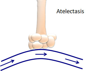
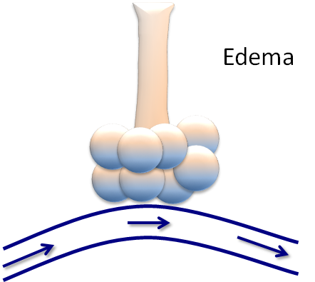
  • In the figure below, the top “lungs” are healthy:
    • The lungs on the left have a resistance problem or impairment in airflow
    • The lungs on the right have a compliance problem or impairment in stretch and recoil
    • In this picture:
      • Both figures could have elevated peak inspiratory pressures (PIP):
        • Due to the excess pressure generated in the system:
          • However:
            • Only the right-hand figure would have an elevate plateau pressure (Pplat):
              • Since this process occurs when there is an absence of airflow
  • Compliance (C) = ∆ volume / ∆ pressure
    • C = Tidal volume / Plateau pressure – Peak inspiratory pressure
      • C = (TV) / (Pplat – PEEP)
  • Therefore, when troubleshooting high-pressures on the ventilators:
    • Two values are needed:
      • The peak inspiratory pressure (PIP):
        • Is the maximum pressure in the system and includes both:
          • Resistance and compliance:
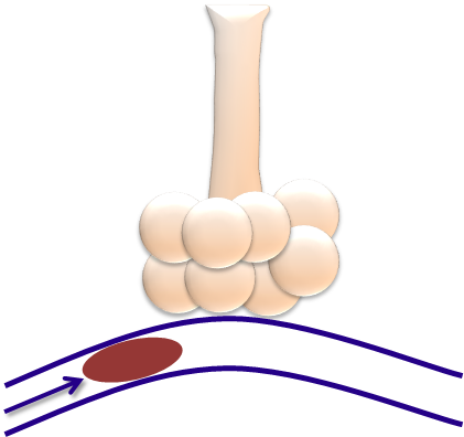
            • An inspiratory pause stops all airflow:
              • Thereby removing resistance, and only leaving compliance, as illustrated in this diagram below:
                • The plateau pressure, or Pplat, is, therefore:
                  • A measure of compliance
  • These values can be displayed on the ventilator screen:
    • On most ventilators:
      • The PIP is always seen
      • While the Pplat is seen by pushing the “inspiratory hold” or “inspiratory pause” button on the ventilator
    • An elevated PIP and normal Pplat is:
      • Indicative of increased airway resistance
    • An elevated PIP and elevated Pplat is:
      • Indicative of abnormal compliance
  • Determining whether the patient has a:
    • Resistance problem or a compliance problem:
      • Can assist in the differential diagnosis of respiratory failure:
        • High Resistance:
          • High PIP, Low / Normal Pplat:
            • Kinked / obstructed ETT
            • Mucus plugging
            • Brochospasm
            • Endotracheal tube to narrow:
              • Small
            • Coughing
            • Bronchospasm:
              • Obstructive lung disease
    • Low Compliance:
      • High PIP, High Pplat:
        • Atelectasis
        • Pulmonary edema
        • ARDS
        • Hemothorax /pneumothorax
        • Pneumonia
        • Pulmonary fibrosis:
          • Restrictive lung disease
        • Air-trapping with accumulated auto-PEEP
        • Obesity
        • Abdominal compartment syndrome
        • Circumferential burns of the chest
        • Scoliosis
        • Supine position
  • Air trapping:
    • Also referred to as breath-stacking:
      • Can lead to the development of auto-PEEP, or intrinsic PEEP (iPEEP):
        • These pressures should be differentiated from the set PEEP, or extrinsic PEEP (ePEEP):
          • ePEEP refers to the additional end-expiratory positive pressure set during mechanical ventilation:
            • To prevent alveolar collapse and derecruitment
      • In contrast:
        • Auto-PEEP, or iPEEP:
          • Is a pathophysiological process:
            • That can occur when the ventilator initiates the next breath prior to complete exhalation:
              • While this is most common in patients with prolonged expiratory phases, such as asthma or COPD:
                • It can also occur in patients:
                  • Who have a fast respiratory rate or
                  • Those who are being ventilated with large tidal volumes
              • The amount of auto-PEEP can be measured by:
                • Pressing the “expiratory hold” or “expiratory pause” button on the ventilator:
                  • When this button is pressed, the ventilator will display the total PEEP:
                    • The auto-PEEP is the difference between the total PEEP and the set PEEP:
                      • Auto-PEEP (iPEEP) = Total PEEP – ePEEP
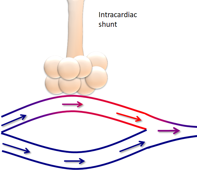
  • The Figure represents the effects of air trapping:
  • Air trapping, or autoPEEP:
    • Can lead to significant adverse cardiopulmonary effects
    • The increased intrathoracic pressure from autoPEEP can:
      • Decrease venous return and lead to hemodynamic instability, even cardiac arrest in severe cases
    • The increased pressures may also result in:
      • A pneumothorax or pneumomediastium
    • Additionally, air trapping can lead to:
      • Ineffective ventilation due to:
        • Collapse of the capillaries responsible for gas exchange:
          • With worsening hypercarbia and hypoxemia
      • While this may seem like a paradox:
        • As one may assume that increasing the minute ventilation, or moving more air:
          • Will improve ventilation, there is a limit to the beneficial effects:
            • Once the lungs are overdistended, gas exchange is ineffective
            • In these circumstances, allowing the patient sufficient time to exhale can decrease CO2 retention

#Arrangoiz #Surgeon #Teacher

Prognostic and Predictive Factors in Breast Cancer

  • Prognostic factors:
    • Provide information regarding:
      • Clinical outcome
    • The prognostic factors in breast cancer that have been validated in clinical testing and related to risk of relapse and survival are:
      • Lymph node status
      • Tumor size
      • Grade
      • Lymphovascular invasion
      • ER/PR status:
        • ER-negative and PR-negative tumors:
          • Are associated with a worse prognosis compared to ER-positive tumors
  • Predictive factors:
    • Provide information on likelihood of benefit from a given therapy
    • An example of a predictive factor:
      • ER/PR receptor expression:
        • Identifies those patients likeliest to benefit from hormonal therapies
      • Of note:
        • Tumor grade:
          • May be predictive of response to therapy, but not necessarily of survival benefit
  • REFERENCES
    • Cianfrocca M, Goldstein LJ. Prognostic and predictive factors in early stage breast cancer. Oncologist. 2004;9(6):606-616.
    • Kleer C, Sabel M. Prognostic and predictive factors in breast cancer. In: Kuerer HM, ed. Breast Surgical Oncology. New York, NY: McGraw Hill; 2010:243-249.

#Arrangoiz #CancerSurgeon #SurgicalOncologist #CancerSurgeon #BreastSurgeon #BreastCancer

NSABP B-35

  • The NSABP B-35:
    • Is a phase III clinical trial:
      • That randomized postmenopausal women with ER-positive DCIS to:
        • 5 years of anastrozole or tamoxifen:
          • Following breast-conserving surgery and radiation
      • The trial sought to determine:
        • How effective anastrozole is compared to tamoxifen in preventing a breast cancer occurrence:
          • As well as the quality of life of patients taking anastrozole
    • The trial is currently closed after meeting its accrual goal of:
      • 3100 patients
    • Median follow-up of 9 years
    • Investigators found:
      • Significantly fewer breast cancer events in the anastrozole group (n = 90) than in the tamoxifen group (n = 122):
        • Hazard ratio, 0.73; confidence interval, 0.56–0.96, P=0.0234
    • The estimated 10-year breast-cancer-free interval rates were:
      • 93.5% for anastrozole versus
      • 89.2% for tamoxifen
        • This recorded difference in breast cancer-free interval:
          • Was attributable almost entirely to younger postmenopausal women:
            • Less than 60 years of age
    • Interestingly:
      • The difference between treatments did not become apparent until after 5 years of follow-up:
        • Likely due to the low number of events in both groups
    • There was no difference in overall survival (OS):
      • Between the two treatment groups

#Arrangoiz #BreastSurgeon #CancerSurgeon #SurgicalOncologist #Teacher #Surgeon

The Parapharyngeal Space

  • The parapharyngeal space:
    • Also known as the:
      • Prestyloid parapharyngeal space:
        • Is one of the seven deep compartments of the head and neck
  • It consists largely of:
    • Fatty areolar tissue
    • Contains branches of the:
      • Trigeminal nerve
    • Deep blood vessels
  • Two naming conventions exist in the literature:
    • In the first definition:
      • Familiar to most head and neck surgeons, the parapharyngeal space is divided into:
        • The prestyloid compartment and
        • Poststyloid (retrostyloid) compartment
    • In the second definition:
      • Introduced by some radiologists:
        • The prestyloid parapharygeal space is simply termed:
          • The parapharyngeal space, and
        • The poststyloid pharapharygeal space is termed:
          • The carotid space
  • Gross anatomy:
    • The parapharyngeal space:
      • Is shaped like a pyramid:
        • An inverted pyramid with:
          • Its base:
            • At the skull base
          • Its apex:
            • Inferiorly:
              • Pointing to the greater cornu of the hyoid bone
    • Contents:
      • Fat:
        • Main component
      • Deeps blood vessels:
        • Internal maxillary artery
        • Ascending pharyngeal artery
        • Pterygoid venous plexus:
          • Only small portion:
            • Because it is mainly within:
              • The masticator space
      • Nerve:
        • Small branch of the mandibular division of the trigeminal nerve (cranial nerve V):
          • Supplying the tensor veli palatini muscle
      • Salivary glands – depends on the definition:
        • Some say that it contains no salivary glands, others
        • Minor or ectopic salivary gland / rests
        • Retromandibular portion of the deep lobe of parotid gland
      • Lymph nodes
  • Boundaries:
    • The parapharyngeal space has complex fascial margins:
      • Occupying the space between the muscles of mastication and the muscles of deglutition:
        • Superior margin:
          • Base of skull
        • Inferior margin:
          • Greater cornu of the hyoid bone:
            • Although some state the space functionally ends higher:
              • With the styloglossus muscle:
                • At the level of the angle of the mandible
        • Medial margin:
          • Middle (pretracheal) layer of the deep cervical fascia:
            • Covering the:
              • Superior pharyngeal constrictor
              • Levator palatini muscle and
              • Tensor veli palatini muscle
        • Lateral margin:
          • Investing fascia (superficial layer) of the deep cervical fascia:
            • Covering the deep lobe of the parotid
        • Anterior margin:
          • Investing fascia (superficial layer) of the deep cervical fascia:
            • Covering the medial pterygoid muscle
        • Posterior margin:
          • Prevertebral layer of the deep cervical fascia
  • Relations:
    • Medial:
      • To the masticator space
    • Lateral:
      • To the pharyngeal mucosal space
    • Anterior:
      • To the prevertebral space
    • Posterior:
      • To the medial pterygoid

#Arrangoiz #HeadandNeckSurgeon #CancerSurgeon #SurgicalOncologist #Teacher

Cervical Fascia

  • Superficial cervical fascia:
    • Primarily includes the:
      • Platysma and
      • Subcutaneous fat and vessels:
        • However:
          • As with other fascia in the body:
            • The use of the terminology of the superficial cervical fascia has declined in favor of:
              • Subcutaneous tissue:
                • Thus, an unspecified reference to cervical fascia:
                  • Mainly refers to the deep cervical fascia
  • The deep cervical fascia:
    • Consists of three separate but related fascial layers:
      • That encircle structures in the neck and
      • Allow anatomic compartmentalisation into:
        • The deep spaces of the head and neck
      • Each layer contributes:
        • To the carotid sheath
    • Layers of the deep cervical fascia:
      • Superficial layer of the deep cervical fascia
      • Middle layer of the deep cervical fascia
      • Deep layer of the deep cervical fascia
  • The superficial layer of the deep cervical fascia:
    • Also known as:
      • The investing layer / fascia
    • Is the one of three layers of the deep cervical fascia:
      • That surrounds all of the neck:
      • That is deep to the platysma
      • The layer includes:
        • The masticator fascia
        • Submandibular fascia, and
        • Sternocleidomastoid-trapezius fascia
        • The fascia that superficially covers the parotid gland:
          • May also be derived from this layer:
            • But this inclusion is controversial
    • Gross anatomy – attachments:
      • Posteriorly:
        • Ligamentum nuchae
        • Cervical vertebral spinous processes
        • External occipital protuberance
      • Superiorly
        • Mandible:
          • The layer thickens to form the extrinsic ligaments of the mandible:
            • Sphenomandibular ligament and
            • Stylomandibular ligament
        • Mastoid process and styloid process
        • Central skull base
        • Zygomatic arch
        • Superior temporal line or temporal ridge
    • Inferiorly
      • Manubrium
      • Clavicle
      • Acromion and spine of the scapula
    • Contents:
      • The superficial layer of the deep cervical fascia:
        • Encircles everything in the neck:
          • Apart from the skin and superficial cervical fascia (subcutaneous tissue)
      • In addition, it splits to enclose the following structures:
        • Two salivary glands
          • Submandibular gland
          • Parotid glands
        • Two spaces:
          • Masticator space
          • Suprasternal space
        • Two muscles (other than those in the masticator space):
          • Sternocleidomastoid muscle
          • Trapezius muscle
    • In addition:
      • All layers of the deep cervical fascia:
        • Contribute to the carotid sheath
  • The middle layer of the deep cervical fascia:
    • Is the one of the three layers of the deep cervical fascia:
      • It most closely surrounds:
        • The visceral organs
    • This layer consists:
      • Anteriorly of the:
        • Strap muscle fascia:
          • Comprised of the:
            • Sterno-omohyoid layer and
            • Sternothyroid layer
            • Thyrohyoid layer
      • Posteriorly:
        • Visceral fascia:
          • Also commonly known as the:
            • Pharyngobasilar and
            • Buccopharyngeal fascia
              • Particularly in the suprahyoid neck, or, less commonly:
                • Pharyngomucosal fascia
          • The alternative term:
            • Pretracheal fascia:
              • May refer to either:
                • The visceral fascia or
                • The sternothyroid-thyrohyoid layer of strap muscle fascia:
                  • Which lies anterior to the trachea
    • Gross Anatomy – attachments:
      • Superiorly:
        • Skull base
      • Anteriorly:
        • Hyoid bone
        • Thyroid cartilage
        • Manubrium
      • Inferiorly:
        • Fibrous pericardium
        • Adventitia of the aortic arch
    • Contents:
      • Infrahyoid (strap) muscles:
        • Sternohyoid
        • Omohyoid
        • Sternothyroid
        • Thyrohyoid
      • Visceral space and pharyngeal mucosal space:
        • Buccinator muscle
        • Pharynx and pharyngeal constrictor muscles:
          • Superior pharyngeal constrictor muscle
          • Middle pharyngeal constrictor muscle, and
          • Inferior pharyngeal constrictor muscle
        • Cervical esophagus
        • Thyroid gland* and parathyroid glands
        • Trachea*
        • Larynx*
        • Visceral lymph nodes
        • Recurrent laryngeal nerve
          • *Some sources consider these structures to have their own fascia not derived from the middle layer of the deep cervical fascia:
            • But they are nevertheless considered part of the visceral space
    • In addition:
      • All layers of the deep cervical fascia:
        • Contribute to the carotid sheath:
          • However:
            • In the suprahyoid neck:
              • Above the carotid bifurcation:
                • The contribution of the middle layer is inconsistent
  • The deep layer of the deep cervical fascia:
    • Is one of the three layers of the deep cervical fascia:
      • It encases the:
        • Paravertebral muscles and
        • Forms the perivertebral space
      • It consists of the:
        • Perivertebral fascia:
          • The anterior part of which is called:
            • The prevertebral fascia and
            • The alar fascia
    • Gross Anatomy – attachments:
    • Medially:
      • Ligamentum nuchae
      • Cervical vertebral spinous processes and transverse processes
    • Laterally:
      • Carotid sheath
      • First rib:
        • From a portion of the layer called:
          • Sibson fascia
      • Axillary sheath
    • Superiorly
      • The skull base
    • Inferiorly
      • Coccyx:
        • For the prevertebral fascia)
      • Endothoracic fascia:
        • For the alar fascia
    • On each side:
      • A flap attaches to the transverse processes of the cervical vertebrae and:
        • Divides the peri-vertebral spaces into:
          • A pre-vertebral compartment:
            • Anteriorly and
          • A para-spinal compartment:
            • Posteriorly
    • Anteromedial to the scalene muscles:
      • The deep layer splits into two leaves:
        • The ventral leaf being:
          • The alar fascia, and
        • The dorsal leaf being:
          • The prevertebral fascia:
            • With the prevertebral space:
              • Space between the prevertebral fascia and the spine
          • The space between the alar fascia and the prevertebral fascia:
            • Is the danger space
          • The space between the alar fascia and the posterior aspect of the middle layer of the deep cervical fascia:
            • Is the retropharyngeal space
    • Contents:
      • Danger space:
        • Space between prevertebral and alar fascia
      • Prevertebral space:
        • Space between the prevertebral fascia and the spine:
          • Anterior component:
            • Of the perivertebral space
      • Longus colli and capitis muscles
      • Rectus capitis anterior and lateralis muscles
      • Scalenus anterior, medius, and posterior muscles
      • Sheath for subclavian artery and vein, brachial plexus
      • Vertebral column
      • Spinal cord and associated thecal sac, nerve roots, and vessels
      • Vertebrae and associated discs and ligaments
      • Paraspinal / paravertebral space:
        • Posterior component:
          • Of the perivertebral space
      • Levator scapulae
      • Deep cervical back muscles
      • Pierced by the four cutaneous branches of the cervical plexus:
        • Greater auricular nerve
        • Lesser occipital nerve
        • Transverse cervical nerve
        • Supraclavicular nerve
    • In addition:
      • All layers of the deep cervical fascia:
        • Contribute to the carotid sheath

#Arrangoiz #HeadandNeckSurgeon #CancerSurgeon #SurgicalOncologist #Teacher

Masticator Space

  • The masticator space:
    • Is one of the seven deep compartments of the head and neck
  • Gross anatomy:
    • The masticator space are:
      • Paired suprahyoid cervical spaces:
        • On each side of the face
    • Each space is enveloped by:
      • The superficial layer of the deep cervical fascia:
        • The superficial layer of deep cervical fascia:
          • Splits into two at the lower border of the mandible:
            • The inner layer:
              • Running deep to the medial pterygoid muscle and attaches to the skull base medial to foramen ovale and
            • The outer layer:
              • Covering the masseter and temporalis muscles and attaches to the parietal calvaria superiorly
  • Contents:
    • Muscles of mastication
    • Ramus and body of mandible
    • Inferior alveolar nerve
    • Inferior alveolar vein and artery
    • Mandibular division of the trigeminal nerve (V3):
      • Enters the masticator space:
        • Via the foramen ovale
    • Pterygoid venous plexus
  • Boundaries and relations:
    • Anteriorly:
      • The buccal space
    • Posterolaterally:
      • Parotid space
    • Medially:
      • Parapharyngeal space
  • Communications:
    • Masticator space malignancies can spread perineurally:
      • Via the mandibular division of the trigeminal (V3) nerve:
        • Into the middle cranial fossa

#Arrangoiz #HeadandNeckSurgeon #CancerSurgeon #surgicalOncologist #Teacher

Atelectasis and Derecruitment

  • Maximizing V/Q matching:
    • By preventing atelectasis:
      • Is a key principle in the management of:
        • Respiratory failure
      • Alveolar derecruitment, or atelectasis:
        • Leads to the creation of shunts
      • Atelectasis has multiple detrimental effects in ventilated patients:
        • First:
          • Atelectasis decreases the surface area for gas exchange
        • Atelectasis on a large scale:
          • Is derecruitment
        • Derecruitment is compounded by:
          • Excessive lung weight:
            • Such as with pulmonary edema
          • Chest wall weight:
            • As with morbid obesity
          • Abdominal contents and distention:
            • As with small bowel obstructions)
          • Cardiac compresses:
            • As with pericardial effusion
          • The addition of sedation and paralysis:
            • To positive pressure ventilation:
              • Can further augment this derecruitment
    • This diagram reflects the pressures leading to compression of the lungs when lying a patient supine:
      • The weight of the heart, the weight of the chest wall, the weight of the abdominal contents, and the weight of the lungs themselves
Atelectasis

#Arrangoiz #Teacher #Surgeon

Issues with Oxygenation

  • Hypoxemia:
    • There are four broad physiologic causes of hypoxemia:
      • Shunting
      • VQ mismatch
      • Alveolar hypoventilation
      • Decreased partial pressure of oxygen
        • Understanding these mechanisms:
          • Allows the clinician at the bedside to quickly develop a differential diagnosis for hypoxemia and target diagnostics to assess for the precise etiology
  • Shunts, or blood bypassing normal gas exchange:
    • Is one of the most common causes of hypoxemia
    • A classic example of a shunt is an intracardiac shunt:
      • In this example:
        • Much of the blood passes by the alveoli, participating in normal gas exchange:
          • However, a small amount is diverted through the heart, bypassing the lungs:
            • This deoxygenated blood mixes with the oxygenated blood:
              • Leading to hypoxemia
  • When an area of the lung is perfused, but not ventilated:
    • That results in an intra-pulmonary shunt:
      • In other words:
        • The inspired oxygen cannot reach the alveoli for gas exchange
      • There are several different causes of intra-pulmonary shunts, including:
        • Atelectasis
        • Pneumonia
        • Pulmonary edema
        • Acute respiratory distress syndrome (ARDS)
        • Hemothorax
        • Pneumothorax
        • Hyperinflation or auto-PEEPing
          • All of these pathological processes:
            • Prevent effective gas exchange at the alveoli
Atelectasis
Edema
  • When an area has ventilation, but no perfusion:
    • This is dead space:
      • In other words:
        • The airways are functioning normally:
          • But there is a disease process in the vasculature
        • The best example would be a patient in cardiac arrest who is intubated and ventilated:
          • But there is an interruption of chest compressions
      • Dead space can be:
        • Anatomic and physiologic:
          • Such as oxygenation but lack of gas exchange:
            • That occurs in the upper airways, like the trachea
        • There can also be pathological causes of dead space:
          • Such as this diagram of microthrombi blocking a capillary
microthrombi
  • Other examples of dead space include:
    • Low cardiac output and hyperinflation:
      • As occurs in obstructive lung disease:
        • In diseases such as chronic obstructive lung disease (COPD):
          • There can be a significant level of hyperinflation or auto-PEEP:
            • Which can lead to vasoconstriction of the capillaries involved in gas exchanged:
              • Thereby leading to impaired gas exchanged
  • Dead space ventilation can lead to both:
    • Hypoxia and hypercapnia:
      • Due to CO2 retention

#Arrangoiz #Surgeon #Teacher

Carotid Space

  • The carotid space:
    • Is one of the seven deep compartments of the head and neck
  • Gross anatomy:
    • The carotid space is a roughly a cylindrical space:
      • That extends from the skull base through to the aortic arch
    • It is circumscribed by all three layers of:
      • The deep cervical fascia:
        • Forming the carotid sheath:
          • Above the carotid bifurcation:
            • The contribution of the middle layer of cervical fascia:
              • Can be inconsistent and the sheath is interrupted
    • The bifurcation of the common carotid:
      • Usually occurs at the boundary of:
        • The suprahyoid and infrahyoid spaces
  • Contents:
    • Common carotid artery:
      • Inferiorly
    • Internal carotid artery:
      • Superiorly
    • Internal jugular vein
    • Ansa cervicalis:
      • Embedded in the anterior wall of the sheath
    • Nerves:
      • Vagus nerve (CN X):
        • Posterior to the vessels:
          • In the posterior notch:
            • Extends below the hyoid to the mediastinum:
              • Within the carotid sheath
      • In the upper part of the carotid sheath, there is also:
        • Glossopharyngeal nerve (CN IX):
          • Anterior to vessels
        • Accessory nerve (CN XI)
        • Hypoglossal nerve (CN XII)
      • Sympathetic nerves:
        • Medial to the vessels and
        • Lateral to retropharyngeal space
    • Deep cervical lymph node chain:
      • Level II, III, IV
  • Relations:
    • Suprahyoid carotid space:
      • Superiorly:
        • Masticator space and
        • Para pharyngeal space
      • Laterally:
        • Parotid space
      • Posteriorly:
        • Peri vertebral space
    • The suprahyoid portion of the carotid space:
      • Is often synonymous with:
        • The post-styloid compartment of the parapharyngeal space
    • Infrahyoid carotid space:
      • Anteriorly:
        • Anterior cervical space
      • Laterally:
        • Posterior cervical space
      • Posteriorly:
        • Perivertebral space
      • Medially:
        • Retropharyngeal and visceral space
  • Boundaries:
    • Superior margin:
      • Lower border of jugular foramen
    • Inferior margin:
      • Aortic arch
    • Anterolateral margin:
      • Sternocleidomastoid muscle

#Arrangoiz #HeadandNeckSurgeon #CancerSurgeon #Teacher #SurgicaOncologist