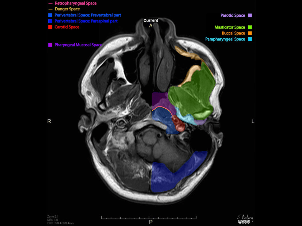
- The parapharyngeal space:
- Also known as the:
- Prestyloid parapharyngeal space:
- Is one of the seven deep compartments of the head and neck
- Prestyloid parapharyngeal space:
- Also known as the:
- It consists largely of:
- Fatty areolar tissue
- Contains branches of the:
- Trigeminal nerve
- Deep blood vessels
- Two naming conventions exist in the literature:
- In the first definition:
- Familiar to most head and neck surgeons, the parapharyngeal space is divided into:
- The prestyloid compartment and
- Poststyloid (retrostyloid) compartment
- Familiar to most head and neck surgeons, the parapharyngeal space is divided into:
- In the second definition:
- Introduced by some radiologists:
- The prestyloid parapharygeal space is simply termed:
- The parapharyngeal space, and
- The poststyloid pharapharygeal space is termed:
- The carotid space
- The prestyloid parapharygeal space is simply termed:
- Introduced by some radiologists:
- In the first definition:
- Gross anatomy:
- The parapharyngeal space:
- Is shaped like a pyramid:
- An inverted pyramid with:
- Its base:
- At the skull base
- Its apex:
- Inferiorly:
- Pointing to the greater cornu of the hyoid bone
- Inferiorly:
- Its base:
- An inverted pyramid with:
- Is shaped like a pyramid:
- Contents:
- Fat:
- Main component
- Deeps blood vessels:
- Internal maxillary artery
- Ascending pharyngeal artery
- Pterygoid venous plexus:
- Only small portion:
- Because it is mainly within:
- The masticator space
- Because it is mainly within:
- Only small portion:
- Nerve:
- Small branch of the mandibular division of the trigeminal nerve (cranial nerve V):
- Supplying the tensor veli palatini muscle
- Small branch of the mandibular division of the trigeminal nerve (cranial nerve V):
- Salivary glands – depends on the definition:
- Some say that it contains no salivary glands, others
- Minor or ectopic salivary gland / rests
- Retromandibular portion of the deep lobe of parotid gland
- Lymph nodes
- Fat:
- The parapharyngeal space:
- Boundaries:
- The parapharyngeal space has complex fascial margins:
- Occupying the space between the muscles of mastication and the muscles of deglutition:
- Superior margin:
- Base of skull
- Inferior margin:
- Greater cornu of the hyoid bone:
- Although some state the space functionally ends higher:
- With the styloglossus muscle:
- At the level of the angle of the mandible
- With the styloglossus muscle:
- Although some state the space functionally ends higher:
- Greater cornu of the hyoid bone:
- Medial margin:
- Middle (pretracheal) layer of the deep cervical fascia:
- Covering the:
- Superior pharyngeal constrictor
- Levator palatini muscle and
- Tensor veli palatini muscle
- Covering the:
- Middle (pretracheal) layer of the deep cervical fascia:
- Lateral margin:
- Investing fascia (superficial layer) of the deep cervical fascia:
- Covering the deep lobe of the parotid
- Investing fascia (superficial layer) of the deep cervical fascia:
- Anterior margin:
- Investing fascia (superficial layer) of the deep cervical fascia:
- Covering the medial pterygoid muscle
- Investing fascia (superficial layer) of the deep cervical fascia:
- Posterior margin:
- Prevertebral layer of the deep cervical fascia
- Superior margin:
- Occupying the space between the muscles of mastication and the muscles of deglutition:
- The parapharyngeal space has complex fascial margins:
- Relations:
- Medial:
- To the masticator space
- Lateral:
- To the pharyngeal mucosal space
- Anterior:
- To the prevertebral space
- Posterior:
- To the medial pterygoid
- Medial:
#Arrangoiz #HeadandNeckSurgeon #CancerSurgeon #SurgicalOncologist #Teacher
