My name is Rodrigo Arrangoiz I am a breast surgeon/ thyroid surgeon / parathyroid surgeon / head and neck surgeon / surgical oncologist that works at Center for Advanced Surgical Oncology in Miami, Florida.
I was trained as a surgeon at Michigan State University from (2005 to 2010) where I was a chief resident in 2010. My surgical oncology and head and neck training was performed at the Fox Chase Cancer Center in Philadelphia from 2010 to 2012. At the same time I underwent a masters in science (Clinical research for health professionals) at the University of Drexel. Through the International Federation of Head and Neck Societies / Memorial Sloan Kettering Cancer Center I performed a two year head and neck surgery and oncology / endocrine fellowship that ended in 2016.
Mi nombre es Rodrigo Arrangoiz, soy cirujano oncólogo / cirujano de tumores de cabeza y cuello / cirujano endocrino que trabaja Center for Advanced Surgical Oncology en Miami, Florida.
Fui entrenado como cirujano en Michigan State University (2005 a 2010 ) donde fui jefe de residentes en 2010. Mi formación en oncología quirúrgica y e n tumores de cabeza y cuello se realizó en el Fox Chase Cancer Center en Filadelfia de 2010 a 2012. Al mismo tiempo, me sometí a una maestría en ciencias (investigación clínica para profesionales de la salud) en la Universidad de Drexel. A través de la Federación Internacional de Sociedades de Cabeza y Cuello / Memorial Sloan Kettering Cancer Center realicé una sub especialidad en cirugía de cabeza y cuello / cirugia endocrina de dos años que terminó en 2016.
Local treatment for ductal carcinoma in situ (DCIS) involves:
Mastectomy or breast-conserving therapy, which consists of partial mastectomy followed in most cases by adjuvant radiation therapy
Sentinel lymph node biopsy (SLNB):
Is not indicated for most patients undergoing breast-conserving therapy for DCIS:
Because DCIS is a preinvasive lesion:
Axillary nodes are rarely involved:
Even in cases of extensive multifocal high-grade disease
If invasive breast cancer is identified after a breast-conserving surgery is performed for DCIS:
SLNB can be performed as a second procedure
If a mastectomy is performed as the initial procedure:
A sentinel node biopsy should be performed in the event of an occult invasive cancer
Radiation therapy alone is not given for DCIS:
Although there are ongoing clinical trials comparing active surveillance to surgery with or without endocrine therapy:
These are reserved for low- and intermediate-grade DCIS and are not yet considered standard of care
References
Virnig BA, Tuttle TM, Shamliyan T, Kane RL. Ductal carcinoma in situ of the breast: a systematic review of incidence, treatment, and outcomes. J Natl Cancer Inst 2010; 102:170.
Lyman GH, Giuliano AE, Somerfield MR, et al. American Society of Clinical Oncology guideline recommendations for sentinel lymph node biopsy in early-stage breast cancer. J Clin Oncol. 2005;23(30):7703-7720.
Intra M, Rotmensz N, Veronesi P, et al. Sentinel node biopsy is not a standard procedure in ductal carcinoma in situ of the breast: the experience of the European Institute of Oncology on 854 patients in 10 years. Ann Surg. 2008;247(2):315-319.
Brennan ME, Turner RM, Ciatto S, et al. Ductal carcinoma in situ at core-needle biopsy: meta-analysis of underestimation and predictors of invasive breast cancer. Radiology. 2011;260(1):119-128.
Youngwirth LM, Boughey JC, Hwang ES. Surgery versus monitoring and endocrine therapy for low-risk DCIS: The COMET Trial. Bull Am Coll Surg. 2017;102(1):62-63.
Women are candidates for breast-conserving therapy:
If the tumor is small enough in relation to the size of the breast to permit resection of the tumor with clear margins and an acceptable cosmetic result
Absolute contraindications to breast-conserving therapy include:
Prior radiation therapy to the breast or chest wall
Breast cancer early in pregnancy that would necessitate radiation therapy during pregnancy
Widespread disease that precludes negative margins with a satisfactory cosmetic result
Retrospective reviews have shown that patients with collagen vascular diseases, specifically scleroderma:
Are at increased risk of radiation toxicities, even with modern techniques:
Therefore, the presence of scleroderma would be a relative contraindication for breast-conserving surgery
There have been attempts to allow for breast-conserving surgery for selected patients with multifocal or multicentric disease:
Retrospective data suggest that although patients with multifocal or multicentric disease have increased risk of local recurrence and lower disease-free survival:
This finding is independent of the type of surgery performed
The Alliance Z11102 study:
Sought to prospectively determine whether patients with multiple ipsilateral breast cancers could safely undergo breast-conserving surgery
Initial report in 2018 showed that breast-conserving surgery:
Was feasible in 93% of patients
And could be accomplished in one operation in 67%
Data regarding local regional recurrence will be forthcoming
Percutaneous ablation techniques:
Such as cryoablation have promising potential for less-invasive management of breast cancer:
However, it is not recommended for multicentric disease
Large multicenter randomized clinical trials are needed to determine long-term efficacy
References
Morrow M, Strom EA, Bassett LW, et al. Standard for breast conservation therapy in the management of invasive breast carcinoma. CA Cancer J Clin. 2002;52(5):277-300.
Zaremba N, Tamkus D, DiCarlo L, Herman J, Martin M, Bumpers HL. The dilemma of breast cancer treatment and existing collagen vascular disease: a case of scleroderma and review of the literature. Breast J. 2016;22(4):451-455.
Alm El-Din MA, Taghian AG. Breast conservation therapy for patients with locally advanced breast cancer. Semin Radiat Oncol. 2009;19(4):229-235.
Rosenkranz KM, Ballman K, McCall L, Kubicky C, Cuttino L, Le-Petross H, et al. The feasibility of breast-conserving surgery for multiple ipsilateral breast cancer: an initial report from ACOSOG Z11102 (Alliance) Trial. Ann Surg Oncol. 2018;25(10):2858-2866.
Lanza E, Palussiere J, Buy X, et al. Percutaneous image-guided cryoablation of breast cancer: a systematic review. J Vasc Interv Radiol. 2015;26(11):1652-1657.
The margin width (distance between the edge of the DCIS and the inked margin):
Reflects the completeness of excision and is an important determinant of local recurrence in DCIS:
Particularly for patients considering omission of radiotherapy after breast-conserving surgery
In 2016, the Society of Surgical Oncology and American Society of Radiation Oncology:
Developed consensus guidelines regarding margins for DCIS
These guidelines were based on:
A meta-analysis of 22 studies enrolling 4,660 women treated with partial mastectomy and radiation therapy:
There was a 64% reduction in local recurrence risk:
In patients with negative margins compared to those with positive margins
Margin thresholds ≥2 mm were associated with fewer local recurrences
For patients with positive margins:
Either re-excision or mastectomy to achieve negative margins should be performed
For patients with close margins:
Multiple factors should be considered:
The volume / extent of DCIS
Its distribution throughout a specimen
The volume of the excision
The volume of DCIS deemed close to the margin (focal or extensive)
After review of pathology:
Re-excision and / or radiation boost should be performed
A post-excision mammogram:
May be considered to rule out residual suspicious calcifications in the partial mastectomy operative bed for targeting during re-excision, and breast-conservation therapy may be re-attempted
If the close margins are extensive:
Mastectomy may be indicated
References
Morrow M, Van Zee KJ, Solin LJ, et al. Society of Surgical Oncology-American Society for Radiation Oncology-American Society of Clinical Oncology consensus guideline on margins for breast-conserving surgery with whole-breast irradiation in ductal carcinoma in situ. Ann Surg Oncol. 2016;23(12):3801-3810.
Van Zee KJ, Subhedar P, Olcese C, Patil S, Morrow M. Relationship between margin width and recurrence of ductal carcinoma in situ: analysis of 2996 women treated with breast-conserving surgery for 30 years. Ann Surg. 2015;262(4):623-631.
Dunne C, Burke JP, Morrow M, Kell MR. Effect of margin status on local recurrence after breast conservation and radiation therapy for ductal carcinoma in situ. J Clin Oncol. 2009;27(10):1615-1620.
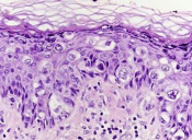
The clinical hallmarks of Paget’s disease of the breast are:
Scaling, erythema, and / or ulceration of the nipple sometimes extending to the areola
Because the main differential diagnosis for this clinical presentation is eczema:
A short course of topical steroids is an appropriate initial step:
Failure to resolve should prompt tissue biopsy by punch or wedge technique and not additional steroid therapy
Pathology revealing adenocarcinoma cells within the epidermis (Paget cells):
Confirms the diagnosis
HER2 amplification:
Is found in 60% to 90% of cases of Paget’s disease of the breast:
But the patient should be fully evaluated prior to making decisions regarding the need for targeted therapy
Appropriate diagnostic imaging includes:
Mammography, ultrasound, and / or breast MRI:
As Paget’s disease is associated with an underlying malignancy 85% of the time
The appropriate surgical management of Paget’s disease is:
Breast conservation with central mastectomy (resection of the nipple-areolar complex) with resection of the primary tumor and irradiation or mastectomy
References
Chen CY, Sun LM, Anderson BO. Paget disease of the breast: changing patterns of incidence, clinical presentation, and treatment in the U.S. Cancer. 2006;107(7):1448-1458.
Killelea BK, Chagpar AB, Horowitz NR, Lannin DR. Characteristics and treatment of human epidermal growth factor receptor 2 positive breast cancer: 43,485 cases from the National Cancer Database treated in 2010 and 2011. Am J Surg. 2017;213(2):426-432.
Caliskan M, Gatti G, Sosnovskikh I, et al. Paget’s disease of the breast: the experience of the European Institute of Oncology and review of the literature. Breast Cancer Res Treat. 2008;112(3):513-521.
Three important conclusions are agreed upon regarding this clinical entity:
Prognosis of occult primary breast cancer is the same or slightly better than women with classic stage IIA disease (T0, N1, M0)
An exhaustive workup for the non-breast primary is usually not fruitful
Treatment of the breast in some manner decreases the risk of local failure over time
Modified radical mastectomy has been the traditional surgical treatment for many years
Previously, the primary breast cancer was found in the mastectomy specimen:
40% to 80% of the time, but with the advent of much better mammography and ultrasound along with breast MRI, this rate is much lower now
However, what was true then and still holds today:
Is that no treatment to the breast itself results in an unacceptably high local recurrence rate
An alternative to a modified radical mastectomy:
Is complete ALND followed by whole-breast irradiation
Axillary dissection:
Provides local control while also fine tuning staging
Theoretically the whole-breast radiation:
Should control any subclinical disease in the breast not detected on imaging
Primary radiation to the breast, axilla, and supraclavicular area without any surgery of the breast or axilla:
Results in higher local and regional recurrence compared to surgery and radiation combined
Axillary node dissection and whole-breast irradiation:
Has been found to have equivalent survival as a modified radical mastectomy
A recent meta-analysis of 7 studies and more than 240 patients with occult primary breast cancers (0.3% to 0.8% of all breast cancers):
Found 39% were treated with ALND and radiation while 47% had modified radical mastectomy and 15% had ALND alone
With a mean follow-up of 5 years:
The study found no difference in local regional recurrence (12.7% vs 9.8%), distant metastasis (7.2% vs 12.7%), or mortality (9.5% vs 17.9%) between ALND and radiation vs modified radical mastectomy (all p>0.16)
ALND with radiation was superior to ALND alone in terms of local regional recurrence (12.7% vs 34.3%, p < 0.01) and trended towards improved survival but this was not statistically significant (P=0.09)
References:
Barton SR, Smith IE, Kirby AM, Ashley S, Walsh G, Parton M. The role of ipsilateral breast radiotherapy in management of occult primary breast cancer presenting as axillary lymphadenopathy. Eur J Cancer. 2011;47:2099-2106. PMID: 21658935. http://www.ncbi.nlm.nih.gov/pubmed/21658935
Macedo FI, Eid JJ, Flynn J, Jacobs MJ, Mittal VK. Optimal surgical management for occult breast carcinoma: a meta-analysis. Ann Surg Oncol. 2016;23:1838-1844. https://www.ncbi.nlm.nih.gov/pubmed/26832884
Rueth NM, Black DM, Limmer AR, et al. Breast conservation in the setting of contemporary multimodality treatment provides excellent outcomes for patients with occult primary breast cancer. Ann Surg Oncol. 2015;22:90-95. [epub ahead of print]. http://www.ncbi.nlm.nih.gov/pubmed/25249256
Walker GV, Smith GL, Perkins GH, et al. Population-based analysis of occult primary breast cancer with axillary lymph node metastasis. Cancer. 2010;116:4000-4006. PMID: 20564117. http://www.ncbi.nlm.nih.gov/pubmed/20564117
Woo SM, Son BH, Lee JW, et al. Survival outcomes of different treatment methods for the ipsilateral breast of occult breast cancer patients with axillary lymph node metastasis: a single center experience. J Breast Cancer. 2013;16:410-416. https://www.ncbi.nlm.nih.gov/pmc/articles/PMC3893343/
The Society of Surgical Oncology – American Society for Radiation Oncology (SSO-ASTRO) 2014 Consensus Guidelines:
Regarding margins of resection for invasive carcinoma of the breast:
Recommend the use of “no ink on tumor” as the standard
Patients with invasive cancer, even with associated ductal carcinoma in situ (DCIS):
Are treated according to these guidelines
In a meta-analysis of 33 studies including 32,363 patients:
Odds of local recurrence were associated with margin status of positive vs. negative:
But not decreased with increasing margin distance for patients with invasive carcinoma
The study reported that rates of in-breast tumor recurrence are twice as high with positive marginsregardless of tumor biology, radiation boost, or endocrine therapy
There was no evidence that wide margins reduce recurrence, even in patients with extensive intraductal component
However, the American Society of Clinical Oncology (ASCO) guidelines recommend consideration of post-excision mammography to document adequate resection in patients with microcalcifications
References
Moran MS, Schnitt SJ, Giuliano AE, et al. SSO-ASTRO consensus guideline on margins for breast-conserving surgery with whole breast irradiation in stage I and II invasive breast cancer. Int J Radiat Oncol Biol Phys. 2014;88(3):553-564.
Houssami N, Macaskill P, Marinovich ML, Morrow M. The association of surgical margins and local recurrence in women with early-stage invasive breast cancer treated with breast-conserving therapy: a meta-analysis. Ann Surg Oncol. 2014;21(3):717-730.
Buchholz TA, Somerfield MR, Griggs JJ, et al. Margins for breast-conserving surgery with whole-breast irradiation in stage I and II invasive breast cancer: American Society of Clinical Oncology endorsement of the Society of Surgical Oncology/American Society for Radiation Oncology consensus guideline. J Clin Oncol. 2014;32(14):1502-1506.
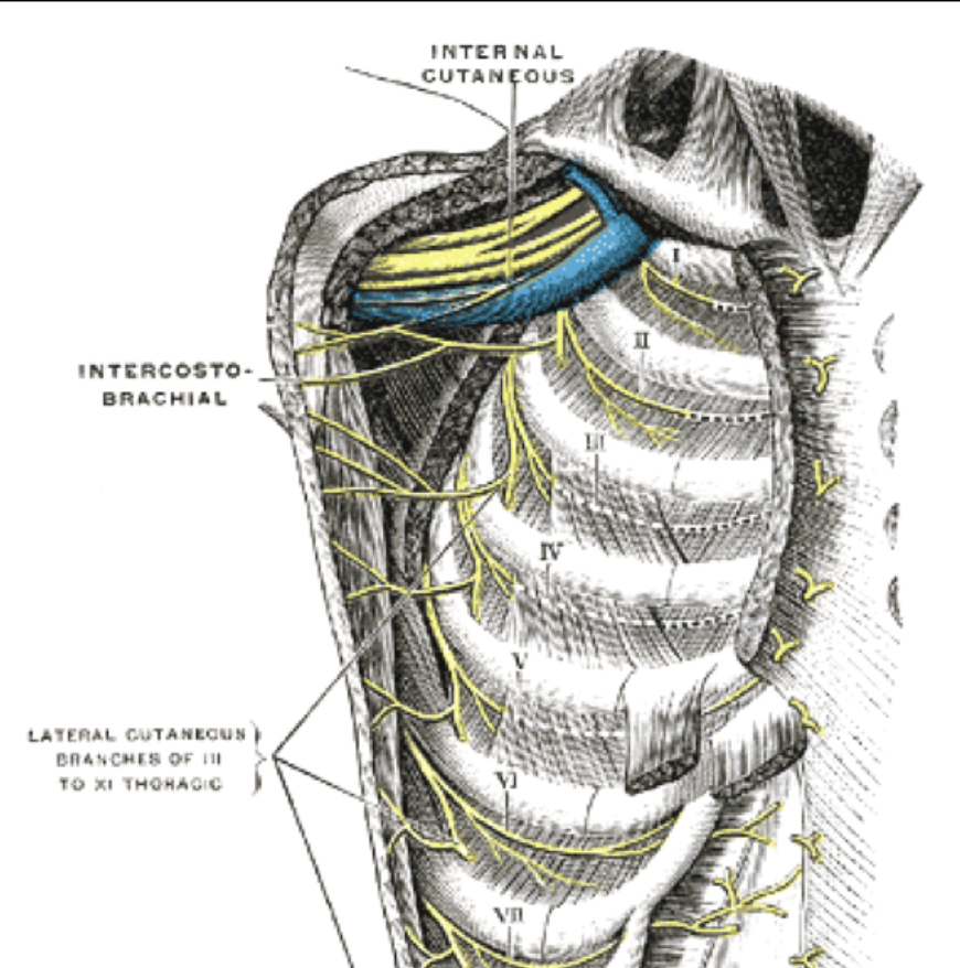
The symptoms of neuropathy are due to injury of the intercostobrachial nerve:
This nerve is a cutaneous branch of the intercostal nerves:
Most commonly the second intercostal nerve:
Which gives off a lateral cutaneous nerve:
Which continues as the intercostobrachial nerve
The intercostal nerves arise from the anterior rami of the thoracic spinal nerves
The intercostobrachial nerve pierces the serratus anterior, and crosses the axilla to the medial side of the upper arm:
The intercostobrachial nerve is commonly in the surgical field during axillary lymph node dissections and may be severed during surgery, or subject to traction or postsurgical inflammation:
Thus leading to intercostobrachial neuralgia
The larger intercostal nerves can be preserved with meticulous dissection
Neuropathic symptoms:
May be limited to numbness or tingling, but may also include a burning sensation
Techniques such as a regional nerve block have been described to alleviate symptoms in severe cases
In a study of 200 patients who underwent axillary dissection:
76% had symptoms of intercostobrachial neuralgia postoperatively:
Of these patients, 82% reported improvement or resolution of these symptoms within 1 year:
Reflecting the richness of the sensory nerve supply to the axilla and upper arm
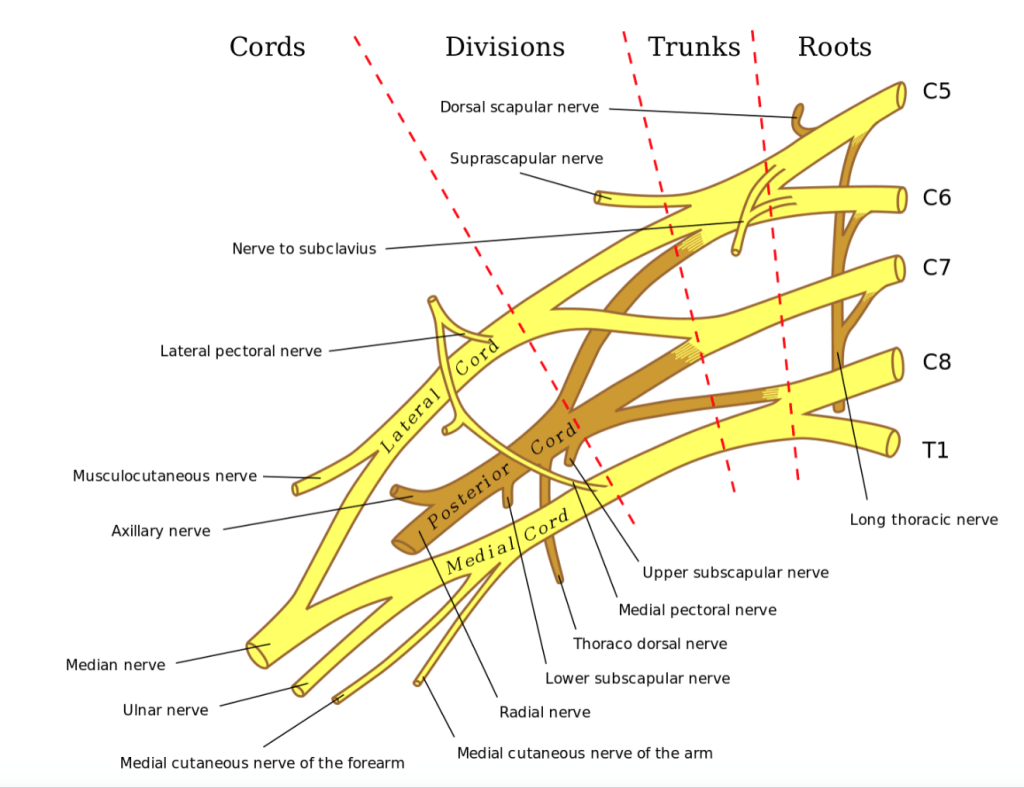
The thoracodorsal nerve:
Is a branch of the posterior cord of the brachial plexus:
Supplies motor function to the latissimus dorsi
If injured:
Patients experience weakness with arm abduction, lateral flexion, and difficulty with activities such as climbing, swimming, and using the arms to pull the body up
The medial cord of the brachial plexus gives rise to the medial pectoral nerve:
Which innervates both the pectoralis minor muscle and the pectoralis major muscle
The medial pectoral nerve typically pierces the pectoralis minor muscle, but may wrap around the lateral aspect of the pectoralis minor before traveling on to innervate the distal pectoralis major muscle
The lateral cord of the brachial plexus:
Gives rise to the lateral pectoral nerve, which innervates the pectoralis major muscle
This nerve travels along the medial border of the pectoralis minor muscle, and then along the undersurface of the pectoralis major muscle along with the pectoral branch of the thoracoacromial artery to supply the proximal pectoralis major muscle
The medial pectoral nerve bundle is often encountered during axillary dissection, as it is located lateral to the lateral pectoral nerve
If either of these nerves is injured, pectoralis muscle atrophy can occur, which can present as a late complication of surgery:
With weakness of shoulder adduction, interior rotation, and flexion
The long thoracic nerve:
Typically arises from anterior rami of the cervical spinal nerve roots C5 to C7:
It courses along the chest wall and supplies the serratus anterior muscle
Injury to this nerve causes a winged scapula
References
Sclafani LM, Baron RH. Sentinel lymph node biopsy and axillary dissection: added morbidity of the arm, shoulder and chest wall after mastectomy and reconstruction. Cancer J. 2008;14(4):216-222.
Wisotzky EM, Saini V, Kao C. Ultrasound-guided intercostobrachial nerve block for intercostobrachial neuralgia in breast cancer patients: a case series. Prev Med Rep, 2016;8(3):273-277.
Roses DF, Brooks AD, Harris MN, Shapiro RL, Mitnick J. Complications of level I and II axillary dissection in the treatment of carcinoma of the breast. Ann Sur. 1999;230(2):194-201.
Porzionato A, Macchi V, Stecco C, Loukas M, Tubbs RS, De Caro R. Surgical anatomy of the pectoral nerves and the pectoral musculature. Clin Anat. 2012;25(5):559-575.
The administration of neoadjuvant chemotherapy (NAC) offers several advantages in locally advanced breast cancer:
It allows for down staging the disease:
Which can potentially allow for less extensive surgery in the breast and axilla
It also provides information regarding the responsiveness of the cancer to systemic therapy while the tumor remains in vivo:
Which can guide the course of therapy
Administering chemotherapy in the neoadjuvant vs. adjuvant setting:
Does not change overall survival:
As demonstrated in the National Surgical Adjuvant Breast and Bowel Project (NSABP) B-18 and NSABP B-27 trials
The patient’s response to chemotherapy:
Does offer prognostic information:
Particularly in patients with hormone receptor negative (HR-) disease
Patients who achieve pathologic complete response (pCR):
Which is typically defined as no residual invasive disease in the breast or axilla:
Appear to have improved event-free survival (EFS) and overall survival (OS) compared with patients with residual disease:
This finding was demonstrated by a recent meta-analysis that included 36 studies including 5,768 patients with HER2 positive breast cancer
This correlation was strongest in patients with HR- disease
Further, among patients with HER2 positive disease that do not have a pCR:
The degree of residual cancer burden appears to correlate with outcomes
Patients with HER2 positive tumors:
May complete up to one year of HER2-targeted therapy:
With trastuzamab ± pertuzamab.
When planning surgery:
The pre-treatment volume does not need to be excised if the tumor has responded to chemotherapy:
However if multifocal disease is present:
The satellite lesion(s) should be localized and excised with the index lesion
When considering the appropriateness for breast conservation following NAC:
The distance between the lesions, location, and breast size must be considered
Placement of clips in the index lesion and any satellite lesions prior to initiation of NAC is critical for appropriate surgical planning post-NAC
References
Rastogi P, Anderson SJ, Bear HD. Preoperative chemotherapy: updates of National Surgical Adjuvant Breast and Bowel Project Protocols B-18 and B-27. J Clin Oncol. 2008; 10;26(5):778-785.
Broglio KR, Quintana M, Foster M, et al. Association of pathologic complete response to neoadjuvant therapy in HER2-positive breast cancer with long-term outcomes: a meta-analysis. JAMA Oncol. 2016;2(6):751-760.
Symmans WF, Wei C, Gould R, et al. Long-term prognostic risk after neoadjuvant chemotherapy associated with residual cancer burden and breast cancer subtype. J Clin Oncol. 2017;35(10):1049-1060.
Boughey JC, Peintinger F, Meric-Bernstam F, et al. Impact of preoperative versus postoperative chemotherapy on the extent and number of surgical procedures in patients treated in randomized clinical trials for breast cancer. Ann Surg. 2006;244(3):464-470.
Multiple ipsilateral breast cancers (MIBC) include:
Both multifocal and multicentric disease
The term multifocal (MF):
Typically refers to two or more foci of disease within a single quadrant of the breast,
Multicentric (MC):
Refers to two or more foci in more than one quadrant of the breast
When staging:
An “m” modifier, is added to TNM classification to identify MF / MC disease
In the setting of MIBC:
The size of the largest focus is used for staging:
Rather than a cumulative measurement of the tumor sizes
Historically, it was believed that multiple ipsilateral breast cancers should be treated surgically with mastectomy:
This thought is based on early retrospective studies (1980s-90s):L
That reported a higher locoregional recurrence (LRR) in patients with MF / MC disease who underwent breast conserving therapy (BCT)
More current studies, which include patients treated in a contemporary multidisciplinary setting:
Have demonstrated a more promising role for BCT in MF / MC disease
A systematic review included six retrospective studies evaluating patients with multiple ipsilateral breast cancers undergoing BCT vs. mastectomy:
The rate of LRR overall was 2% to 23% for BCT:
With similar rates of LRR for BCS compared to mastectomy
The largest of these compared 887 patients who underwent mastectomy vs. 300 who underwent BCT:
And found that BCT was not inferior to mastectomy:
With respect to:
5-year (2.5% vs. 4.5%) LRR
10-year (6.5% vs 5.7%) LRR
In a study reviewing surgical management of 6,134 patients undergoing neoadjuvant chemotherapy:
1,401 (23%) were found to have MF / MC disease
617 patients (44%) underwent BCT
Local recurrence-free survival, disease-free survival (DFS), and overall survival (OS):
We’re not inferior in patients with MF / MC compared with unifocal disease:
If negative margins or a pathologic complete response was obtained
In a study evaluating 110 patients with MF / MC disease compared to 263 matched-case controls with unifocal disease:
MF / MC disease had worse local control and DFS:
But was not impacted by the type of surgery performed
The heterogeneity of findings related to the management of multiple ipsilateral breast cancers underscores the need for a prospective clinical trial to address this issue:
The Alliance Z11102 prospective trial aimed to evaluate the feasibility and safety of breast conservation in women with multiple ipsilateral breast cancers:
Defined as having tumors separated by 2 cm or more of normal breast tissue:
The authors found that of the 198 patients enrolled:
93% underwent successful lumpectomy:
67% underwent lumpectomy in a single operation
Conversion to mastectomy occurred in 7.1% of patients due to positive margins
Primary endpoint is LRR at 5 years, and these results will be forthcoming as the data mature
Thus, based on available data:
MF / MC is not an absolute contraindication to BCT
Oncoplastic techniques:
Can be considered as needed for an improved cosmetic outcome, with high rates with disease-free survival and low risk of local recurrence
Chemotherapy can be given as her primary treatment:
Which may improve her surgical options
References
Kurtz JM, Jacquemier J, Amalric R, et al. Breast-conserving therapy for macroscopically multiple cancers. Ann Surg. 1990;212(1):38-44.
Winters ZE, Horsnell J, Elvers KT et al. Systematic review of the impact of breast-conserving surgery on cancer outcomes of multiple ipsilateral breast cancers. BJS Open. 2018;2(4):162-174. https://www.ncbi.nlm.nih.gov/pmc/articles/PMC6069349/. Accessed August 25, 2019.
Yerushalmi R, Tyldesley S, Woods R, et al. Is breast-conserving therapy a safe option for patients with tumor multicentricity and multifocality? Ann Oncol. 2012;23(4):876-881.
Ataseven B, Lederer B, Blohmer JU, et al. Impact of multifocal or multicentric disease on surgery and locoregional, distant and overall survival in 6134 breast cancer patients treated with neoadjuvant chemotherapy. Ann Surg Oncol. 2015;22(4):1118–1127.
Shaikh T, Tam T, Li T, et al. Multifocal and multicentric breast cancer is associated with increased local recurrence regardless of surgery type. Breast J. 2015;21(2):121-126.
Rosenkranz, K.M., Ballman, K., McCall, L. et al. The feasibility of breast-conserving surgery for multiple ipsilateral breast cancer: an initial report from ACOSOG Z11102 (Alliance) Trial. Ann Surg Oncol. 2018;25(10):2858-2866.
De La Cruz L, Blakenship SA, Chatterjee A, et al. Outcomes after oncoplastic breast-conserving surgery in breast cancer patients: a systematic literature review. Ann Surg Oncol. 2016;23(10):3247-3258.
Rastogi P, Anderson SJ, Bear HD. Preoperative chemotherapy: updates of National Surgical Adjuvant Breast and Bowel Project Protocols B-18 and B-27. J Clin Oncol. 2008;10;26(5):778-785.
Fisher B, Anderson S, Bryant J, et al. Twenty-year followup of a randomized trial comparing total mastectomy, lumpectomy, and lumpectomy plus irradiation for the treatment of invasive breast cancer. New Engl J Med. 2002;347(16):1233-1241.
Litiere S, Werutsky G, Fentiman IS, et al. Breast-conserving therapy versus mastectomy for stage I-II breast cancer: 20 year followup of the EORTC 10801 phase 3 randomized trial. Lancet Oncol. 2012;13(4):412-419.
Veronesi U, Cascinelli N, Mariani L, et al. Twenty-year follow-up of a randomized study comparing breast-conserving surgery with radical mastectomy for early breast cancer. New Engl J Med. 2002;347(16):1227-1232.