My name is Rodrigo Arrangoiz I am a breast surgeon/ thyroid surgeon / parathyroid surgeon / head and neck surgeon / surgical oncologist that works at Center for Advanced Surgical Oncology in Miami, Florida.
I was trained as a surgeon at Michigan State University from (2005 to 2010) where I was a chief resident in 2010. My surgical oncology and head and neck training was performed at the Fox Chase Cancer Center in Philadelphia from 2010 to 2012. At the same time I underwent a masters in science (Clinical research for health professionals) at the University of Drexel. Through the International Federation of Head and Neck Societies / Memorial Sloan Kettering Cancer Center I performed a two year head and neck surgery and oncology / endocrine fellowship that ended in 2016.
Mi nombre es Rodrigo Arrangoiz, soy cirujano oncólogo / cirujano de tumores de cabeza y cuello / cirujano endocrino que trabaja Center for Advanced Surgical Oncology en Miami, Florida.
Fui entrenado como cirujano en Michigan State University (2005 a 2010 ) donde fui jefe de residentes en 2010. Mi formación en oncología quirúrgica y e n tumores de cabeza y cuello se realizó en el Fox Chase Cancer Center en Filadelfia de 2010 a 2012. Al mismo tiempo, me sometí a una maestría en ciencias (investigación clínica para profesionales de la salud) en la Universidad de Drexel. A través de la Federación Internacional de Sociedades de Cabeza y Cuello / Memorial Sloan Kettering Cancer Center realicé una sub especialidad en cirugía de cabeza y cuello / cirugia endocrina de dos años que terminó en 2016.
Rebbeck TR, Kauff ND, Domchek SM. Meta-analysis of risk reduction estimates associated with risk-reducing salpingo-oophorectomy in BRCA1 or BRCA2 mutation carriers. J Natl Cancer Inst. 2009;101(2):80-87.
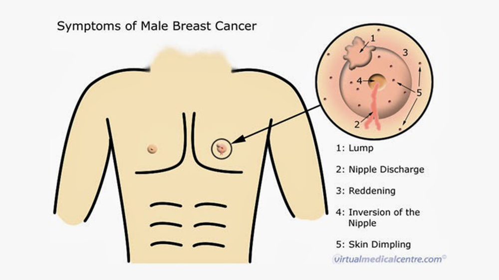
Male breast cancer accounts for less than 1% of all breast cancers:
There have been no randomized control trials for surgical management of breast cancer in men
Although breast conservation is increasing:
The current surgical management remains simple mastectomy with sentinel lymph node biopsy
Sentinel lymph node biopsy has been demonstrated to be accurate in men
There are no data to support staging studies:
Such as positron emission tomography (PET) or computed tomography (CT):
In early-stage breast cancer in either men or women
The role of the 21-gene signature assay is an emerging field in male breast cancer
Approximately 10% of male breast cancers are associated with genetic mutations:
BRCA2 is the most common among the mutations:
With a lifetime risk of 5% to 10% among BRCA2 carriers
Klinefelter’s is also associated with an increased risk of male breast cancer:
With an incidence between 3% and 7%
All male breast cancer patients should be referred for genetic counseling and testing
Endocrine therapy is indicated for ER+ breast cancer:
Aromatase inhibitors can be used but have not been well-studied in men
Tamoxifen is the best-studied drug for male breast cancer
The role of hormonal therapy in male breast cancer has not been evaluated by a prospective, randomized trial
Nevertheless, the rationale for estrogen blockade in male breast cancer is the same as that for female breast cancer:
With numerous prospective, randomized trials supporting the role of endocrine therapy:
To reduce risk of breast cancer recurrence and death
Anti-hormone therapy may also provide chemoprevention for the contralateral breast in men treated by ipsilateral mastectomy
The majority of male breast cancer is ER+ and thus eligible for targeted antiestrogen therapy
Men who have breast cancer:
Should not take testosterone therapy:
Because it may stimulate breast cancer growth by modulating increased estrogenic activity
The balance between estrogen and testosterone in men may have physiologic effects on breast tissue
References
Fentiman IS. Surgical options for male breast cancer. Breast Cancer Res Treat. 2018;172(3):539-544.
Gentilini O, Chagas E, Zurrida S, Intra M, De Cicco C, Gatti G, et al. Sentinel lymph node biopsy in male patients with early breast cancer. Oncologist. 2007;12(5):512-515.
Massarweh SA, Sledge GW, Miller DP, McCullough D, Petkov VI, Shak S. Molecular characterization and mortality from breast cancer in men. J Clin Oncol 2018;36:1396-1404.
Giordano SH. Breast cancer in men. N Engl J Med. 2018;378(24):2311–2320.
Korde LA, Zujewski JA, Kamin L, et al. Multidisciplinary meeting on male breast cancer: summary and research recommendations. J Clin Oncol. 2010;28(12):2114-2122.
Greif JM, Pezzi CM, Klimberg VS, Bailey L, Zuraek M. Gender differences in breast cancer: analysis of 13,000 breast cancers in men from the National Cancer Data Base. Ann Surg Oncol. 2012;19:3199-3204.
Kiluk JV, Lee MC, Park CK, Meade T, et al. Male breast cancer: management and follow-up recommendations. Breast J. 2011;17:503-509.
Mendez, JE, ter Meulen D, Padussis J, et al. Tissue compression is not necessary for needle-localized lesion identification. Amer J Surg. 2005;190(4):580-582.
Graham RA, Homer MJ, Katz J, Rothschild J, Safaii H, Supran S. The pancake phenomenon contributes to the inaccuracy of margin assessment in patients with breast cancer. Am J Surg. 2002;184(2):89-93.
Graham RA, Homer MJ, Sigler CJ et al. The efficacy of specimen radiography in evaluating the surgical margins of impalpable breast carcinoma. AJR Am J Roentgenol. 1994;162(1):33-36.
As the primary method of axillary staging for patients with early stage breast cancer
Changes in patient presentation and advancements in systemic therapy:
Have led clinicians to question the utility of ALND even in the presence of involved nodes
The American College of Surgeons Oncology Group (ACOSOG) Z0011 trial:
Randomized women with T1 / T2 tumors undergoing breast conservation with one or two positive sentinel nodes to undergo ALND vs. no additional axillary surgery
Results showed no difference in local recurrence, disease-free survival (DFS), or overall survival (OS) between the groups
The authors concluded that ALND was not indicated in this setting
One of the major advantages of SLNB compared to ALND:
Is the ability to stage the axilla with reduced rates of lymphedema
A recent meta-analysis of five randomized controlled trials (including the Z0011 trial):
Reported a 70% reduction in risk of lymphedema with SLNB compared to ALND
Multi-gene assays such as the 21-gene recurrence score (RS):
Have provided prognostic information regarding risk of distant recurrence:
For patients with node-negative, ER+ breast cancers
Although evidence suggests that adding chemotherapy to endocrine therapy does result in improved DFS and OS for node-positive patients:
Exploratory data suggest that this may not be true for all patients
A retrospective analysis of the RS performed on 367 specimens from the SWOG 8814 trial:
Showed that RS was prognostic for DFS and OS in node-positive patients
The National Comprehensive Cancer Network:
Allows patients with 1 to 3 positive nodes to consider the 21-gene recurrence score to determine benefit from chemotherapy
References
Giuliano AE, Ballman K, McCall L, et al. Locoregional recurrence after sentinel lymph node dissection with or without axillary dissection in patients with sentinel lymph node metastases: long-term follow-up from the American College of Surgeons Oncology Group (Alliance) ACOSOG Z0011 randomized trial. Ann Surg. 2016; 264(3):413-420.
Glechner A, Wockel A, Gartlehner G, et al. Sentinel lymph node dissection only versus complete axillary lymph node dissection in early invasive breast cancer: a systematic review and meta-analysis. Eur J Cancer. 2013;49(4):812-825.
Albain KS, Barlow WE, Shak S, et al. Prognostic and predictive value of the 21-gene recurrence score assay in postmenopausal women with node-positive, estrogen-receptor-positive breast cancer on chemotherapy: a retrospective analysis of a randomised trial. Lancet Oncol. 2010;11(1):55-65.
Several definitions of resectability of pancreatic adenocarcinoma (PDAC) have been approved for determining the possibility for complete clearance (R0 resection) by surgery:
Taking into account oncological and general aspects
Surgical resectability of PDAC is assessed by:
The evaluation of local tumor extension to vessels and distant metastases
Excluding tumor with distant metastases:
Which is defined as unresectable with metastases (UR‐M:
Local resectability is classified in three categories:
Resectable (R)
Borderline resectable (BR)
Unresectable (UR‐LA)
R PDAC shows:
No vascular infiltration to major vessels
Complete clearance of R tumor is required in standard pancreatectomy without combined vascular resection
BR PDAC is sub‐classified into two categories:
BR‐PV showing PV distortion or narrowing
BR‐A showing semi‐circumferential abutment with a major artery
There is a theoretical “borderline” between BR‐PV and BR‐A:
Whereas PV resection is currently recommended for achieving R0 resection:
Arterial resection remains controversial due to significantly increased rates of morbidity
From the surgical perspective, BR‐PV PDAC is borderline resectable:
Whereas BR‐A PDAC is borderline unresectable
Considering surgical feasibility:
R and BR‐PV PDAC should be considered as:
Candidates for “PDAC that is planned for resection (potentially resectable PDAC):
Potentially resectable PDAC has been treated by upfront surgery, although neoadjuvant for BR PDAC might be considered given the poor oncological outcomes
Multiple studies, including a meta-analysis of six studies:
Show significant benefit with use of bilateral prophylactic mastectomies (BPM) in BRCA carriers:
With up to 90% risk reduction
The need for axillary staging in the context of prophylactic mastectomy:
Pooled results from a meta-analysis and systematic review report similar findings:
The risk of occult disease at time of mastectomy:
Is low (less than 2%)
There is an extremely low risk of nodal involvement:
Less than 2%
Only 2.8% of cases have a change in management as a result of the SLNB
As a result, the routine use of SLNB is not recommended
Data have evolved that show the benefit of MRI screening in patients at highest risk for breast cancer development
McLaughlin and colleagues questioned whether preoperative breast MRI could be used to determine the need for SLNB at time of prophylactic mastectomy:
In their series of 178 patients with MRI and SLNB at time of prophylactic mastectomy:
Six occult cancers were found
All six patients had abnormal findings preoperatively on MRI, with negative predictive value of 100% for invasive disease
The researchers concluded that MRI could be utilized to select low-risk patients who may avoid SLNB at time of BPM
The study by McLaughlin et al. included BRCA mutation carriers, but only one study specifically evaluated the use of SLNB in BRCA carriers:
This trial reported similar results, with occult invasive disease detected in 2.5%
No patients in this cohort had node-positive disease, and there were no axillary recurrences at median 34-month followup
There are case reports of successful SLNB after mastectomy:
But currently the routine use is discouraged by the National Comprehensive Cancer Network guidelines
References
Zhou WB, Liu XA, Dai JC, Wang S. Meta-analysis of sentinel lymph node biopsy at the time of prophylactic mastectomy of the breast. Can J Surg 2011;54(5):300-306.
Nagaraja V, Edirimanne S, Eslick GD. Is sentinel lymph node biopsy necessary in patients undergoing prophylactic mastectomy? a systematic review and meta-analysis. Breast J. 2016;22(2):158-165.
McLaughlin SA, Stempel M, Morris EA, Liberman L, King TA. Can magnetic resonance imaging be used to select patients for sentinel lymph node biopsy in prophylactic mastectomy? Cancer. 2008;112(6):1214-1221.
Câmara S, Pereira D, André S, et al. The use of sentinel lymph node biopsy in brca1/2 mutation carriers undergoing prophylactic mastectomy: a retrospective consecutive case-series study. Int J Breast Cancer. 2018:1426369
Does not prolong survival compared to adjuvant chemotherapy:
However, it does result in decreased disease burden, and can be beneficial at time of surgery:
Providing increased opportunity to perform breast-conserving surgery and reducing need for axillary lymph node dissection
A meta-analysis of 14 prospective randomized trials of neoadjuvant vs. adjuvant chemotherapy:
In 5,500 patients with breast cancer demonstrated that:
NAC was associated with an absolute decrease in the mastectomy rate of 16.6% (95% CI 15.1–18.1%)
Patients with ER– and HER2 positive breast cancers:
Are more likely to experience complete pathologic response than those with ER+ cancers
Patients with a clinically positive axilla after neoadjuvant chemotherapy:
Should undergo axillary dissection at the time of breast surgery
Patients with no residual adenopathy on clinical exam:
May be considered for sentinel lymph node biopsy (SLNB):
Accuracy of SLNB after NAC can be improved with:
Localization of previously-clipped nodes
Use of dual tracer
Increasing the number of sentinel nodes retrieved:
SLNB after NAC has a false-negative rate of less than 10% only when ≥ 3 sentinel nodes were identified
References
Mieog, JS, van der Hage JA, van de Velde CJ. Neoadjuvant chemotherapy for operable breast cancer. Br J Surg. 2007;94(1):1189-1200.
van der Hage JA, van de Velde CJH, Julien JP, Tubiana-Hulin M, Vandervelden C, Duchateau L. Preoperative chemotherapy in primary operable breast cancer: results from the European Organization for Research and Treatment of Cancer trial 10902. J Clin Oncol. 2001;19(22):4224-4237.
Fisher B, Brown A, Mamounas E, et al. Effect of preoperative chemotherapy on local-regional disease in women with operable breast cancer: findings from National Surgical Adjuvant Breast and Bowel Project B-18. J Clin Oncol. 1997;15(7):2483-2493.
Rouzier R, Perou CM, Symmans WF, et al. Breast cancer molecular subtypes respond differently to preoperative chemotherapy. Clin Cancer Res. 2005;11(16):5678-5685.
Boughey JC, Ballman KV, Le-Petross HT, et al. Identification and resection of clipped node decreases the false-negative rate of sentinel lymph node surgery in patients presenting with node-positive breast cancer (T0-T4, N1-N2) who receive neoadjuvant chemotherapy: results from ACOSOG Z1071 (Alliance). Ann Surg. 2016;263(4):802-807.
Boileau, JF, Poirier B, Basik M, et al. Sentinel node biopsy after neoadjuvant chemotherapy in biopsy-proven node-positive breast cancer: the SN FNAC study. J Clin Oncol. 2015;33(3):258-264.
Rebbeck TR, Friebel T, Lynch HT, et al. Bilateral prophylactic mastectomy reduces breast cancer risk in BRCA1 and BRCA2 mutation carriers: the PROSE Study Group. J Clin Oncol. 2004;22(6):1055-1062.
Domchek SM, Friebel TM, Singer CF, et al. Association of risk-reducing surgery in BRCA1 or BRCA2 mutation carriers with cancer risk and mortality. JAMA 2010;304(9):967-975.
They are the most common gene alterations seen in the hereditary breast cancer population
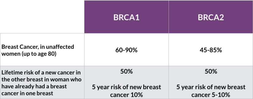
They are associated with an increased risk of breast cancer estimated to be:
55% to 70% for BRCA 1 carriers by age 70
45% to 70% in BRCA 2 carriers by age 70
While both BRCA 1 and BRCA 2 mutations are associated with an increased risk of breast cancer:
BRCA 1 breast cancers more commonly occur in:
Younger
Premenopausal women
Are more likely to be triple negative
BRCA 1 is associated with a higher risk of ovarian cancer compared to BRCA 2:
With a lifetime risk of 40% to 45% in BRCA 1 carriers compared to 15% to 20% in BRCA 2 carriers
BRCA 2 breast cancers more closely resemble the sporadic breast cancer pattern:
With a predominance of hormone receptor positive cancers in women greater than 50 years
CHEK 2 and PALB 2 are moderate penetrance genes:
That are less common than BRCA mutations
Similar to BRCA 2 deleterious mutations:
CHEK 2 and PALB 2 mutations are associated with:
Hormone receptor positive postmenopausal breast cancer
Imaging surveillance for BRCA mutation carriers begins at:
Age 25 with annual breast MRI with contrast, with addition of mammography after age 30
Although use of screening ultrasound is evolving in women with dense breast tissue:
Its use in BRCA carriers has not been defined
Surveillance strategies have significantly improved early detection but do not prevent breast cancer
Bilateral salpingo-oophorectomy:
Has been shown to provide approximately 50% relative reduction in breast cancer risk:
But ultimately prophylactic mastectomy provides the greatest reduction
The optimal timing of surgery depends on multiple factors, including:
The patient’s desire for future breastfeeding
Ages of family members at diagnosis
Several retrospective series and meta-analyses of four prospective studies:
Have supported prophylactic mastectomy in BRCA mutation carriers
While the data demonstrate a 93% relative risk reduction in breast cancers:
They do not demonstrate a survival benefit in this population
Recent non-randomized studies have evaluated use of nipple-sparing mastectomy (NSM) and demonstrated its feasibility and safety in patients with BRCA mutation:
Jakub et al. reported data from 548 risk-reducing NSMs in 346 patients treated at 9 institutions
This study included both women opting for prophylactic mastectomies concurrent with treatment for a contralateral breast cancer, and women undergoing bilateral prophylactic mastectomies for risk reduction
With median and mean follow up of 34 and 56 months, respectively:
No ipsilateral breast cancers occurred after prophylactic NSM
Breast cancer did not develop in any patients undergoing bilateral risk-reducing NSMs
References
Heemskerk-Gerritsen BA, Menke-Pluijmers MB, Jager A, et al. Substantial breast cancer risk reduction and potential survival benefit after bilateral mastectomy when compared with surveillance in healthy BRCA1 and BRCA2 mutation carriers: a prospective analysis. Ann Oncol. 2013;24(8):2029-2035.
De Felice F, Marchetti C, Musella A, et al. Bilateral risk-reduction mastectomy in BRCA1 and BRCA2 mutation carriers: a meta-analysis. Ann Surg Oncol. 2015;22(9):2876-2880.
Ludwig KK, Neuner J, Butler A. Risk reduction and survival benefit of prophylactic surgery in BRCA mutation carriers: a systematic review. Am J Surg. 2016;212(4):660-669
Jakub JW, Peled AW, Gray RJ. Oncologic Safety of Prophylactic Nipple-Sparing Mastectomy in a Population With BRCA Mutations: A Multi-institutional Study. JAMA Surg. 2018;153:123-129.
Loi M, Desideri I, Olmetto E, Francolini G, Greto D, Bonomo P, et al. BRCA mutation in breast cancer patients: Prognostic impact and implications on clinical management. Breast J. 2018;24(6):1019-1023.
The standard adequate margin for patients with DCIS treated with breast-conserving surgery followed by whole-breast radiation is:
2 mm
Negative margins:
Halve the risk of ipsilateral breast tumor recurrence (IBTR) compared with positive margins (defined as ink on DCIS)
A 2 mm margin minimizes the risk of IBTR relative to narrower negative margin widths:
However, larger margins (>2 mm) do not significantly decrease IBTR
In 2016, margin guidelines related to the treatment of non-invasive breast cancer (e.g., DCIS) in the setting of breast-conservation therapy:
Were developed by the Society of Surgical Oncology, American Society for Radiation Oncology, and the American College of Surgeons in a similar manner
A consensus statement released by a multidisciplinary panel included:
The optimal margins for:
Pure DCIS and mixed tumors (invasive and non-invasive components within the same tumor) in the setting of breast conservation
Results from the meta-analysis showed:
That a 2 mm margin decreases the risk of IBTR in pure DCIS compared to closer negative margins
This differs from the previous margin recommendation for invasive cancer:
Which remains no ink on tumor:
However, in the setting of mixed tumors (invasive cancer with a DCIS component):
The recommendation for negative margins remains no ink on tumor, as patients with mixed disease are treated as invasive cancer and therefore receive systemic therapy more often than pure DCIS patients
In the setting of DCIS with micro-invasion (no focus of invasive disease larger than 1 mm):
The multidisciplinary panel recommends a 2 mm margin:
As these lesions have similar rates of IBTR as pure DCIS
Patients with positive margins after breast-conserving surgery:
Should undergo re-excision
Patients for whom adequate surgical margins cannot be achieved with lumpectomy:
Total mastectomy should be performed
Complete axillary lymph node dissection should not be performed:
In the absence of evidence of invasive cancer or proven axillary metastatic disease in women with apparent pure DCIS
However, a small proportion of patients with apparent pure DCIS will be found to have invasive cancer at the time of their definitive surgical procedure:
Therefore, a sentinel lymph node biopsy should be strongly considered if the patient with apparent pure DCIS is to be treated with mastectomy or with excision in an anatomic location compromising the performance of a future sentinel lymph node procedure
References
Morrow M, Van Zee KJ, Solin LJ, et al. Society of Surgical Oncology-American Society for Radiation Oncology-American Society of Clinical Oncology consensus guideline on margins for breast-conserving surgery with whole-breast irradiation in ductal carcinoma in situ. J Clin Oncol. 2016;34(33):4040-4046.