My name is Rodrigo Arrangoiz I am a breast surgeon/ thyroid surgeon / parathyroid surgeon / head and neck surgeon / surgical oncologist that works at Center for Advanced Surgical Oncology in Miami, Florida.
I was trained as a surgeon at Michigan State University from (2005 to 2010) where I was a chief resident in 2010. My surgical oncology and head and neck training was performed at the Fox Chase Cancer Center in Philadelphia from 2010 to 2012. At the same time I underwent a masters in science (Clinical research for health professionals) at the University of Drexel. Through the International Federation of Head and Neck Societies / Memorial Sloan Kettering Cancer Center I performed a two year head and neck surgery and oncology / endocrine fellowship that ended in 2016.
Mi nombre es Rodrigo Arrangoiz, soy cirujano oncólogo / cirujano de tumores de cabeza y cuello / cirujano endocrino que trabaja Center for Advanced Surgical Oncology en Miami, Florida.
Fui entrenado como cirujano en Michigan State University (2005 a 2010 ) donde fui jefe de residentes en 2010. Mi formación en oncología quirúrgica y e n tumores de cabeza y cuello se realizó en el Fox Chase Cancer Center en Filadelfia de 2010 a 2012. Al mismo tiempo, me sometí a una maestría en ciencias (investigación clínica para profesionales de la salud) en la Universidad de Drexel. A través de la Federación Internacional de Sociedades de Cabeza y Cuello / Memorial Sloan Kettering Cancer Center realicé una sub especialidad en cirugía de cabeza y cuello / cirugia endocrina de dos años que terminó en 2016.
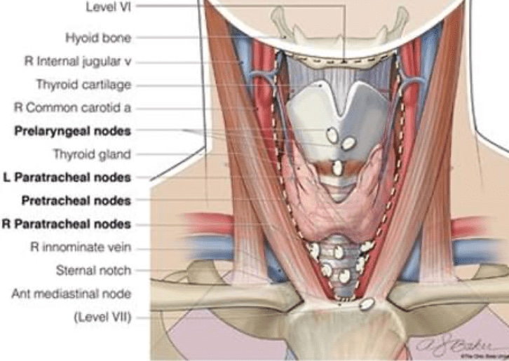
👉Not all thyroid cancer recurrences require surgery.
👉Small lymph node recurrences in the neck can often be observed, however recurrent nodes greater than 0.8 cm in the central neck and greater than 1 cm in the lateral neck are typically removed.
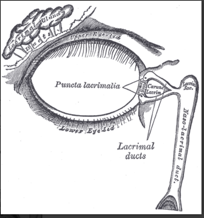
Drain tears from the ocular surface to the lacrimal sac and, ultimately, the nasal cavity
Blockage of the nasolacrimal system:
Can cause tears to flow over the eyelid and down the cheek:
This condition is epiphora
Structure and Function:
Both the upper eyelid and the lower eyelid have a small opening on the surface of the eyelid margin near the medial canthus:
These are called puncta:
Each puncta leads to a drainage canal that eventually flows into the lacrimal sac and then the nasal cavity
The drainage canal connecting the ocular surface to the nasal cavity consists of multiple parts:
Within the lower eyelid:
The punctum leads to a 2 mm long ampulla:
Which runs perpendicular to the eyelid margin
The ampulla turns 90 degrees medially:
Becoming the inferior canaliculus and travels 8 to 10 mm before reaching the common canaliculus
The upper canaliculus travels 2 mm superiorly in the eyelid before turning 90 degrees medially and moving 8 to 10 mm before connecting to the common canaliculus
The common canaliculus:
Drains into the lacrimal sac
Within the junction between the common canaliculus and the lacrimal sac:
Is the valve of Rosenmuller:
This apparatus is a one-way valve that prevents reflux from the lacrimal sac to the puncta
The lacrimal sac drains:
Inferiorly to the nasolacrimal duct:
Which is bordered:
Medially by:
Palatine bone and the inferior turbinate in the nose
Laterally by:
Maxillary bone
The nasolacrimal duct:
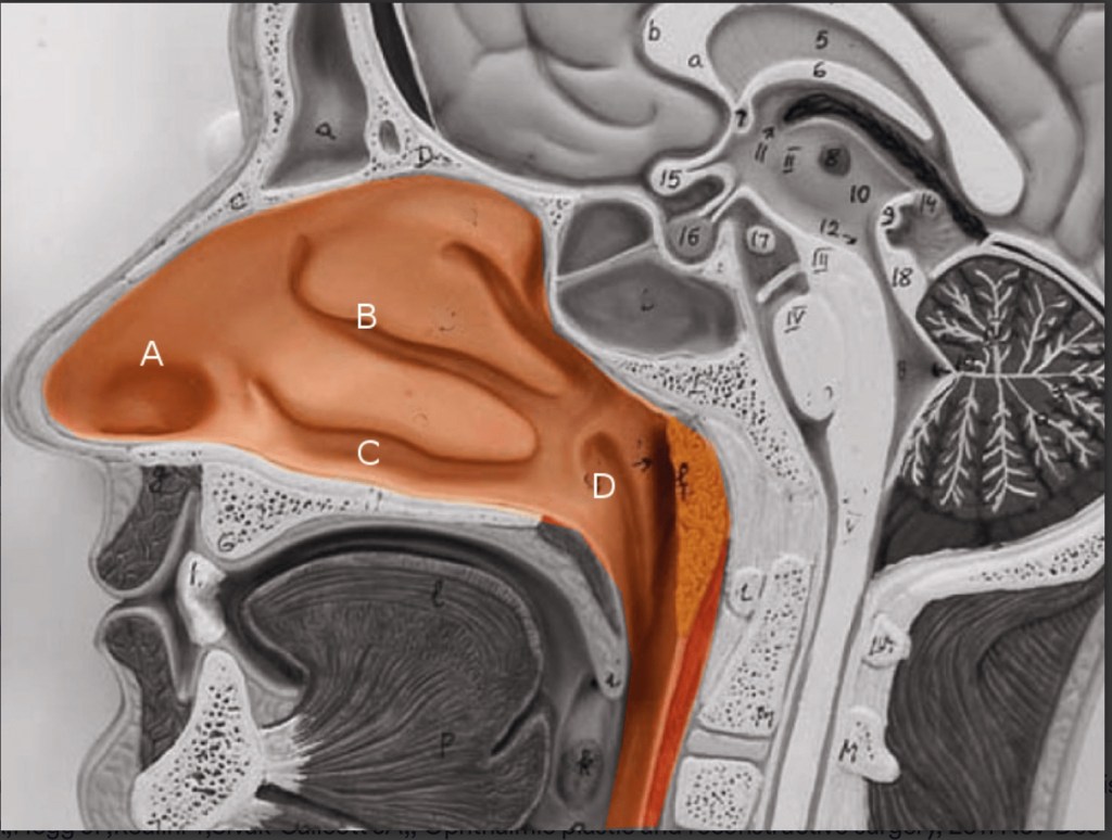
Opens at the inferior meatus:
Located underneath the inferior nasal turbinate
The lacrimal sac is:
Approximately 10 to 15 mm in axial length and 13 to 20 mm in corneal length
The nasolacrimal duct is:
12 to 18 mm long
The inferior nasal meatus is partially covered by a mucosal fold:
Known as the valve of Hasner
Embryology:
The nasolacrimal duct:
Starts forming around five weeks of gestation
It starts out as a linear thickening of ectoderm:
Located in a groove between the nasal and maxillary prominences
This thickening:
Eventually separates into a solid cord and sinks into the surrounding mesenchyme
Over time the cord canalizes:
Forming the lacrimal sac and the beginning of the nasolacrimal duct
The nasolacrimal duct extends:
Intranasally until it exits under the inferior turbinate
The lacrimal sac extends caudally:
To complete the canalicular system
The inside of the canal breaks down and forms a lumen:
So that the nasolacrimal system is patent:
This process is generally complete by the time of birth
Blood Supply and Lymphatics:
Blood supply to the nasolacrimal area of the face:
Is generally from the angular artery:
The angular artery is considered a branch of the facial artery:
However, some studies have shown that it can originate from the ophthalmic artery in some individuals
It terminates in anastomosis with the dorsal nasal branch of the ophthalmic artery
The angular artery and vein:
Appear alongside the nose near the medial orbit
A correlating angular vein drains this region
The medial and lateral portions of the eyelids have different lymphatic drainage systems:
The medial one-third of the upper eyelid and the medial two-thirds of the lower eyelid:
Drain to the submandibular lymph nodes
The lateral two-thirds of the upper eyelid and the lateral one-third of the lower eyelid:
Drain to the pre-auricular lymph nodes
Nerves:
Cranial nerve VII:
Supplies the motor innervation to the muscles of the face
The movement of these muscles:
Aid in proper drainage of the tears through the nasolacrimal system:
By what is known as the lacrimal pump mechanism
Cranial nerve III and cranial nerve VII:
Innervate the muscles that control the blinking of the eyelids:
This action is the primary driver of the lacrimal pump mechanism
Irritation of the ocular surface:
Stimulates the ophthalmic branch of cranial nerve five:
Which begins the reflex tear arc pathway:
The efferent pathway involves cranial nerve VII and parasympathetic fibers
The role of the sympathetic nervous system in tear production:
Is not well understood
Muscles:
The action of the orbicularis muscle and surrounding tissues:
Helps propel the flow of tears from the canaliculi to the nasolacrimal duct:
Via the lacrimal pump mechanism
References:
Computed tomography dimensions of the lacrimal gland in normal Caucasian orbits., Tamboli DA,Harris MA,Hogg JP,Realini T,Sivak-Callcott JA,, Ophthalmic plastic and reconstructive surgery, 2011 Nov-Dec.
An Unusual Case of Nasolacrimal Obstruction Caused by Foodstuffs., Matsumoto H,Matsumoto A,, Case reports in ophthalmology, 2015 Sep-Dec.
Incidence of neoplasia in patients with unilateral epiphora., Bewes T,Sacks R,Sacks PL,Chin D,Mrad N,Wilcsek G,Tumuluri K,Harvey R,, The Journal of laryngology and otology, 2015 Jul.
There is robust literature to support the use of CPM as a cost-effective strategy:
In patients with hereditary breast cancer syndromes
Anderson et al. demonstrated that the most cost-effective strategy:
With and without quality adjustment:
For women with BRCA1 or BRCA2 mutations was:
Prophylactic bilateral salpingo-oophorectomy with bilateral mastectomy
Simulation models analyzing costs for CPM versus surveillance in patients with sporadic breast cancer reveal disparate findings:
An initial Markov model study found that CPM was cost effective compared with surveillance for:
Patients younger than 70 years:
But this finding was highly dependent on the quality of life assumptions
A second study that included operative complications and breast reconstruction costs used a decision-tree model and concluded that:
Although CPM resulted in a cost savings over surveillance for women younger than 50 years:
It also reduced quality of life years
When MRI was inserted in the model as the primary method of screening:
The cost-effectiveness of CPM increased
Loss of quality of life years was largely attributed to complications from reconstructive procedures
The two models differ in the assumptions regarding quality of life:
If we assume an improvement in quality of life after CPM:
Then CPM could be cost effective
Alternatively, if quality of life is decreased,:
CPM would not be a cost-effective strategy
The available data on cost effectiveness for CPM is limited
Summary:
CPM is a cost-effective strategy for women with BRCA mutations
At this time, there is insufficient evidence to support the concept of superior cost effectiveness for CPM in women with sporadic breast cancer and the cost effectiveness is highly dependent on the quality of life assumptions
Reference:
Contralateral Prophylactic Mastectomy Consensus Statement from the American Society of Breast Surgeons: Additional Considerations and a Framework for Shared Decision Making. Ann Surg Oncol (2016) 23:3106–3111
Diagnosis is always established by the measurement of:
Sensitive TSH and thyroid hormone levels (free T3 and free T4)
Thyrotoxicosis caused by TNG or Graves’ disease is usually characterized by:
A suppressed TSH level with either:
Normal (subclinical) or elevated (overt) free thyroid hormone levels
It is insufficient to rely on the measurement of TSH or free thyroid hormones:
Alone to diagnose TNG or Graves’ disease:
Because suppression of TSH or elevation of thyroid hormones can be associated with clinical conditions other than TNG and Graves’ disease
Low TSH
High TSH
Secondary hypothyroidism
TSH-secreting pituitary tumor
Non-thyroidal illness
Thyroid hormone resistance
Glucocorticoid therapy
Amiodarone use
Excessive thyroid hormone therapy
Other serologic findings:
Such as antithyroid antibodies – antithyroid peroxidase and antithyroglobulin:
That support the diagnosis of autoimmune thyroid disease:
May be detected in patients with Graves’ disease:
However, serum TSHR-Ab:
Is occasionally helpful in the diagnosis of Graves’ disease:
Though there is no consensus regarding its routine measurement in Graves’ disease
Imaging:
Radionuclide Imaging:
Radionuclide scanning and radioactive iodine uptake (RAIU):
Are useful tests to elucidate the cause of hyperthyroidism
In toxic nodular goiter (TNG):
The radioactive iodine (RAI) concentration is in the nodule(s), and uptake is inhibited in the surrounding tissue:
Giving the appearance of “patchy uptake”
Consequently, the total RAIU may be either:
Slightly raised or at the upper limit of normal
In Graves’ disease:
Because of the diffuse thyroid involvement:
The RAIU is always intense and increased
Thyroid radionuclide imaging may not be necessary in every case:
When the diagnosis is obvious:
But it is helpful in the differentiation of other clinical conditions associated with hyperthyroidism but with low RAIU
Radionuclide scan of toxic nodular goiter demonstrating intense focal uptake of several hot nodules with different degrees of suppression of adjacent thyroid tissue (A) compared with the scan from a patient with non-toxic multi-nodular goiter, showing less intense patchy radioactive iodine uptake (B).
Radionuclide scan in Graves’ hyperthyroidism demonstrating the diffuse and homogeneous nature of increased uptake in both lobes of the thyroid.
Clinical Conditions Associated with Low Radioactive Iodine Uptake and Hyperthyroidism
Thyroiditis
Iodine-induced thyrotoxicosis
Exogenous thyrotoxicosis (factitia)
Ectopic functional thyroid tissue
Computed tomography scan:
In any patient with compressive or obstructive symptoms and an MNG:
Chest radiography and chest computed tomography (CT) are often informative
Chest CT is particularly valuable to define the size and extent of the goiter:
Especially into the mediastinum
Care to avoid iodinated contrast:
Until the patient’s thyroid functional status must be taken into account or the significant iodine load CT contrast agent may acutely induce or worsen hyperthyroidism
A, Chest radiography showing a huge solid goiter (horizontal arrow) displacing the trachea without compression (vertical arrow).B, Neck computed tomography of the same goiter (arrows). Thyroidectomy revealed a 290-g benign thyroid gland.
Associated metabolic abnormalities:
Altered glucose metabolism:
Reversible hyperglycemia
Elevated C-peptide
Elevated intact proinsulin
Insulin resistance
Increased bone turnover:
Elevated markers of bone formation and resorption
Are hallmarks of untreated hyperthyroidism
Untreated hyperthyroidism is associated with an elevated chromogranin A level:
That changes in parallel with thyroid status
Metabolic Abnormalities Associated with Hyperthyroidism
The benefit of performing sentinel lymph node (SLN) surgery at the time of CPM is:
That the lymph nodes have been evaluated in the event that an occult malignancy is found
The downside is:
Increased surgical morbidity such as lymphedema
By meta-analysis:
The risk of lymphedema after SLN alone is:
5.6 % (95 % CI 6.1–7.9 %) and increases with longer follow-up
The chance of finding occult invasive disease in a prophylactic mastectomy is:
1.8 %
An additional small percent of CPM specimens harbor noninvasive disease:
That would not require nodal evaluation
The rate of nodal positivity in patients with occult malignancy in CPM is:
Only 1.3 %
Considering these data:
Routine SLN surgery at time of CPM:
Places more patients at risk of lymphedema:
Than would be expected from the 1% to 2 % of patients with occult disease undergoing axillary dissection
Therefore the consensus group:
Does not recommend routine SLN for CPM
Patients at higher risk of contralateral occult malignancy are:
Postmenopausal patients
Those with triple-negative
Locally advanced
Inflammatory breast cancer
Invasive lobular disease
MRI at the time of breast cancer diagnosis:
Identifies occult contralateral disease:
2% to 4 % of the time
Suspicious lesions in the contralateral breast should be biopsied:
But if a biopsy is not done:
SLN surgery should be considered for highly suspicious lesions
Summary:
Sentinel lymph node surgery on the CPM side should not be routinely performed
References:
Contralateral Prophylactic Mastectomy Consensus Statement from the American Society of Breast Surgeons: Additional Considerations and a Framework for Shared Decision Making. Ann Surg Oncol (2016) 23:3106–3111
Is more responsive to preoperative chemotherapy compared to ER/PR+, HER2neu negative breast cancer:
Pathologic complete response is seen in approximately 30% to 40% of patients undergoing treatment with a third-generation regimen:
A pathologic complete response:
Is highly prognostic in this subset
While ER negative breast cancers:
Have a lower propensity for regional nodal metastasis compared to ER+ tumors:
The difference is relatively small (2% to 5%):
Therefore, nodal staging is still a standard practice recommendation
The Choosing Wisely guideline:
For omission of routine use of sentinel node biopsy in clinically node-negative women ≥ 70 years of age applies to hormone receptor positive breast cancer
Sentinel node biopsy may be successfully performed after neoadjuvant chemotherapy and should be performed patients with a clinically negative axilla
References
Cortazar P, Zhang L, Untch M, et al. Pathologic complete response and long term clinical benefit in breast cancer: the CTNeoBC pooled analysis. Lancet. 2014;384(9938):164-172.
Symmans WF, Peintinger F, Hatzis C, et al. Measurement of residual breast cancer burden to predict survival after neoadjuvant chemotherapy. J Clin Oncol. 2007;25(28):4414-4422.
von Minckwitz G, Untch M, Blohmer JU, et al. Definition and impact of pathologic complete response on prognosis after neoadjuvant chemotherapy in various intrinsic breast cancer subtypes. J Clin Oncol. 2012;30(15):1796-1804.
Viale G, Zurrida S, Maiorano E, et al. Predicting the status of axillary sentinel lymph nodes in 4351 patients with invasive breast carcinoma treated in a single institution. Cancer. 2005;103(3):492-500.
Hughes KS, Schnaper LA, Bellon JR, et al. Lumpectomy plus tamoxifen with or without irradiation in women age 70 years or older with early breast cancer: long-term follow-up of CALGB 9343. J Clin Oncol. 2013;31(19):2382-2387.
Synchronous and metachronous bilateral breast cancers:
Appear in 1% to 20% of patients with breast cancer
Improvements in screening and the increased use of MRI:
Often diagnose more early-stage synchronous bilateral cancer
The role of MRI in the preoperative planning:
Is controversial and may be partly responsible for increasing mastectomy rates in the United States
MRI may identify additional lesions in both the ipsilateral and contralateral breast in many women diagnosed with unifocal breast cancer:
Many of these additional lesions are often found to be benign once additional diagnostic imaging is performed and biopsies are completed
Additional MRI findings should not prompt surgeons to recommend mastectomy:
Unless they are biopsy-proven to represent additional sites of malignancy not amenable to breast conservation, and/or the patient was inclined toward mastectomy prior to MRI findings
Retrospective studies evaluating the outcomes of synchronous bilateral breast cancer:
Are limited by small cohort sizes, differing definitions, and non-matched unilateral patients as controls
Most retrospective studies show no differences in local recurrence or survival for bilateral breast cancers:
Making bilateral breast-conserving treatment a safe option for early-stage synchronous cancers
References
Heron DE, Komarnicky LT, Hyslop T, Schwartz GF, Mansfield CM. Bilateral breast carcinoma: risk factors and outcomes for patients with synchronous and metachronous disease. Cancer. 2000;88(12):2739-2750.
Intra M, Rotmensz N, Viale G, et al. Clinicopathologic characteristics of 143 patients with synchronous bilateral invasive breast carcinomas treated in a single institution. Cancer. 2004;101(5):905-912.
Current management strategies for primary breast lymphoma are largely based on:
Results published in small, single-institution series
Historically, primary breast lymphoma was treated with:
Modified radical mastectomy with or without adjuvant chemotherapy or radiotherapy:
Treatment strategies had focused on anthracycline-based chemotherapy with or without consolidative radiotherapy
Current treatment guidelines dictate:
That surgery should be reserved for
Obtaining adequate tissue for diagnosis, if needed, and:
Should not be regarded as a therapeutic modality in the treatment of this disease:
In several series, surgery has been associated with worse outcomes
Some histologies may be amenable to localized surgery so understanding the disease pathology is important in decision making
While axillary nodal status is an important prognosticator:
There are no definitive guidelines regarding how to stage the axilla:
In addition to CT scan, axillary ultrasound with percutaneous biopsy is frequently used
Sentinel lymph node biopsy has not been studied in this malignancy and currently has no role in its workup
References
Aviles A, Delgado S, Nambo MJ, Neri N, Murillo E, Cleto S. Primary breast lymphoma: results of a controlled clinical trial. Oncology. 2005;69(3):256-260.
Aviv A, Tadmor T, Polliack A. Primary diffuse large B-cell lymphoma of the breast: looking at pathogenesis, clinical issues and therapeutic options. Ann Oncol. 2013;24(9):2236-2244.
el-Ghazawy IM, Singletary SE. Surgical management of primary lymphoma of the breast. Ann Surg. 1991;214(6):724-726.
Jennings WC, Baker RS, Murray SS, et al. Primary breast lymphoma: the role of mastectomy and the importance of lymph node status. Ann Surg. 2007;245(5):784-789.
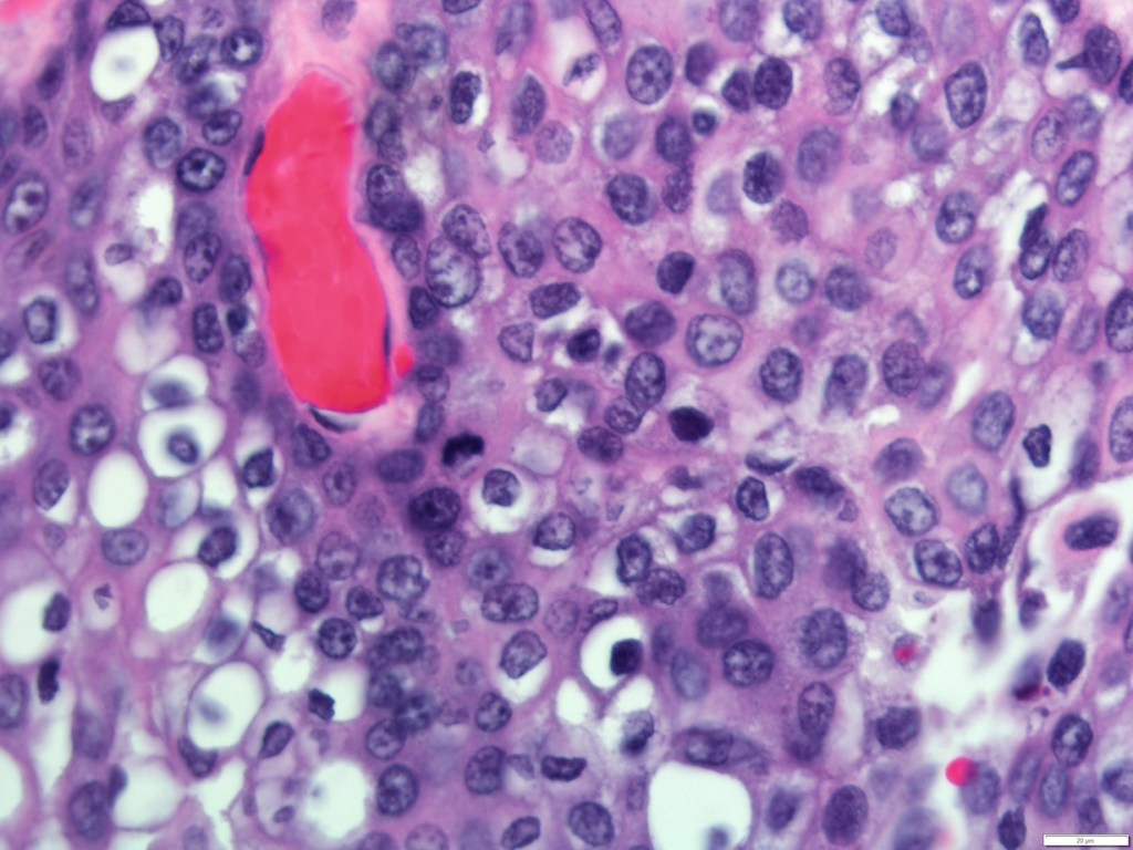
Tends to present in patients age 60 years and older
Is rare in young or premenopausal women
Compared to IDC:
It has been shown to present in a higher proportion of African Americans and Hispanics
Metaplastic breast cancer (MBC):
Is more likely to be high grade but axillary node negative at presentation
The mean tumor size:
Is about 4 cm
Patients with this diagnosis are also more likely to receive chemotherapy and undergo mastectomy
Recurrence tends to be locoregional or pulmonary:
And is associated with a high mortality rate
Future directions may include immunotherapies:
As MBC has a unique histology, demonstrating increased PDL-1:
Which may make it a good candidate for targeted therapy:
More research is needed on this unique tumor phenotype
References
Pezzi CM, Patel-Parekh L, Cole K, Frank J, Klimberg VS, Bland K. Characteristics and treatment of metaplastic breast cancer: analysis of 892 cases from the National Cancer Data Base. Ann Surg Oncol. 2006;14(1):166-173.





 Rodrigo Arrangoiz MS, MD, FACS, FSSO