- The purpose of the nasolacrimal system is to:
- Drain tears from the ocular surface to the lacrimal sac and, ultimately, the nasal cavity
- Blockage of the nasolacrimal system:
- Can cause tears to flow over the eyelid and down the cheek:
- This condition is epiphora
- Can cause tears to flow over the eyelid and down the cheek:
- Structure and Function:
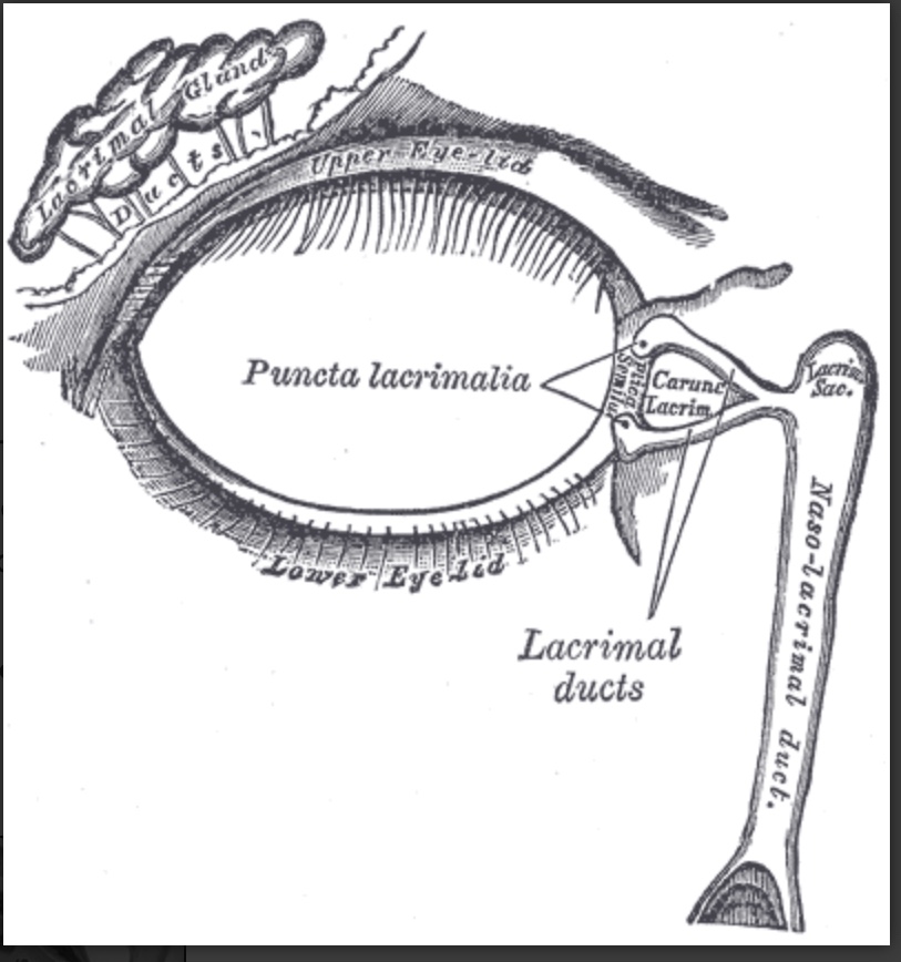
- Both the upper eyelid and the lower eyelid have a small opening on the surface of the eyelid margin near the medial canthus:
- These are called puncta:
- Each puncta leads to a drainage canal that eventually flows into the lacrimal sac and then the nasal cavity
- These are called puncta:
- The drainage canal connecting the ocular surface to the nasal cavity consists of multiple parts:
- Within the lower eyelid:
- The punctum leads to a 2 mm long ampulla:
- Which runs perpendicular to the eyelid margin
- The ampulla turns 90 degrees medially:
- Becoming the inferior canaliculus and travels 8 to 10 mm before reaching the common canaliculus
- The upper canaliculus travels 2 mm superiorly in the eyelid before turning 90 degrees medially and moving 8 to 10 mm before connecting to the common canaliculus
- The punctum leads to a 2 mm long ampulla:
- The common canaliculus:
- Drains into the lacrimal sac
- Within the junction between the common canaliculus and the lacrimal sac:
- Is the valve of Rosenmuller:
- This apparatus is a one-way valve that prevents reflux from the lacrimal sac to the puncta
- Is the valve of Rosenmuller:
- Within the lower eyelid:
- The lacrimal sac drains:
- Inferiorly to the nasolacrimal duct:
- Which is bordered:
- Medially by:
- Palatine bone and the inferior turbinate in the nose
- Laterally by:
- Maxillary bone
- Medially by:
- Which is bordered:
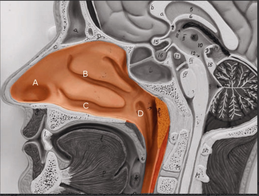
- The nasolacrimal duct:
- Opens at the inferior meatus:
- Located underneath the inferior nasal turbinate
- Opens at the inferior meatus:
- The lacrimal sac is:
- Approximately 10 to 15 mm in axial length and 13 to 20 mm in corneal length
- The nasolacrimal duct is:
- 12 to 18 mm long
- The inferior nasal meatus is partially covered by a mucosal fold:
- Known as the valve of Hasner
- Inferiorly to the nasolacrimal duct:
- Both the upper eyelid and the lower eyelid have a small opening on the surface of the eyelid margin near the medial canthus:
- Embryology:
- The nasolacrimal duct:
- Starts forming around five weeks of gestation
- It starts out as a linear thickening of ectoderm:
- Located in a groove between the nasal and maxillary prominences
- This thickening:
- Eventually separates into a solid cord and sinks into the surrounding mesenchyme
- Over time the cord canalizes:
- Forming the lacrimal sac and the beginning of the nasolacrimal duct
- The nasolacrimal duct extends:
- Intranasally until it exits under the inferior turbinate
- The lacrimal sac extends caudally:
- To complete the canalicular system
- The inside of the canal breaks down and forms a lumen:
- So that the nasolacrimal system is patent:
- This process is generally complete by the time of birth
- So that the nasolacrimal system is patent:
- The nasolacrimal duct:
- Blood Supply and Lymphatics:
- Blood supply to the nasolacrimal area of the face:
- Is generally from the angular artery:
- The angular artery is considered a branch of the facial artery:
- However, some studies have shown that it can originate from the ophthalmic artery in some individuals
- It terminates in anastomosis with the dorsal nasal branch of the ophthalmic artery
- The angular artery and vein:
- Appear alongside the nose near the medial orbit
- A correlating angular vein drains this region
- The angular artery is considered a branch of the facial artery:
- Is generally from the angular artery:
- The medial and lateral portions of the eyelids have different lymphatic drainage systems:
- The medial one-third of the upper eyelid and the medial two-thirds of the lower eyelid:
- Drain to the submandibular lymph nodes
- The lateral two-thirds of the upper eyelid and the lateral one-third of the lower eyelid:
- Drain to the pre-auricular lymph nodes
- The medial one-third of the upper eyelid and the medial two-thirds of the lower eyelid:
- Blood supply to the nasolacrimal area of the face:
- Nerves:
- Cranial nerve VII:
- Supplies the motor innervation to the muscles of the face
- The movement of these muscles:
- Aid in proper drainage of the tears through the nasolacrimal system:
- By what is known as the lacrimal pump mechanism
- Aid in proper drainage of the tears through the nasolacrimal system:
- Cranial nerve III and cranial nerve VII:
- Innervate the muscles that control the blinking of the eyelids:
- This action is the primary driver of the lacrimal pump mechanism
- Innervate the muscles that control the blinking of the eyelids:
- Irritation of the ocular surface:
- Stimulates the ophthalmic branch of cranial nerve five:
- Which begins the reflex tear arc pathway:
- The efferent pathway involves cranial nerve VII and parasympathetic fibers
- The role of the sympathetic nervous system in tear production:
- Is not well understood
- Which begins the reflex tear arc pathway:
- Stimulates the ophthalmic branch of cranial nerve five:
- Cranial nerve VII:
- Muscles:
- The action of the orbicularis muscle and surrounding tissues:
- Helps propel the flow of tears from the canaliculi to the nasolacrimal duct:
- Via the lacrimal pump mechanism
- Helps propel the flow of tears from the canaliculi to the nasolacrimal duct:
- The action of the orbicularis muscle and surrounding tissues:
- References:
- Computed tomography dimensions of the lacrimal gland in normal Caucasian orbits., Tamboli DA,Harris MA,Hogg JP,Realini T,Sivak-Callcott JA,, Ophthalmic plastic and reconstructive surgery, 2011 Nov-Dec.
- An Unusual Case of Nasolacrimal Obstruction Caused by Foodstuffs., Matsumoto H,Matsumoto A,, Case reports in ophthalmology, 2015 Sep-Dec.
- Lacrimal Gland Volume Changes in Unilateral Primary Acquired Nasolacrimal Obstruction., Yazici A,Bulbul E,Yazici H,Sari E,Tiskaoglu N,Yanik B,Ermis S,, Investigative ophthalmology & visual science, 2015 Jul.
- Incidence of neoplasia in patients with unilateral epiphora., Bewes T,Sacks R,Sacks PL,Chin D,Mrad N,Wilcsek G,Tumuluri K,Harvey R,, The Journal of laryngology and otology, 2015 Jul.
- Ducasse A,Arndt C,Brugniart C,Larre I, [Lacrimal traumatology]. Journal francais d’ophtalmologie. 2016 Feb.
- Modified External Dacryocystorhinostomy in Primary Acquired Nasolacrimal Duct Obstruction., Sharma HR,Sharma AK,Sharma R,, Journal of clinical and diagnostic research : JCDR, 2015 Oct.
#Arrangoiz #CancerSurgeon #HeadandNeckSurgeon #SurgicalOncologist #CASO #Miami #CenterforAdvancedSurgicalOncology
