Medullary Thyroid Carcinoma – Recurrent or Persistent Disease Management

  • Kinase inhibitors:
    • May be appropriate for select patients with recurrent or persistent MTC that is not resectable
    • Although kinase inhibitors may be recommended for patients with MTC:
      • It is important to note that kinase inhibitors:
        • May not be appropriate for patients with stable or slowly progressing indolent disease
    • Vandetanib and cabozantinib:
      • Are oral receptor kinase inhibitors:
        • That increase progression free survival (PFS):
          • In patients with metastatic MTC
  • RET inhibitors that are FDA-approved for RET-mutated MTC include:
    • Selpercatinib and pralsetinib
  • Vandetanib:
    • Is a multi-targeted kinase inhibitor
    • It inhibits:
      • RET
      • Vascular endothelial growth factor receptor (VEGFR)
      • Endothelial growth factor receptor (EGFR)
    • In a phase III randomized trial in patients with unresectable, locally advanced, or metastatic MTC (n = 331):
      • Vandetanib increased PFS when compared with placebo (HR, 0.46; 95% CI, 0.31– 0.69; P < .001)
      • OS data are not yet available
      • A post-hoc subgroup analysis including 184 patients with symptomatic and progressive disease at baseline:
        • Also showed increased PFS (HR, 0.43; 95% CI, 0.28–0.64; P < .001) in patients who received vandetanib, compared to the placebo:
          • Although time to worsening pain was not significantly different between the two groups (HR, 0.67; 95% CI, 0.43–1.04; P = .07)
          • In this subgroup, the overall response rate (ORR) was:
            • 37% in the patients who received vandetanib and 2% in patients who received the placebo (P < .001)
    • The FDA approved the use of vandetanib for patients with locally advanced or metastatic MTC:
      • Who are not eligible for surgery and whose disease is causing symptoms or growing:
        • However, access is restricted through a vandetanib Risk Evaluation and Mitigation Strategy (REMS) program:
          • Because of potential cardiac toxicity
    • The NCCN Panel recommends vandetanib (category 1):
      • As a preferred option for patients with recurrent or persistent MTC
  • Cabozantinib:
    • Is a multi-targeted kinase inhibitor:
      • That inhibits:
        • RET
        • VEGFR2
        • MET
    • In a phase 3 randomized trial (EXAM) in patients with locally advanced or metastatic MTC (n = 330):
      • Cabozantinib increased median PFS when compared with placebo:
        • 11.2 vs. 4.0 months:
          • HR, 0.28; 95% CI, 0.19–0.40; P < .001
      • Following long-term follow-up:
        • The median OS for patients treated with cabozantinib was:
          • 26.6 months compared to 21.1 months for placebo:
            • Although this difference was not statistically significant (stratified HR, 0.85; 95% CI, .64–1.12, P = .24)
    • Exploratory analyses have suggested that cabozantinib:
      • May have a greater clinical benefit:
        • For medullary thyroid cancers harboring:
          • RET M918T or RAS mutations:
            • Although prospective trials are needed to confirm
    • In 2012:
      • The FDA approved the use of cabozantinib for patients with progressive, metastatic MTC
    • The NCCN Panel recommends cabozantinib (category 1):
      • As a preferred option based on the phase III randomized trial and FDA approval
    • Rare adverse events with cabozantinib include:
      • Severe bleeding and gastrointestinal perforations or fistulas:
        • Severe hemorrhage is a contraindication for cabozantinib
  • RET mutations account for a significant percentage of MTC cases:
    • Supporting investigation into the impact of recently developed RET inhibitors on RET-mutated MTC
    • The phase I–II LIBRETTO-001 study:
      • Evaluated the efficacy of the RET inhibitor selpercatinib in 143 patients with RET-mutant MTC in patients previously treated with vandetanib or cabozantinib (n = 55):
        • The ORR and 1-year PFS rates were:
          • 69% (95% CI, 55%–81%) and 82% (95% CI, 69%–90%), respectively
      • In patients with no previous vandetanib or cabozantinib treatment (n = 88):
        • The ORR and 1year PFS rates were:
          • 73% (95% CI, 62%–82%) and 92% (95% CI, 82%– 97%), respectively
      • The most commonly reported toxicities (grade 3 and 4) were:
        • Hypertension (21%)
        • Increased alanine aminotransferase (11%)
        • Increased aspartate aminotransferase (9%)
        • Hyponatremia (8%)
        • Diarrhea (6%)
      • Dose reductions due to treatment-related adverse events were reported in 30% of patients
    • Pralsetinib, another RET inhibitor, was evaluated in the phase I–II ARROW study:
      • Which included 92 patients with RET-mutant MTC
      • The ORR was:
        • 60% (95% CI, 46%–74%) in patients previously treated with vandetanib or cabozantinib (n = 61)
        • 74% (95% CI, 49%–91%) in patients with no previous vandetanib or cabozantinib treatment (n = 22)
      • Pralsetinib was generally well-tolerated, with the most commonly reported grade 3–4 treatment-related adverse events being:
        • Hypertension (11%)
        • Neutropenia (10%)
      • These results are currently reported in abstract form, and the ARROW study is ongoing and continuing to enroll patients
  • In 2020:
    • The FDA approved both of these RET inhibitors for RET-mutated MTC requiring systemic therapy
    • Based on the data and the FDA approvals:
      • The NCCN Panel recommends selpercatinib and pralsetinib:
        • As preferred options for patients with RETmutant disease
      • RET somatic genotyping may be done:
        • In patients who are germline wild-type or if germline status is unknown
  • When locoregional disease is identified in the absence of distant metastases:
    • Surgical resection is recommended with (or without) postoperative EBRT or IMRT
  • For unresectable locoregional disease that is symptomatic or progressing by Response Evaluation Criteria in Solid Tumors (RECIST) criteria:
    • The following options can be considered:
      • RT (EBRT or IMRT)
      • Systemic therapy
  • Treatment can be considered for symptomatic distant metastases (eg, those in bone):
    • Recommended options include:
      • Palliative resection, ablation (eg, radiofrequency, embolization), or other regional treatment
      • Vandetanib (category 1)
      • Cabozantinib (category 1)
    • These interventions may be considered for asymptomatic distant metastases (especially for progressive disease):
      • But disease monitoring is acceptable given the lack of data regarding alteration in outcome
      • If systemic therapy is indicated, then vandetanib and cabozantinib are category 1 preferred options
      • Selpercatinib or pralsetinib are preferred options for patients with RETmutation positive disease
  • Pembrolizumab is also an option for patients with:
    • TMB-H (metastatic tumor mutational burden-high [≥ 10 mutations/megabase (mut/Mb)]) disease:
      • Based on results of the phase II KEYNOTE-158 trial:
        • Which included two patients with thyroid cancer
  • The NCCN Panel does not recommend treatment with systemic therapy:
    • For increasing calcitonin or CEA alone
  • In the setting of symptomatic disease or progression:
    • The NCCN Panel recommends systemic therapy or enrollment in a clinical trial:
      • As stated above for locoregional disease:
        • Preferred systemic therapy options include:
          • Vandetanib (category 1)
          • Cabozantinib (category 1)
          • Selpercatinib:
            • For patients with RET-mutation positive disease
          • Pralsetinib:
            • For patients with RET-mutation positive disease
          • Other small-molecule kinase inhibitors (ie, sorafenib, sunitinib, lenvatinib, pazopanib):
            • May be considered if clinical trials or the NCCN-preferred systemic therapy options are not available or are not appropriate
  • If the patient progresses on a preferred option:
    • Then systemic chemotherapy can be administered:
      • Using dacarbazine or combinations including dacarbazine
    • Pembrolizumab is also an option for patients with TMB-H (≥10 mut/Mb) disease (useful in certain circumstances)
    • EBRT or IMRT can be used for local symptoms
    • Intravenous bisphosphonate therapy or denosumab can be considered for bone metastases
    • Best supportive care is also recommended
  • Results from clinical trials have shown:
    • The effectiveness of novel multi-targeted therapies have shown promise in the management of MTC, these include:
      • Sunitinib
      • Sorafenib
      • Lenvatinib
      • Pazopanib
  • Severe or fatal side effects from kinase inhibitors include:
    • Bleeding, hypertension, and liver toxicity:
      • However, many side effects can be managed
  • Because some patients may have indolent and asymptomatic disease:
    • Potentially toxic therapy may not be appropriate
  • Calcitonin levels decreased dramatically after vandetanib therapy:
    • Which did not directly correlate with changes in tumor volume:
      • Thus, calcitonin may not be a reliable marker of tumor response in patients receiving RET inhibitor therapy
  • A phase 2 trial in patients with progressive metastatic MTC:
    • Assessed treatment using pretargeted anti–CEA radioimmunotherapy with iodine-131:

      • OS was improved in the subset of patients with increased calcitonin doubling times

#Arrangoiz #HeadandNeckSurgeon #CancerSurgeon #ThyroidSurgeon #ThyroidExpert #CASO #Miami #CenterforAdvancedSurgicalOncology
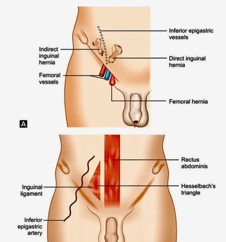
Busoga hernias, also known as Gill-Ogilvie hernias

🔴They are a variant of the direct inguinal hernia involving the conjoint tendon.

🔴Busoga herniae have been noted to occur most commonly in young, athletic men with a well-developed abdominal musculature. There is an increased incidence of Busoga hernia in Uganda.

🔴The conjoint tendon medially reinforces the posterior wall of the inguinal canal. It is protrusion here, at the medial aspect of the posterior aspect of the inguinal canal were weakening can result in herniation. Weakening in the conjoint tendon is thought to be secondary to rigorous training, with kicking, running, and sharp turns

#Arrangoiz #Miami #Surgeon #CASO #CenterforAdvancedSurgicalOncology

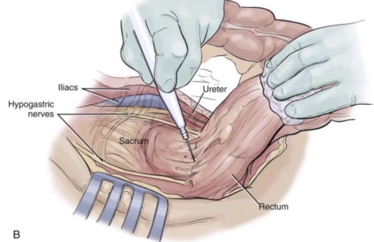
📙📜Holy plane of Heald📜📙

🔴Dissection is along the avascular alveolar plane between the presacral and mesorectal fascia, described as holy plane (Heald’s “holy plane”).

🔴The mesorectum refers to a fatty connective tissue layer, measuring 2–3 cm in thickness, with associated vessels, lymphatics and lymph nodes, which surrounds the rectum and is enveloped by fascia.

🔴mesorectal dissection occurs in a total avascular plane that, once identified, can be easily dissected, exposing ‘the white side of the yellow’ as Heald first described in 1979. As the posterior dissection continues downward, the mesorectum looks more and more bilobate (indicating a good quality of the dissection) until it thins out and disappears.
Once again traction and counter traction are essential in lateral and anterior resection to identify the dissection plane.

🔴The relationship of the rectum and pelvic autonomic nerves during open surgery when standing on the patient’s left. The ligation of the inferior mesenteric artery should be performed 1.5–2 cm from its origin from the aorta to avoid damaging the superior hypogastric plexus. At the pelvis, for posterior and lateral tumours, dissection should be directed below the Denonvillliers fascia to avoid damaging the neurovascular bundles that run along the tip of the seminal vesicle (2 and 10 o’clock directions).

#Arrangoiz #Surgeon #Teacher #CancerSurgeon #CASO #Miami #CenterforAdvancedSurgicalOncology
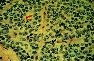
Surgical specimen showing typical histologic appearance of medullary cancer.

Surgical specimens from patients with MTC show spindle-shaped and frequently pleomorphic cells without follicle development because these cells originate from the calcitonin-producing parafollicular C cells of the thyroid 

Medullary Thyroid Carcinoma Pathology

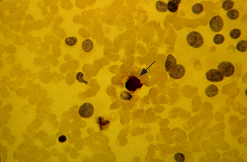
Fine-needle aspirate with immunostaining for calcitonin in medullary cancer of the thyroid. The nuclei of the tumor cells are placed eccentrically and are larger and more pleomorphic than those of normal follicular cells. Immunocytologic staining for calcitonin is positive (brown staining, which is best seen at the arrow). The background contains many red cells that nonspecifically take up the stain.

Hereditary Medullary Thyroid Carcinoma (MTC)

MTC

  • Hereditary MTC:
    • Is unique in many ways among inherited cancer syndromes:
      • MTC is founded on the following findings:
        • The hereditary variant:
          • Affecting as many as 25% of MTC patients:
            • Is more frequent than many other common hereditary tumors.
        • Unlike many other hereditary tumors:
          • Hereditary MTC features a strong genotype-phenotype correlation:
            • That is utilized worldwide for risk assessment
            • This genotype-dependent, age-related tumor progression:
              • Not only underlies the development of:
                • Hereditary MTC
                • But also the formation of:
                  • MEN 2–associated pheochromocytoma and hyperparathyroidism (HPT).
      • Disease progression from C-cell hyperplasia to MTC:
        • Requiring the acquisition of somatic mutations for malignant progression:
          • Is a stochastic sequence of events not fully under the control of a gene carrier’s genetic makeup.
      • Serum calcitonin:
        • A sensitive diagnostic marker of MTC, better reflects a gene carrier’s stage of C-cell disease than his or her underlying germline mutation in the RET (REarranged during Transfection) proto-oncogene:
          • We may now tailor the timing and extent of surgical intervention in the neck to the gene carrier’s stage of disease through consideration of serum calcitonin levels.
      • Lymph node metastases are indicative of progressive disease:
        • Portending a worse prognosis for sporadic and hereditary MTC alike.
      • Although multifocal tumor growth:
        • More common in hereditary than sporadic MTC:
          • 65% versus 8%:
            • No difference in biochemical cure and survival between hereditary and sporadic disease has been found after adjusting for extent of disease.
      • MEN 2B is a special and virulent variant of MEN type 2:
        • Characterized by the presence of MTC in early infancy.
        • More than 90% of MEN 2B RET gene carriers:
          • Harbor de novo germline mutations.
          • Such de novo germline mutations are rare in other MTC syndromic settings such as familial MTC (FMTC) and MEN 2A.
          • DNA-based screening of families for the MEN 2B trait for early detection of hereditary MTC hence is rarely an option.
          • Intriguingly, there are some early clues in MEN 2B infants:
            • Notably tearless crying and pseudo-Hirschsprung’s disease:
              • These clinical signs may help identify gene carriers before they develop the more characteristic MEN 2B stigmata:
                • Prompting rapid DNA-based screening and immediate surgical intervention.

cropped-18403652_10206829497335208_5004404657991480104_n1.jpg

  • What is Head and Neck Surgery?:
    • It is a surgical sub-specialty that deals mainly with benign and malignant tumors of the head and neck region, including:
      • The scalp, facial region, eyes, ears, nose, nasal fossae, paranasal sinuses, oral cavity, pharynx (nasopharynx, oropharynx, hypopharynx), larynx (supraglotic larynx, glottis larynx, subglotic larynx), thyroid gland, parathyroid gland, salivary glands (parotid glands, submandibular glands, sublingual glands, minor salivary glands), soft tissues of the neck, skin of the head and neck region.
        • The head and neck surgeon’s work area:
          • Does not cover tumors or diseases of the brain and other areas of the central nervous system or those of the cervical spine:
            • This is the neurosurgeon field.
    • Among the diagnostic procedures performed by the head and neck surgeon,  are the following:
      • Nasopharyngolaryngoscopy:
        • Performed to examine, evaluate and, possibly perform a biopsy, of oral cavity, pharyngeal and laryngeal lesions.
    • The surgeries most commonly performed by the head and neck surgeon are:
      • Total or near total thyroidectomies
      • Hemithryoidectomies (lobectomies)
      • Comprehensive neck dissections
      • Selective neck dissections
      • Maxillectomies:
        • Total maxillectomy
        • Subtotal maxillectomy
        • Infrastructure maxillectomy
        • Suprastructure maxillectomy
        • Medial maxillectomy
      • Mandibulectomy:
        • Segmental
        • Marginal
      • Tracheostomy
      • Salivary gland surgeries:
        • Parotid gland operations:
          • Limited superficial parotidectomy with identification and preservation of the facial nerve
          • Superficial parotidectomy with identification and preservation of the facial nerve
          • Near total parotidectomy with identification and preservation of the facial nerve
          • Total parotidectomy
        • Submandibular gland resection
        • Sublingual gland resection
      • Resection of tumors of the oral cavity:
        • Glossectomy
        • Resection of the floor of the mouth tumors
      • Resection of tumors of the pharynx
      • Resection of tumors of the larynx
      • Split-thickness skin grafts
      • Full-thickness skin grafts
      • Sentinel lymph node mapping and sentinel lymph node biopsy
      • Resection of malignant skin tumors (BCC, SCC, melanoma) of the head and neck region
  • The formation of the head and neck surgeon includes mastering the following subjects:
    • Surgical Anatomy
    • History and Basic Principles of Head and Neck Surgery
    • Epidemiology, Etiology, and Pathology of Head and Neck Diseases
    • Diagnostic Radiology of the Head and Neck Region
    • Tumors of the Scalp, Skin and Melanoma
    • Eyelids and Orbit
    • Nasal Cavity and Paranasal Sinuses
    • Skull Base and Temporal Bone
    • Lips and Oral Cavity
    • Pharynx and Esophagus
    • Larynx and Trachea
    • Cervical Lymph Nodes
    • Thyroid and Parathyroid Glands
    • Salivary Glands
    • Neurogenic Tumors and Paragangliomas
    • Soft Tissue Tumors
    • Bone Tumors and Odontogenic Lesions
    • Reconstructive Surgery
    • Oncologic Dentistry and Maxillofacial Prosthetics
    • Principles of Radiation Oncology
    • Principles of Chemotherapy
    • Molecular Oncology, Genomics and Immunology
    • Nutrition
    • Biostatistic


  • Rodrigo Arrangoiz MS, MD, FACS a head and neck surgeon / endocrine surgeon / surgical oncologist and is a member of Sociedad Quirúrgica S.C at the America British Cowdray Medical Center in Mexico City:


prof_739_20190417135234

  • Rodrigo Arrangoiz MS, MD, FACS:
    • Is a member of the American Head and Neck Society

img_4750

    • He is a member of the American Thyroid Association:

Unknown

Training:

• General surgery:

• Michigan State University:

• 2004 al 2010

• Surgical Oncology / Head and Neck Surgery / Endocrine Surgery:

• Fox Chase Cancer Center (Filadelfia):

• 2010 al 2012

• Masters in Science (Clinical research for health professionals):

• Drexel University (Filadelfia):

• 2010 al 2012

• Surgical Oncology / Head and Neck Surgery / Endocrine Surgery:

• IFHNOS / Memorial Sloan Kettering Cancer Center:

• 2014 al 2016

#Arrangoiz

#Teacher

#Surgeon

#Cirujano

#ThyroidExpert

#ThyroidSurgeon

#CirujanodeTiroides

#ExpertoenTiroides

#ExpertoenParatiroides

#Paratiroides

#Hiperparatiroidismo

#CancerdeTiroides

#ThyroidCancer

#PapillaryThyroidCancer

#SurgicalOncologist

#CirujanoOncologo

#CancerSurgeon

#CirujanodeCancer

#HeadandNeckSurgeon

#CirugiaEndocrina

#CirujanodeTumoresdeCabezayCuello

#OralCavityCancer

#Melanoma

    Medullary Thyroid Carcinoma Part 3

    • Staging:
      • TNM staging:
        • The pathological tumor, node, metastasis (pTNM) criteria for clinical / pathologic tumor staging (eighth edition) adopted by the Union for International Cancer Control (UICC) and the American Joint Committee on Cancer (AJCC) are based upon:
          • Tumor size
          • The presence or absence of extra-thyroidal invasion
          • Local and regional nodal metastases
          • Distant metastases 
      • Stage I :
        • Medullary thyroid cancers (MTCs) that are equal or less than 2 cm in diameter without evidence of disease outside of the thyroid gland
      • Stage II:
        • Tumors  greater than 2 cm confined to the thyroid or tumors of any size without lymph node metastasis that demonstrate gross extrathyroidal extension invading only the strap muscles (sternohyoid, sternothyroid, thyrohyoid, or omohyoid muscles)
      • Stage III:
        • Tumors of any size demonstrating metastatic lymph node involvement in the central neck (levels VI or VII; pretracheal, paratracheal, or prelaryngeal/Delphian, or upper mediastinal lymph nodes) with or without gross invasion into the strap muscles (sternohyoid, sternothyroid, thyrohyoid, or omohyoid muscles)
      • Stage IV :
        • Any distant metastases, or lymph node involvement outside of the central neck (level VI/VII), or gross invasion into other structures of the neck (beyond just strap muscle involvement)
    • One study evaluated the prognostic significance of a previous TNM staging scheme in patients with MTC:
      • Most of whom were treated by total thyroidectomy and then followed for a median of four years:
        • Although the follow-up was short:
          • Mortality due to MTC was:
            • 0% stage I 
            • 13% in stage II
            • 56% in stage III
            • 100% in stage IV
    • A subsequent analysis of MTC patients using the National Cancer Database and the SEER (Surveillance, Epidemiology, and End Results) data set demonstrated that the seventh and eighth editions of the AJCC staging system:
      • Were associated with five-year overall survival rates of:
        • 95% in stage I
        • 91% in stage II
        • 89% in stage III
        • 68% in stage IV
      • Furthermore:
        • Disease-specific survival rates were:
          • 100% in stage I
          • 99%  in stage II
          • 97%  in stage III
          • 82% in stage IV
    • Dynamic risk stratification:
      • Using the same concepts that were initially developed for differentiated thyroid cancer:
        • Dynamic risk stratification for MTC allows clinicians to modify initial AJCC staging risk estimates over time based on:

          • The biological behavior the tumor and the response to therapy in individual patients
        • For application in MTC, the definitions of the response to therapy categories needed to be modified to utilize calcitonin and carcinoembryonic antigen (CEA) as tumor markers (rather than thyroglobulin):
          • At each follow-up visit, patients are classified as having one of the following clinical outcomes:
            • Excellent response:
              • An undetectable calcitonin and normal-range CEA in the absence of structurally identifiable disease
            • Biochemical incomplete response:
              • A detectable calcitonin or elevated CEA in the absence of structurally identifiable disease
            • Structural incomplete response:
              • The presence of recurrent or persistent structurally identifiable disease
      • In two retrospective studies examining MTC patients with a median of 5 to 7 years of follow-up:
        • An excellent response to therapy was associated with:
          • Structural disease recurrence rate of 1% to 4%
          • Biochemical recurrence rate of 11% to 15%
          • Disease-specific mortality of less than 3%
        • Patients with a biochemical incomplete response demonstrated a:
          • Structural disease recurrence rate of 32% to 37%
          • Biochemical recurrence rate of 51% to 53%
          • Disease-specific mortality of 11%
        • The poorest outcomes were seen in those patients with a structural incomplete response to initial therapy with:
          • Disease-specific mortality rates of 38% to 56%
    • The calcitonin and CEA doubling times:
      • Can also provide meaningful insights into:
        • Prognosis
        • Expected course of disease progression that can further refine these response to therapy assessments


    cropped-18403652_10206829497335208_5004404657991480104_n1.jpg

    • What is Head and Neck Surgery?:
      • It is a surgical sub-specialty that deals mainly with benign and malignant tumors of the head and neck region, including:
        • The scalp, facial region, eyes, ears, nose, nasal fossae, paranasal sinuses, oral cavity, pharynx (nasopharynx, oropharynx, hypopharynx), larynx (supraglotic larynx, glottis larynx, subglotic larynx), thyroid gland, parathyroid gland, salivary glands (parotid glands, submandibular glands, sublingual glands, minor salivary glands), soft tissues of the neck, skin of the head and neck region.
          • The head and neck surgeon’s work area:
            • Does not cover tumors or diseases of the brain and other areas of the central nervous system or those of the cervical spine:
              • This is the neurosurgeon field.
      • Among the diagnostic procedures performed by the head and neck surgeon,  are the following:
        • Nasopharyngolaryngoscopy:
          • Performed to examine, evaluate and, possibly perform a biopsy, of oral cavity, pharyngeal and laryngeal lesions.
      • The surgeries most commonly performed by the head and neck surgeon are:
        • Total or near total thyroidectomies
        • Hemithryoidectomies (lobectomies)
        • Comprehensive neck dissections
        • Selective neck dissections
        • Maxillectomies:
          • Total maxillectomy
          • Subtotal maxillectomy
          • Infrastructure maxillectomy
          • Suprastructure maxillectomy
          • Medial maxillectomy
        • Mandibulectomy:
          • Segmental
          • Marginal
        • Tracheostomy
        • Salivary gland surgeries:
          • Parotid gland operations:
            • Limited superficial parotidectomy with identification and preservation of the facial nerve
            • Superficial parotidectomy with identification and preservation of the facial nerve
            • Near total parotidectomy with identification and preservation of the facial nerve
            • Total parotidectomy
          • Submandibular gland resection
          • Sublingual gland resection
        • Resection of tumors of the oral cavity:
          • Glossectomy
          • Resection of the floor of the mouth tumors
        • Resection of tumors of the pharynx
        • Resection of tumors of the larynx
        • Split-thickness skin grafts
        • Full-thickness skin grafts
        • Sentinel lymph node mapping and sentinel lymph node biopsy
        • Resection of malignant skin tumors (BCC, SCC, melanoma) of the head and neck region
    • The formation of the head and neck surgeon includes mastering the following subjects:
      • Surgical Anatomy
      • History and Basic Principles of Head and Neck Surgery
      • Epidemiology, Etiology, and Pathology of Head and Neck Diseases
      • Diagnostic Radiology of the Head and Neck Region
      • Tumors of the Scalp, Skin and Melanoma
      • Eyelids and Orbit
      • Nasal Cavity and Paranasal Sinuses
      • Skull Base and Temporal Bone
      • Lips and Oral Cavity
      • Pharynx and Esophagus
      • Larynx and Trachea
      • Cervical Lymph Nodes
      • Thyroid and Parathyroid Glands
      • Salivary Glands
      • Neurogenic Tumors and Paragangliomas
      • Soft Tissue Tumors
      • Bone Tumors and Odontogenic Lesions
      • Reconstructive Surgery
      • Oncologic Dentistry and Maxillofacial Prosthetics
      • Principles of Radiation Oncology
      • Principles of Chemotherapy
      • Molecular Oncology, Genomics and Immunology
      • Nutrition
      • Biostatistic


    • Rodrigo Arrangoiz MS, MD, FACS a head and neck surgeon / endocrine surgeon / surgical oncologist and is a member of Sociedad Quirúrgica S.C at the America British Cowdray Medical Center in Mexico City:


    prof_739_20190417135234

    • Rodrigo Arrangoiz MS, MD, FACS:
      • Is a member of the American Head and Neck Society

    img_4750

      • He is a member of the American Thyroid Association:

    Unknown

    Training:

    • General surgery:

    • Michigan State University:

    • 2004 al 2010

    • Surgical Oncology / Head and Neck Surgery / Endocrine Surgery:

    • Fox Chase Cancer Center (Filadelfia):

    • 2010 al 2012

    • Masters in Science (Clinical research for health professionals):

    • Drexel University (Filadelfia):

    • 2010 al 2012

    • Surgical Oncology / Head and Neck Surgery / Endocrine Surgery:

    • IFHNOS / Memorial Sloan Kettering Cancer Center:

    • 2014 al 2016

    #Arrangoiz

    #Teacher

    #Surgeon

    #Cirujano

    #ThyroidExpert

    #ThyroidSurgeon

    #CirujanodeTiroides

    #ExpertoenTiroides

    #ExpertoenParatiroides

    #Paratiroides

    #Hiperparatiroidismo

    #CancerdeTiroides

    #ThyroidCancer

    #PapillaryThyroidCancer

    #SurgicalOncologist

    #CirujanoOncologo

    #CancerSurgeon

    #CirujanodeCancer

    #HeadandNeckSurgeon

    #CirugiaEndocrina

    #CirujanodeTumoresdeCabezayCuello

    #OralCavityCancer

    #Melanoma

    Internal Mammary Lymph Node and SNLB After Neoadjuvant Chemotherapy

    • As reported by Veronesi in 1999,:
      • 737 patients were randomized to either undergo:
        • Halsted mastectomy or extended mastectomy with IM node dissection:
          • After 30 years of follow-up, there was no difference in overall survival or disease-specific survival:
            • For the patients eligible with T1, T2, T3, N0, and N1 disease who underwent IM node dissection vs. no IM dissection
    • 2019 retrospective review of 95 breast cancer patients with clinically detected IM nodes (IMNs) at diagnosis:
      • Were treated with surgery and radiation:
        • With median follow-up of 43 months
      • 77 received neoadjuvant chemotherapy:
        • With IMN normalization in 67.5%, and partial IMN response in 24.6%
      • The 5-year IMN failure-free survival, disease-free survival, and overall survival were:
        • 96%, 70%, and 84%, respectively
      • IMN failure-free survival was significantly affected by:
        • Resection margin status
        • Size of IMN
        • Receipt of IMN boost radiation
    • A recently published meta-analysis in the Annals of Surgery found that axillary staging following neoadjuvant chemotherapy:
      • Is best performed with a combination approach of:
        • Sentinel lymph node biopsy with excision of the pre-chemotherapy-marked positive node:
          • With a false negative rate of 2% to 4%:
            • The identification rate was 100%
        • ACOSOG Z1071 reported an overall false negative rate of 12.6%:
          • When sentinel node biopsy was performed after neoadjuvant chemotherapy with documented node-positive disease prior to treatment
          • The false-negative rate decreased to 6.8%:
            • When both sentinel node(s) and the clipped node were retrieved at the time of surgery
    • References
      • Veronesi U, Marubini E, Mariani L, Valagussa P, Zucali R. The dissection of internal mammary nodes does not improve the survival of breast cancer patients. 30-year results of a randomised trial. Eur J Cancer. 1999;35(9):1320-1325.
      • Kim J, Chang JS, Choi SH, et.al. Radiotherapy for initial clinically positive internal mammary nodes in breast cancer. Radiat Oncol J. 2019;37(2):91-100.
      • Simons JM, van Nijnatten TJA, van der Pol CC, Luiten EJT, Koppert LB, Smidt ML. Diagnostic accuracy of different surgical procedures for axillary staging after neoadjuvant systemic therapy in node-positive breast cancer: a systematic review and meta-analysis. Ann Surg. 2019;269(3):432-442.
      • Boughey JC, Ballman KV, Le-Petross HT, et al. Identification and resection of clipped node decreases the false-negative rate of sentinel lymph node surgery in patients presenting with nodepositive breast cancer (T0-T4, N1-N2) who receive neoadjuvant chemotherapy: results from ACOSOG Z1071 (Alliance). Ann Surg. 2016;263(5):802-807.

    #Arrangoiz #BreastSurgeon #CancerSurgeon #SurgicalOncology #BreastCancer #CASO #Miami #CenterforAdvancedSurgicalOncology
    Tubular Carcinoma of the Breast

    • Tubular carcinoma:
      • Is a distinct histopathologic subtype of breast cancer:
        • Representing 1% to 2% of breast cancers diagnosed
      • It is a distinct entity:
        • From low-grade ductal carcinoma
      • The literature continues to suggest that is has an excellent prognosis:
        • With a very low likelihood of distant metastasis and excellent disease-free survival.
      • In select patients:
        • Adjuvant therapies may be omitted:
          • However, there is a still a risk of axillary nodal metastasis:
            • Sentinel lymph node biopsy is still recommended due to a 10% to 20% risk of lymphatic spread
            • Despite this finding, there is likely to be only one node involved:
              • This histopathologic subtype still conveys an excellent prognosis
      • Tubular carcinoma is more likely to be identified on:
        • Screening mammography and is more common in Caucasians than blacks
      • Emerging data suggest that adjuvant systemic therapy can likely be safely omitted, although it should still be discussed with the multidisciplinary team
    • References
      • Rakha EA, Lee AH, Evans AJ, et al: Tubular carcinoma of the breast: further evidence to support its excellent prognosis. J Clin Oncol. 2010;28(1):99-104.
      • Fedko MG, Scow JS, Shah SS, et al. Pure tubular carcinoma and axillary nodal metastases. Ann Surg Oncol. 2010;17(Suppl 3):338-342.
      • Anderson WF, Chu KC, Chang S, Sherman ME. Comparison of age-specific incidence rate patterns for different histopathologic types of breast carcinoma. Cancer Epidemiol Biomarkers Prev. 2004;13(7): 1128-1135.

    #Arrangoiz #CancerSurgeon #BreastSurgeon #SurgicalOncologist #BreastCancer #TubularBreastCancer #CASO #Miami #CenterforAdvancedSurgicalOncology

    Oncotype DX 21 Gene Recurrence Score is Better in Predicting Cancer Recurrence Than Traditional Clinical Parameters

    • ER positive tumors:
      • Can be very sensitive to endocrine therapy:
        • Which may allow some patients to safely avoid chemotherapy
    • However:
      • The presence of ER receptors on immunohistochemistry:
        • Does not necessarily mean that the tumor’s growth:
          • Is being driven by ER-related pathways
      • Additionally, other molecular features may influence the tumor cells’ sensitivity to hormonal therapy
    • The development of predictive molecular assays has been a major advancement in the field:
      • The assay measures mRNA expression of 21 genes:
        • Using reverse transcriptase-polymerase chain reaction techniques
      • It can be performed on formalin-fixed paraffin-embedded tumor specimens:
        • Obtained by core biopsy or surgery
      • It has been validated in ER+, node-negative women:
        • Who have not received any prior therapy
      • This assay is more reliable in predicting cancer recurrence than such clinical parameters as:
        • Size
        • Hormone receptor status
        • Nuclear grade
        • Ki-67
      • The assay measures:
        • Downstream ER-regulated genes:
          • To assess the functionality of the ER receptor
      • Patients with low scores (< 18) are considered at low risk for disease recurrence:
        • May not receive any benefit from adjuvant chemotherapy:
          • These patients are now treated with hormonal therapy alone without cytotoxic chemotherapy
        • In fact, the recently published subset analysis of the prospective validation of the 21-gene expression assay in breast cancer:
          • Confirmed that 98.7% of women:
            • With 21-gene signature scores of less than 10:
              • Managed with endocrine therapy alone had no evidence of local, regional, or distant recurrence at 5 years
            • Patients with a high score (> 31):
            • Have been shown to gain a large benefit from the addition of chemotherapy
          • While the assay is not performed on HER2-overexpressing tumors:
            • It does measure HER2 and other proliferative genes
      • It is currently only validated for:
        • Node-negative patients:
          • Although the RxPONDER (SWOG 1007) trial:
            • Is currently evaluating women with:
              • 1 to 3 positive lymph nodes and an 21-gene signature score of less than 25
            • These patients were randomized to receive chemotherapy and endocrine therapy to endocrine therapy alone:
              • We are awaiting the results of this trial
    • References
    • Paik S. Development and clinical utility of a 21-gene recurrence score prognostic assay in patients with early breast cancer treated with tamoxifen. Oncologist. 2007;12(6):631-635.
    • Paik S, Shak S, Tang G, et al. A multigene assay to predict recurrence of tamoxifen-treated, node-negative breast cancer. N Engl J Med. 2004;351(27):2817-2826.
    • Paik S, Tang G, Shak S, et al. Gene expression and benefit of chemotherapy in women with node-negative, estrogen receptor-positive breast cancer. J Clin Oncol. 2006;24(23):3726-3734.
    • Sparano JA, Gray RJ, Makower DF, et al. Prospective validation of a 21-gene expression assay in breast cancer. New Engl J Med. 2015;373(21):2005-2014.

    #Arrangoiz #BreastSurgeon #CancerSurgeon #SurgicalOncologist #BreastCancer #OncotypeDx #CASO #Miami #CenterforAdvancedSurgicalOncology