Blog

Evidence-Based Approach to a “Missing” Parathyroid Gland during Thyroid or Parathyroid Surgery.

Arrangoiz, R., Cordera, F. and Zambrano, M. (2026) Evidence-Based Approach to a “Missing” Parathyroid Gland during Thyroid or Parathyroid Surgery. International Journal of Otolaryngology and Head & Neck Surgery , 15, 261-283.

https://doi.org/10.4236/ijohns.2026.154024

Abstract

Failure to identify a parathyroid gland during thyroidectomy or parathyroidec-

tomy represents a well-recognized intraoperative challenge and is most com-

monly explained by ectopic gland location rather than true agenesis. A com-

prehensive understanding of parathyroid embryology, migration patterns, and

cervical anatomy is therefore essential for safe endocrine neck surgery and for

minimizing failed explorations, hypoparathyroidism, and recurrent disease.

This review provides a practical and surgically oriented overview of the “miss-

ing” parathyroid gland, integrating embryologic development, anatomic vari-

ability, operative identification strategies, and contemporary nomenclature sys-

tems. The embryologic descent of the superior and inferior parathyroid glands

from the fourth and third pharyngeal pouches, respectively, explains the pre-

dictable distribution of ectopic glands encountered during surgery. Superior

glands typically maintain relatively constant locations near the posterior as-

pect of the upper thyroid pole, whereas inferior glands demonstrate greater po-

sitional variability because of their longer migratory course with the thymus.

Common ectopic locations include the tracheoesophageal groove, retroesopha-

geal space, thyrothymic ligament, cervical thymus, carotid sheath, retropha-

ryngeal region, mediastinum, and intrathyroidal or subcapsular locations. This

review emphasizes operative pearls for gland identification, including recog-

nition of key landmarks such as the inferior thyroid artery, recurrent laryngeal

nerve, prevertebral fascia, cervical thymus, and characteristic parathyroid fat

pads. Strategies for systematic exploration are discussed in the context of both

thyroidectomy and parathyroidectomy. Additionally, the manuscript reviews

the Perrier classification system for ectopic parathyroid glands and proposes

DOI: 10.4236/ijohns.2026.154024 Jul. 9, 2026 261 Int’l J. of Otolaryngology and Head & Neck SurgeryR. Arrangoiz et al.

its practical application during cervical exploration to facilitate structured lo-

calization of missing glands. By combining embryologic principles with oper-

ative anatomy and contemporary surgical nomenclature, this review aims to

provide endocrine surgeons, trainees, and head and neck surgeons with a clin-

ically relevant framework for identifying orthotopic and ectopic parathyroid

glands, reducing operative failure, and improving surgical outcomes in thy-

roid and parathyroid surgery.

Keywords

Parathyroid Gland, Ectopic Parathyroid Gland, Missing Parathyroid Gland,

Parathyroid Embryology, Thyroidectomy, Parathyroidectomy, Mediastinal

Parathyroid Gland, Intrathyroidal Parathyroid Gland, Perrier Classification,

Surgical Anatomy, Parathyroid Localization

Signs of Hyperthyroidism

#Arrangoiz #ThyroidSurgeon #CancerSurgeon #HeadandNeckSurgeon #SurgicalOncologist #Hyperthyroidism #EndocrineSurgery #MountSinaiMedicalCenter #MSMC #Miami #Mexico #Florida #Doctor #Surgeon #Thyroid #ThyroidDisease

Symptoms and Signs of Hyperthyroidism

#Arrangoiz #ThyroidSurgeon #CancerSurgeon #HeadandNeckSurgeon #SurgicalOncologist #Hyperthyroidism #EndocrineSurgery #MountSinaiMedicalCenter #MSMC #Miami #Mexico #Florida #Doctor #Surgeon #Thyroid #ThyroidDisease

Dermopathy of Graves Disease

  • Pretibial myxedema occurs in 0.5% to 4.3% of patients with Graves’ disease:
    • It is an infiltrative dermopathy:
      • Most often involving the skin of the legs
    • It is manifested by painful, pruritic, raised plaque-like violaceous and hyperpigmented lesions:
      • That have the texture of an orange peel
  • Acropachy occurs in less than 1% of patients with Graves’ disease:
    • It is manifested by clubbing and periosteal new bone formation of the metacarpal bones and the phalanges
Pre Tibial Myxedema
Clubbing of fingers as the most common form of thyroid acropachy.

#Arrangoiz #ThyroidSurgeon #CancerSurgeon #HeadandNeckSurgeon #SurgicalOncologist #Hyperthyroidism #EndocrineSurgery #MountSinaiMedicalCenter #MSMC #Miami #Mexico #Florida #Doctor #Surgeon #Thyroid #ThyroidDisease

Evaluation of Hyperthyroidism

  • The symptoms and signs of hyperthyroidism are also quite varied as with hypothyroidism:
    • But often more characteristic of the condition
  • Signs and Symptoms of Hyperthyroidism:
    • Common signs and symptoms of hyperthyroidism include:
      • Weight loss
      • Heat intolerance
      • Tremors
      • Palpitations
      • Anxiety
      • Menstrual abnormalities
      • Atrial fibrillation
    • Older patients with hyperthyroidism may have more cardiac symptoms:
      • But less systemic manifestations of hyperthyroidism
    • The most common symptoms are:
      • Palpitations, weakness, heat intolerance, and disturbed sleep
    • The most frequent physical findings are:
      • Tachycardia, tremor of the extremities, and weight loss
  • In clinical practice:
    • The symptoms and signs of hyperthyroidism are often not necessarily correlated with the biochemical severity of the thyroid dysfunction and can be variable and less prevalent in the elderly
  • General symptoms:
    • Weight loss
    • Heat intolerance
    • Anxiety / nervousness
    • Insomnia
    • Muscle weakness
  • Cardiovascular symptoms and signs:
    • Tachycardia
    • Palpitations
    • Dyspnea on exertion
    • Bounding pulses
    • Atrial fibrillation
  • Head and neck symptoms and signs:
    • Ophthalmopathy (in Graves’ disease only)
    • Goiter
  • Skin symptoms and signs:
    • Excess perspiration
    • Palmer erythema
  • Nervous System symptoms and signs:
    • Tremor
    • Anxiety / Nervousness
    • Hyperkinesis
  • Gastrointestinal symptoms and signs:
    • Frequent stools / Diarrhea
  • Reproductive symptoms and signs:
    • Irregular menstrual periods / amenorrhea
    • Light menstrual flow
    • Infertility
    • Gynecomastia (males)
Screenshot

Concurrent Chemoradiotherapy with Cisplatin for Head and Neck Cancers

  • Concurrent chemoradiotherapy with cisplatin:
    • Is the standard of care for patients with locally advanced head and neck squamous cell carcinoma (HNSCC):
      • With the most widely accepted regimen being cisplatin 100 mg/m² administered intravenously every 3 weeks for up to three cycles during radiotherapy
    • This approach is supported by multiple large randomized trials demonstrating survival benefit in patients with good performance status and is endorsed by major guidelines, including those from the American Society of Clinical Oncology and NCCN
  • The rationale for exploring low-dose weekly cisplatin (20 to 50 mg/m²):
    • Stems from the significant acute and chronic toxicities associated with the high-dose regimen, including:
      • Nephrotoxicity, ototoxicity, and myelosuppression:
        • Which can limit compliance and preclude delivery of the intended cumulative dose
    • Weekly regimens are hypothesized to:
    • Improve tolerability and allow more patients to achieve a cumulative cisplatin dose of at least 200 mg/m²:
      • Which is considered important for optimal tumor control
  • Efficacy, Survival and Disease Control:
    • Meta-analyses and large retrospective studies indicate that overall survival and response rates are similar between high-dose and low-dose cisplatin regimens in the definitive chemoradiation setting:
      • For example, a large population-based study in US veterans found no significant difference in overall survival between high-dose (100 mg/m² every 3 weeks) and low-dose (40 mg/m² weekly) cisplatin:
        • Though high-dose was associated with greater toxicity
      • Similarly, systematic reviews and meta-analyses have not demonstrated a meaningful survival difference between the two dosing strategies in either definitive or postoperative settings
      • However, some studies and clinical guidelines suggest that high-dose cisplatin may offer superior locoregional control and overall survival:
        • Particularly in altered fractionation or postoperative settings
      • For instance, randomized trials and meta-analyses have reported improved locoregional control and, in some analyses, overall survival with high-dose regimens, especially when combined with altered fractionation radiotherapy
  • The American Society of Clinical Oncology recommends the every-3-week high-dose regimen as the standard:
    • Noting that weekly regimens lack level I evidence and may be associated with inferior outcomes in some studies
  • Achieving a cumulative cisplatin dose of ≥ 200 mg/m² is consistently associated with better survival and locoregional control:
    • Regardless of the dosing schedule
  • Toxicity and Compliance:
    • High-dose cisplatin regimens are associated with increased rates of severe hematologic, renal, and ototoxic toxicities compared to weekly low-dose regimens
    • In contrast, weekly low-dose cisplatin is generally better tolerated and associated with improved compliance:
      • With more patients able to complete the planned cumulative dose
    • Notably, patients with low skeletal muscle mass are at higher risk for dose-limiting toxicity with high-dose cisplatin and may particularly benefit from weekly regimens to improve compliance and reduce toxicity
    • However, some studies have reported increased rates of grade 3 to 4 dysphagia and weight loss with weekly regimens in the postoperative setting
  • Guideline Recommendations and Ongoing Controversies:
    • Current US and international guidelines, including those from the American Society of Clinical Oncology:
      • Continue to endorse high-dose cisplatin (100 mg/m² every 3 weeks) as the standard regimen for concurrent chemoradiation in eligible patients
    • Weekly low-dose cisplatin is widely used in clinical practice, particularly for patients with comorbidities or poor performance status:
      • But is considered investigational pending results from ongoing prospective trials
    • The optimal dosing schedule remains an area of active research, and further adequately powered randomized trials are needed to clarify the relative efficacy and toxicity of these regimens, especially in specific subgroups such as HPV-positive, elderly, or comorbid patients
  • In summary, high-dose cisplatin every 3 weeks remains the standard of care for concurrent chemoradiation in locally advanced HNSCC, with weekly low-dose regimens offering a reasonable alternative for selected patients, particularly those at higher risk for toxicity or with difficulty tolerating high-dose therapy
  • References:
    • Head and Neck Cancer. Chow LQM. The New England Journal of Medicine. 2020;382(1):60-72. doi:10.1056/NEJMra1715715.
    • Management of the Neck in Squamous Cell Carcinoma of the Oral Cavity and Oropharynx: ASCO Clinical Practice Guideline. Koyfman SA, Ismaila N, Crook D, et al. Journal of Clinical Oncology : Official Journal of the American Society of Clinical Oncology. 2019;37(20):1753-1774. doi:10.1200/JCO.18.01921.
    • Low-Dose vs. High-Dose Cisplatin: Lessons Learned From 59 Chemoradiotherapy Trials in Head and Neck Cancer. Szturz P, Wouters K, Kiyota N, et al. Frontiers in Oncology. 2019;9:86. doi:10.3389/fonc.2019.00086.
    • Concurrent Chemoradiotherapy With Cisplatin Given Once-a-Week Versus Every-Three Weekly in Head and Neck Squamous Cell Carcinoma: Non-Inferior, Equivalent, or Superior?. Gupta T, Kannan S, Ghosh-Laskar S, Agarwal JP. Oral Oncology. 2022;134:106130. doi:10.1016/j.oraloncology.2022.106130.
    • Cisplatin Every 3 Weeks Versus Weekly With Definitive Concurrent Radiotherapy for Squamous Cell Carcinoma of the Head and Neck. Bauml JM, Vinnakota R, Anna Park YH, et al. Journal of the National Cancer Institute. 2019;111(5):490-497. doi:10.1093/jnci/djy133.
    • Weekly Low-Dose Versus Three-Weekly High-Dose Cisplatin for Concurrent Chemoradiation in Locoregionally Advanced Non-Nasopharyngeal Head and Neck Cancer: A Systematic Review and Meta-Analysis of Aggregate Data. Szturz P, Wouters K, Kiyota N, et al. The Oncologist. 2017;22(9):1056-1066. doi:10.1634/theoncologist.2017-0015.
    • Low Dose Cisplatin Weekly Versus High Dose Cisplatin Every Three Weeks in Primary Chemoradiotherapy in Head and Neck Cancer Patients With Low Skeletal Muscle Mass: The CISLOW-study Protocol. Schaeffers AWMA, Devriese LA, van Gils CH, et al. PloS One. 2023;18(11):e0294147. doi:10.1371/journal.pone.0294147.
    • Comparison of Standard-Dose 3-Weekly Cisplatin and Low-Dose Weekly Cisplatin for Concurrent Chemoradiation of Patients With Locally Advanced Head and Neck Squamous Cell Cancer: A Multicenter Retrospective Analysis. Lee SY, Choi YS, Song IC, et al. Medicine. 2018;97(21):e10778. doi:10.1097/MD.0000000000010778.
    • Altered Fractionation Radiotherapy Combined With Concurrent Low-Dose or High-Dose Cisplatin in Head and Neck Cancer: A Systematic Review of Literature and Meta-Analysis. Szturz P, Wouters K, Kiyota N, et al. Oral Oncology. 2018;76:52-60. doi:10.1016/j.oraloncology.2017.11.025.
    • Comparison of Weekly Administration of Cisplatin Versus Three Courses of Cisplatin 100 mg/M(2) for Definitive Radiochemotherapy of Locally Advanced Head-and-Neck Cancers. Rades D, Seidl D, Janssen S, et al. BMC Cancer. 2016;16:437. doi:10.1186/s12885-016-2478-8.
#Arrangoiz #Doctor #Surgeon #CancerSurgeon #SurgicalOncologist #HeadandNeckSurgeon #MountSinaiMedicalCenter #MSMC #BramanComprehensiveCancerCenter #BCCC #Miami #Mexico

Cancer of the Retromolar Trigone (RMT)

– The retromolar trigone (RMT) is a small triangular shaped subsite of the oral cavity:

  • It is the portion of mucosa that lies behind the third inferior molar tooth:

    • Covering the anterior ramus of the mandible

  • The base of the triangle:

    • Is posterior to the last inferior molar tooth (3rd)

  • The apex is in continuity with the tuberosity of the maxilla:

    • Behind the last upper molar tooth

  • It is bounded laterally by the gingival buccal sulcus and medially by the anterior tonsillar pillar

early_stage_oral_cavity_cancer-fig_1-enAnnMaxillofacSurg_2016_6_2_304_200349_f1

– Cancerous lesions involving the RMT are almost always squamous cell carcinomas (SCC):

  • Minor salivary gland tumors:

    • Are sometimes diagnosed in this subsite

  • Because of its continuity with the oral cavity mucosa and its close relation to the mandible:

    • Spreading to adjacent structures can easily occur

    • Because squamous cell carcinoma of the retromolar trigone results in bone infiltration:

      • Lymph node metastasis and infiltration to the infratemporal fossa in the early stages is common:

        • Its prognosis is poorer than that of squamous cell carcinoma in neighboring areas:

          • Such as the tonsils, oral floor and gums

#Arrangoiz #Doctor #Surgeon #CancerSurgeon #SurgicalOncologist #HedandNeckSurgeon #MountSinaiMedicalCenter #MSMC #BramanComprehensiveCancerCenter #BCCC #Miami #Mexico

Rodrigo Arrangoiz MS, MD, FACS a head and neck surgeon and is a member of Sociedad Quirúrgica S.C at the America British Cowdray Medical Center.

He is first author on some publications on oral cavity cancer:

Oral Tongue Cancer: Literature Review and Current Management

https://www.oatext.com/pdf/CRR-2-153.pdf

Understand Cancer: Research and Treatment Oral Cavity Cancer: Literature Review and Current Management.

https://www.researchgate.net/publication/303366031_Understand_Cancer_Research_and_Treatment_Oral_Cavity_Cancer_Literature_Review_and_Current_Management

Training:

• General surgery:

• Michigan State University:

• 2004 al 2010

• Surgical Oncology / Head and Neck Surgery / Endocrine Surgery:

• Fox Chase Cancer Center (Filadelfia):

• 2010 al 2012

• Masters in Science (Clinical research for health professionals):

• Drexel University (Filadelfia):

• 2010 al 2012

• Surgical Oncology / Head and Neck Surgery / Endocrine Surgery:

• IFHNOS / Memorial Sloan Kettering Cancer Center:

• 2014 al 2016


Buccal Squamous Cell Carcinoma (SCC)

A. Introduction:

Carcinoma of the buccal mucosa is relatively uncommon in North America, compared with other oral cavity cancers such as carcinomas of the oral tongue or floor of the mouth:

  • Squamous cell carcinoma (SCC) is the most common pathology (greater than 90% of all oral cavity cancers) and more prevalent in those who use tobacco and alcohol

As the orifice of the upper aerodigestive tract, the oral cavity plays a critical role in breathing, speech, and swallowing:

  • The buccal region is particularly important in bolus formation, preventing food from spilling into the lateral oral gutters or extra-orally during the oral preparatory phase of swallowing:

    • Cancer of the buccal mucosa and subsequent treatment of the disease may interfere with these functions

Buccal carcinoma has the propensity to become aggressive, with high rates of local and regional recurrence

  • Diagnosis and treatment at an early stage leads to significantly improved prognosis and function over advanced disease

08_buccal

B. Epidemiology:

  • SCC of the buccal mucosa accounts for approximately 5% to 10% of all cancers of the oral cavity in North America and Western Europe

  • It occurs more often in men:

    • With a male to female ratio of 3:1 to 4:1

  • It is diagnosed most commonly in the 7th or 8th decade of life (in the USA)

  • The incidence of buccal carcinoma is much higher in Asia:

    • In Southeast Asia, the disease is the most common form of oral cavity cancer (in the USA it is tongue cancer)

    • In India, buccal carcinoma is the most common cancer in men and the third most common cancer in women

      • The higher rate of buccal carcinoma in Asia is likely related to the widespread practice of betel nut chewing:

        • Betel nut, composed mainly of the fruit of the Areca Palm and often mixed with tobacco, is placed along the buccal mucosa to induce a feeling of euphoria:

          • Buccal carcinoma related to betel nut chewing tends to develop at an earlier age, with most cases occurring between the ages of 40 to 70

C. Etiology:

  • Tobacco and alcohol use are the main etiologic agents associated with the development of buccal carcinoma:

    • In North America:

      • A history of using tobacco is documented in 70% of patients

  • Although alcohol by itself is not thought to be a significant risk factor:

    • Tobacco and alcohol have a well-recognized synergistic effect in the development of carcinoma

  • In Asia, betel nut is a significant etiologic agent:

    • In addition to tobacco and alcohol

  • In India:

    • Over 90% of patients with buccal carcinoma have a history of using betel nut

  • Other suspected but not confirmed etiologic agents include:

    • Poor oral hygiene, and chronic irritation

  • Premalignant conditions include:

    • Submucous fibrosis and lichen planus:

      • The latter has a reported transformation rate of 0.5% to 3%, whereas the former has a malignant transformation rate of 0.5%

images

D. Presentation:

  • Buccal SCC commonly presents as a slow-growing mass on the buccal mucosa

Philadelphia Illustration Dept./Elsevier
Synchronous SCC of the buccal mucosa
Philadelphia Illustration Dept./Elsevier
Papillary SCC of the buccal mucosa
  • Small lesions tend to be asymptomatic and are often noted incidentally on dental examination

  • Pain commonly occurs as the lesion enlarges and ulceration develops

  • Oral intake may worsen the pain and lead to malnutrition and dehydration

  • Associated symptoms include:

    • Bleeding, poor denture fit, facial weakness or sensory changes, dysphagia, odynophagia, and trismus

Rodrigo Arrangoiz MS, MD, FACS, FSSO the Head and Neck Department at the Braman Comprehensive Cancer Center at Mount Sinai Medical Center in Miami, Florida

He is first author on some publications on oral cavity cancer:

Training:

• General surgery:

• Michigan State University:

• 2004 al 2010

• Surgical Oncology / Head and Neck Surgery / Endocrine Surgery:

• Fox Chase Cancer Center (Filadelfia):

• 2010 al 2012

• Masters in Science (Clinical research for health professionals):

• Drexel University (Filadelfia):

• 2010 al 2012

• Surgical Oncology / Head and Neck Surgery / Endocrine Surgery:

• IFHNOS / Memorial Sloan Kettering Cancer Center:

• 2014 al 2016

Gingivobuccal Carcinoma

Incidence and Mortality of Oral Cavity and Pharyngeal SCC
  • Squamous cell carcinoma (SCC) of the oral cavity:
    • Ranks as the 15th most common cancer in the world and 8th most frequent in males
    • There is not only a marked variation in the incidence and mortality from oral cancer between various countries:
      • But also between ethnic groups and regions of one country:
        • This is primarily attributable to variations in the prevalence of major risk factors between populations:
          • However, other dietary and genetic influences may also contribute
  • The lower gingivobuccal complex:
    • Is comprised of buccal mucosa, gingivobuccal sulcus, inferior alveolar ridge, lower gingiva and retromolar trigone
    • It is the most common site for oral cancer in the Indian subcontinent due:
      • To the habit of chewing tobacco
  • The goal of evaluating a patient with gingivobuccal complex cancer:
    • Is to assess the extent of disease and to define the tumor type histologically
    • Patients usually present with:
      • A persistent ulcer or an exophytic growth in the gingivobuccal complex
      • Loosening of teeth
      • Ill-fitting denture
      • Trismus
      • Pain:
        • Is a late feature
    • Patients with advanced disease present with:
      • Orocutaneous fistula
      • Severe trismus
      • Lymph node metastasis
    • Many patients have associated premalignant lesions like:
      • Leukoplakia and erythroplakia or premalignant condition like submucous fibrosis
    • An asymptomatic lesion, with a history of tobacco and / or alcohol consumption:
      • Should raise suspicion of oral cancer and biopsy should be done
  • Examination assesses the extent of involvement of important structures such as:
    • Mandible, floor of the mouth musculature, and cervical nodes
    • The presence of trismus:
      • May suggest deep invasion
      • It is important to determine whether this trismus is due to:
        • Associated submucous fibrosis or malignant disease
    • The clinician should evaluate any medical and nutritional problems
      • Common problems in patients with cancer of the oral cavity are:
        • Hepatic disease, pulmonary disease and malnutrition
  • Knowledge of the mode of spread of gingivobuccal complex cancer:
    • Is important for developing a rational therapeutic approach
    • Local spread to adjacent structures:
      • May lead to invasion of the underlying soft tissue, muscles, bone and neurovascular structures
    • Gingivobuccal complex cancer extends:
      • Along surface mucosa and the submucosal soft tissue:
        • To approach the buccal or labial gingiva
      • From this point onwards:
        • The tumor does not extend directly through the intact periosteum and cortical bone towards the cancellous part:
          • Because the periosteum acts as a significant protective barrier
      • Instead:
        • The tumor advances along the attached gingiva towards the alveolus
      • Subsequently:
        • The mandible is involved by infiltration through the dental sockets or the dental pores (in edentulous patients) on the alveolar ridge:
          • These cells proceed along the root of the tooth into the cancellous part of the mandible and then along the mandibular canal:
            • This understanding has led to the development of mandible-sparing, surgical resections
    • Cervical lymph nodes:
      • Are the most commonly involved metastatic site
    • The neck has been divided into five nodal levels:
      • For planning treatment of gingivobuccal complex cancer
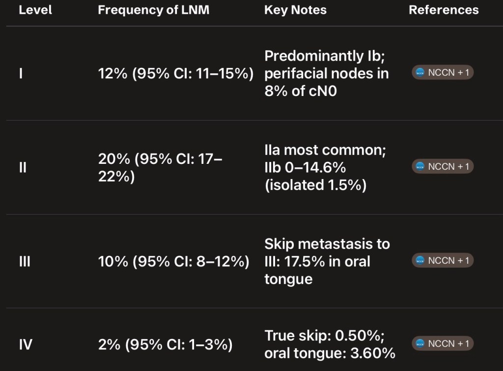
  • The gingivobuccal complex has a predictable lymphatic drainage:
    • The first echelon lymph nodes are in the:
      • Supraomohyoid triangle of neck:
        • Levels I, II, III
    • Spread to lymph nodes in posterior triangle:
      • In the absence of metastasis at other levels:
        • Is rare
    • Skip metastasis from gingivobuccal complex carcinomas:
      • Are rare
    • Distant metastasis at the time of initial diagnosis is exceedingly rare:
      • When it does occur it is to lung and bones
  • Biopsy of the lesion is mandatory before treatment:
    • Often, this can be done under local anaesthesia
    • The biopsy should be deep and encompass a portion of the tumor as well as adjacent normal appearing mucosa
    • Superficial biopsies are inconclusive and yield negative results
    • In suspected verrucous carcinomas:
      • Where the basement membrane is intact:
        • A deep biopsy is mandatory to reach a diagnosis
  • Imaging work-up depends on the extent of the disease:
    • Patients with early lesions do not need an extensive evaluation:
      • An orthopantomogram or oblique radiograph of the mandible is a cost-effective initial investigation to assess mandibular involvement
    • The accuracy of clinical examination, perioperative periosteal stripping and imaging techniques have been compared:
      • Clinical examination alone was not shown to be accurate:
        • But periosteal stripping at the time of resection was extremely accurate
      • No single imaging technique will accurately predict mandibular invasion:
        • However, a combination of orthopantomogram and bone scintigraphy is recommended in early invasion
        • Magnetic resonance imaging (MRI) is more sensitive than computerised tomography (CT) for mandibular invasion
        • CT scanning gives additional information regarding the extent of mandibular involvement, malignant infiltration and cervical nodal disease
        • MRI can be used to determine soft tissue and perineural involvement
        • However, all patients do not need CT scan or MRI:
          • These are especially indicated in patients with large lesions having trismus and lesions abutting the mandible where marginal mandibulectomy is being planned
        • It is also used to evaluate patients with a clinically negative neck or those with large nodes for presence of carotid involvement
        • Ultrasound guided fine needle aspiration cytology (FNAC) has the highest accuracy in diagnosing cervical nodal metastasis in the clinically negative neck compared to ultrasonography, CT scan and MRI:
          • However, none of the imaging methods can determine occult metastatic nodal disease
    • In view of the risk of multifocal changes:
      • Endoscopic evaluation (panendoscopy) of the upper aerodigestive tract is recommended to evaluate the presence of second primaries:
        • Direct laryngoscopy, bronchoscopy, esophagoscopy and examination under general anesthesia may be done for accurate assessment of the disease and the upper aerodigestive tract
    • Depending on the specific site in the gingivobuccal complex (alveolus, gingivobuccal sulcus or buccal mucosa alone):
      • The extent of the primary tumor and the status of lymph nodes:
        • The treatment of these cancers may be by surgery or radiation therapy used alone or in combination, with or without chemotherapy
    • Early lesions (T1, T2) can be effectively treated with either surgery or radiation as a single modality:
      • Certain factors influence this decision:
        • For example, in the presence of associated submucous fibrosis:
          • Surgery is preferable to radiation therapy
        • Lesions located in lower gingivobuccal sulcus or involving mandible are usually not treated with radiation:
          • Because of proximity to bone and chances of osteo-radionecrosis
      • Advanced lesions require a combined modality treatment
  • Treatment of gingivobuccal complex cancer:
    • Is primarily surgical:
      • The aim of surgical treatment is to excise the entire primary lesion with clear margins (1 cm to 2 cm) three-dimensionally, and also effectively treat the regional lymph nodes
      • This ablative surgery is followed by primary reconstruction:
        • To provide rapid healing, restore function and appearance and thereby improve patient’s quality of life
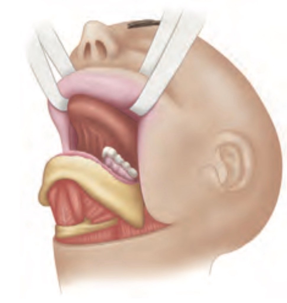
      • These lesions are resected either by an upper or lower cheek flap with a lip split, visor flap or per-orally:
        • Depending upon the size and location of the primary
      • These cancers often abut or involve the mandible
      • Most of these cancers are not amenable to per-oral resection:
        • Owing to inadequate access:
          • Which may jeopardise the oncological resection
      • Per-oral resection is possible in:
        • Small lesions (usually, 2 cm or less), situated anteriorly, with no or minimal mandibular involvement, and with good mouth opening
      • Radical ablative surgery is followed by reconstructive surgery:
        • Surgical defects may be reconstructed by primary closure, skin graft, locoregional flaps or free tissue transfer from different sites
      • The decision to resect the mandible as part of the management of oral cancer:
        • Should be taken on the evidence of clinical examination, periosteal stripping and at least two imaging techniques that complement each other in terms of specificity and sensitivity
      • Lesions that directly invade the bone:
        • Should undergo a segmental or hemi-mandibulectomy
      • Resection of the posterior part of the body or ramus of mandible leaves very little aesthetic deformity, but there is always functional compromise with segmental resection of any part of mandible
      • Resection of anterior arch of mandible:
        • Results in significant functional and cosmetic deformity and immediate reconstruction should be done by an osteomyocutaneous flap or composite free tissue transfer
          • If immediate reconstruction is not feasible or desirable, the mandibular stirrups should be immobilised by internal, external or interdental fixation
          • Heavy reconstruction plates may also be used in this situation
    • Mandibular sparing techniques like marginal mandibulectomy have gained popularity for lesions with no, or minimal, cortical mandibular invasion:
      • Marginal mandibulectomy has been used for along time in cancer of the floor of the mouth and can also be used for cancers of gingivobuccal complex
      • Mandibular continuity is maintained and a much better cosmetic and functional end result is achieved with marginal mandibulectomy:
        • At least, a 1-cm thick segment of bone must be left inferiorily after a marginal mandibulectomy
      • Marginal mandibulectomy is contraindicated in patients with:
        • Gross clinical and radiological involvement of mandible
        • Invasion of mandibular canal by cancer
        • In deeply infiltrating lesions of gingivobuccal sulcus where there is paramandibular infiltration:
          • As the margin of resection may pass through infiltrated paramandibular tissue
        • It is also contraindicated in previously irradiated mandible
        • Reduced vertical height of the bone in an edentulous mandible is a relative contraindication for marginal mandibulectomy
        • Marginal mandibulectomy is also usually not done in lesions of the retromolar trigone as clearance of pterygoid region is possible only if ascending ramus of the mandible is resected:
          • However, some studies have also reported satisfactory results of marginal mandibulectomy in lesions of the retromolar trigone
        • Results of marginal mandibulectomy for gingivobuccal complex carcinoma show an:
          • Overall local recurrence-free survival rate of 79% and 70% at 2 and 5 years, respectively
        • Other studies have also demonstrated the oncological safety of marginal mandibulectomy in carefully selected patients with oral cancers
  • The management of neck for gingivobuccal complex cancers:
    • Depends on whether the neck is clinically node-negative or node-positive
    • In patients with clinically positive lymph nodes (N1, N2, N3):
      • Radical neck dissection (RND) has been the gold standard
    • However, there is mounting evidence that RND should not be the only therapeutic option for the clinically positive neck:
      • In patients with clinical N1 disease and selected N2 disease:
        • A modified radical neck dissection may be done for better cosmetic and functional results
        • Preservation of the spinal accessory nerve, internal jugular vein (IJV) and sternocleidomastoid muscle:
          • Is done in the form of a modified radial neck dissection (MRND)
  • RND, however, is still appropriate for patients with:
    • Massive lymphadenopathy (N3 disease)
    • Multiple positive nodes involving the spinal accessory nerve and / or the IJV
    • Residual or recurrent neck disease after radiotherapy
    • Gross extranodal spread
  • A supraomohyoid neck dissection (SOHND):
    • Clearance of level I, II and III nodes plus postoperative radiation therapy has been advocated by a few authors for N1, level I disease:
      • There are still no prospective, randomised trials comparing SOHND with RND / MRND in clinically positive neck and it is unlikely that studies large enough to answer this question will be carried out in the future
  • Nodal spread can occur to both sides of neck:
    • Especially in lesions:
      • Close to mid-line
    • In patients with bilateral nodal metastasis:
      • A bilateral neck dissection with preservation of IJV on at least one side (the less affected side) is indicated:
        • An alternative to this is to do a staged RND:
          • The IJV resection is done on both the sides with an interval of 4 weeks between them
  • Occult nodal metastatic disease:
    • Is present in 5% to 26% of gingivobuccal complex cancers:
      • Depending on the T-status and grade
    • Management of the clinically negative is thus an important issue:
      • Patients with T1 / T2 cancers (low risk, less than 20% risk of nodal metastasis):
        • Do not require elective neck treatment
      • SOHND should be performed in patients with:
        • T3 / T4 primary (high risk, greater than 20% risk of nodal metastasis)
        • If entering the neck to resect the primary
        • Short-necked individuals:
          • Who require a bulky flap for oral reconstruction:
            • To create space in neck
        • Patients who are unreliable for follow-up
      • Patients who are found to have pathologically positive neck nodes after SOHND:
        • Should receive additional treatment
      • If detected positive intra-operatively (on frozen section):
        • Then SOHND should be changed to RND or MRND
      • Patients with positive lymph nodes, diagnosed on histopathology following SOHND:
        • Should either undergo RND / MRND or postoperative radiotherapy
      • Patients with a single positive, level I node only, without extra-capsular spread:
        • May not need additional treatment
      • A randomized trial comparing SOHND with comprehensive neck dissection in patients with clinically negative nodes:
        • Found no difference in the regional control and overall survival rates between the two groups:
          • However, SOHND alone is inadequate treatment for patients with pathologically confirmed or clinically positive nodes
      • Patients undergoing MRND need adjuvant radiation therapy to the neck if N2 disease is present:
        • Adjuvant radiation provides good regional control
      • The role of ultrasound-guided FNAC of the N0 neck in the decision for elective neck dissection has been reported:
        • However, its application to gingivobuccal complex cancers in particular is uncertain
    • Recently, lymphatic mapping with sentinel lymph node biopsy has been used in N0 oral cancer patients but further refinement of technique and larger studies are needed before this can be recommended as standard treatment
  • Radiation therapy and surgery:
    • Have equal success in controlling early lesions of the oral cavity
    • Radiation is given either as:
      • External beam, brachytherapy or acombination of both
    • For gingivobuccal complex cancers:
      • Radiotherapy is usually not the preferred modality of treatment for early cancers (T1, T2):
        • Due to the close proximity of the tumor to bone and risk of radionecrosis
      • Radiotherapy is used for treatment of early lesions of buccal mucosa and gingivobuccal sulcus:
        • Where the patient is not medically fit or is unwilling for surgery
      • It is also used as an adjuvant treatment for the primary tumor in patients with histologically positive margins on resection:
        • And has been shown to decrease the local recurrence rate
    • In patients with advanced lesions (T3, T4):
      • A combination of surgery and radiation therapy provides a better chance of cure than either modality alone
      • The 3-year survival for stages III and IV disease treated with radiation therapy or surgery alone:
        • Is 41% and 15%, respectively
      • These rates increase to 60% and 35%, respectively:
        • When surgery is combined with postoperative radiation therapy
      • In a randomized trial, Mishra et al:
        • Have reported a significant improvement in disease-free survival in patients with T3 / T4 carcinoma of the buccal mucosa
      • Postoperative radiation therapy is indicated in all patients with:
        • T3 primary tumors
        • T4 primary tumors
        • Patients with positive or close surgical margins
        • Pathologically positive lymph nodes after SOHND
        • Two or more positive lymph nodes after RND / MRND
        • Lymph nodes showing extracapsular spread
      • There is evidence for the use of adjuvant concurrent chemoradiotherapy:
        • In patients of head and neck cancers with poor prognostic factors
    • Definitive radiation or concurrent chemoradiotherapy:
      • Is used in advanced (stage III / IV) disease, if the disease is inoperable or the patient is unfit or unwilling for surgery
    • Concurrent chemoradiotherapy:
      • Has evolved as the standard of care for such locally advanced head and neck cancers
    • The toxicity of concurrent chemoradiotherapy is more than radiation therapy alone and requires aggressive supportive measures:
      • However, evidence for use of concurrent chemoradiotherapy for oral cavity subsite is sparse
  • In clinically node-negative neck cancer, elective neck irradiation is done if the primary is being treated with radiation therapy