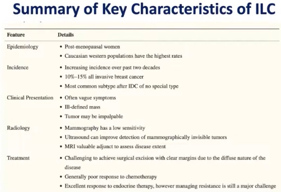
Blog

Early Breast Cancer New Information from San Antonio Breast Cancer Conference 2025

  • Adjuvant endocrine therapy is evolving: 
    Oral SERD in early HR+ / HER2− disease:
    • lidERA (giredestrant vs standard endocrine therapy):
      • What it tested:
        • Adjuvant giredestrant (oral SERD) vs standard endocrine therapy after surgery in ER+ /HER2− early breast cancer
    • Headline result:
      • Significant iDFS improvement:
        • They reported ~30% relative risk reduction (HR ~0.70) vs standard ET
      • Why it matters:
        • First credible signal in a long time that “better endocrine backbone” (beyond AI / tamoxifen) could become standard for selected early HR+ patients:
          • Especially if subgroup and safety data remain favorable
      • Surgical implication:
        • Expect more conversations about systemic escalation / de-escalation:
          • For example who truly needs CDK4/6 inhibitors vs a more potent endocrine option
  • Post-neoadjuvant HER2+ residual disease: 
    T-DXd moves earlier:
    • DESTINY-Breast05 (T-DXd vs T-DM1):
      • Population:
        • HER2+ early breast cancer with residual invasive disease after neoadjuvant therapy:
          • High-risk post-NAC setting
      • Key outcome:
        • T-DXd superior to T-DM1 for invasive disease-free outcomes
      • Reported data (NEJM / PubMed):
        • Events 6.4% vs 12.6%, HR 0.47:
          • With 3-year DFS 92.3% vs 83.5%
      • Why it matters:
        • This is a direct “KATHERINE-successor” story:
          • If adopted into guidelines / pathways, it could reset the post-neoadjuvant standard for residual HER2+ disease 
      • Surgical implication:
        • Reinforces the importance of accurate residual disease documentation (pathology, RCB, nodal status) because this is what triggers the post-NAC systemic pathway
  • Axillary de-escalation: 
    Omitting SLNB in selected cN0 patients:
    • Dutch randomized trial (BOOG 2013-08):
      • Design:
        • Clinically node-negative (cT1 to cT2), treated with BCS + whole-breast RT, randomized to SLNB vs omission
      • Message:
        • Regional control / oncologic outcomes were not worse with omission in carefully selected patients, supporting a further step in axillary de-escalation
      • SABCS:
        • Also highlighted interpretation alongside RT fields (since RT contributes to axillary control)
    • Practical “surgeon filter”:
      • This is not “no axillary surgery for everyone,” but it strengthens the discussion for:
        • Older / low-risk, cN0, BCS+RT patients in centers that can replicate selection / imaging rigor
  • Pre-op staging MRI:
    • Routine use questioned in specific early BC subsets:
      • Alliance A011104 / ACRIN 6694:
        • Population:
          • Stage I / II HR-negative early breast cancer
        • Result:
          • Routine pre-op MRI did not improve key oncologic outcomes
          • MD Anderson reported 5-yr locoregional recurrence 6.8% with MRI vs 4.3% without (not favoring MRI)
        • Message:
          • More evidence that routine MRI may not improve outcomes and can drive additional procedures
        • Surgical implication:
          • Helps justify a more selective MRI strategy, dense breasts, lobular carcinoma, occult primary, discordant imaging, suspected multicentric disease, rather than reflex MRI for all
  • Immune priming concept: 
    • Preoperative RT + pembrolizumab
       in HR+ / HER2− (early signal):
      • TBCRC-053 (P-RAD):
        • Concept:
          • Short-course preoperative RT (e.g., 24 Gy / 3 fractions) added to pembrolizumab + chemotherapy
      • Signal:
        • Increased tumor T-cell infiltration and “immune activation” endpoints:
          • Hypothesis-generating but compelling
      • Clinical implication:
        • Not practice-changing yet, but it’s a blueprint for future trials aiming to convert HR+ tumors into more immunogenic disease
  • DCIS de-escalation reality check: 
    Tamoxifen-only (no surgery) did not meet safety bar:
    • LORETTA (JCOG1505):
      • Design:
        • Single-arm tamoxifen without surgery for selected low-risk ER+ / HER2− DCIS
      • Outcome:
        • Reported 5-year ipsilateral invasive events exceeded prespecified safety threshold (widely summarized from SABCS)
      • Takeaway:
        • Supports continued caution:
          • Endocrine-only, surgery-free DCIS management should remain investigational / exceptional rather than broadly adopted
  • What to say at tumor board (tight synthesis) HR+/HER2−:
    • LidERA:
      • Suggests a credible pathway toward next-generation endocrine therapy (oral SERD) in early disease
    • HER2+ residual disease post-NAC:
      • T-DXd > T-DM1 in DESTINY-Breast05 – likely a major sequencing shift as pathways update 
    • Surgery de-escalation:
      • Growing evidence that less axillary surgery and less routine MRI can be safe in well-defined populations
    • Translational front:
      • Preoperative RT + IO is an emerging strategy to watch, not a standard yet
    • DCIS:
      • Surgery-free endocrine-only strategies still struggle with invasive recurrence risk thresholds

Destiny – Breast 02 Trial

  • Is a randomized phase III trial:
    • That established trastuzumab deruxtecan (T-DXd) as the preferred therapy for patients with HER2-positive metastatic breast cancer:
      • Previously treated with trastuzumab emtansine (T-DM1)
  • Population:
    • HER2-positive unresectable or metastatic breast cancer
    • Prior therapy:
      • Trastuzumab + taxane and T-DM1
    • Randomization: T
      • DXd vs Investigator’s choice (trastuzumab + capecitabine or lapatinib + capecitabine)
    • Primary endpoint:
      • Progression-free survival (PFS, BICR)
    • Key secondary endpoints:
      • Overall survival (OS), ORR, duration of response, safety
  • Mechanism of Action (Why T-DXd Is Different):
    • Trastuzumab deruxtecan is a next-generation antibody–drug conjugate (ADC) with:
      • High drug-to-antibody ratio (≈8:1)
      • Cleavable linker membrane-permeable topoisomerase I payload bystander effect:
        • Enabling killing of adjacent tumor cells with heterogeneous HER2 expression:
          • This design explains its activity after T-DM1 failure, where resistance commonly develops.
  • Efficacy Results:
    • Progression-Free Survival (Primary Endpoint):
      • Median PFS:
        • T-DXd:
          • ~17.8 months
        • Control:
          • ~6.9 months
        • Hazard ratio:
          • ~0.36
        • Risk reduction:
          • ~64% reduction in progression or death
      • Clinically transformative improvement in disease control
    • Overall Survival:
      • Median OS:
        • T-DXd:
          • ~39.2 months
        • Control:
          • ~26.5 months
        • Hazard ratio:
          • ~0.66
      • Statistically significant and clinically meaningful OS benefit, uncommon in heavily pretreated HER2-positive MBC trials
    • Objective Response Rate:
      • T-DXd:
        • ~69%
      • Control:
        • ~29%
      • Complete responses:
        • Observed with T-DXd
  • Safety Profile:
    • Common Adverse Events (T-DXd):
      • Nausea
      • Fatigue
      • Alopecia
      • Vomiting
      • Neutropenia
      • Anemia
      • Interstitial Lung Disease (ILD) / Pneumonitis:
        • Any-grade ILD:
          • ~10%
        • Grade ≥3 ILD:
          • ~1–2%
        • Fatal events:
          • Rare but reported
  • Key clinical takeaway:
    • Early recognition, prompt drug interruption, and steroid initiation are essential
    • Patient education and routine symptom monitoring are mandatory
  • How DESTINY-Breast 02 Changed Practice:
    • Before DESTINY-Breast 02:
      • Post-T-DM1 options relied on capecitabine-based combinations:
        • Limited durability and modest survival benefit
    • After DESTINY-Breast 02:
      • T-DXd is the standard of care after T-DM1:
        • Supported by PFS + OS superiority:
          • Endorsed by NCCN, ASCO, ESMO
    • Represents a paradigm shift in the HER2-positive metastatic sequence
  • Treatment Sequencing (Current Standard):
    • First line:
      • Trastuzumab + pertuzumab + taxane
    • Second line:
      • T-DXd
    • Later lines:
      • Tucatinib-based regimens
      • Clinical trials
      • Other HER2-targeted agents
  • Surgical and Multidisciplinary Relevance:
    • Durable systemic control increases:
      • Consideration of local therapies for oligoprogression
      • Delayed need for palliative surgery
    • Highlights importance of:
      • Early referral to medical oncology
      • Coordinated surveillance for pulmonary toxicity
  • Key Take-Home Messages:
    • DESTINY-Breast 02 firmly establishes T-DXd as best-in-class post-T-DM1 therapy:
      • Demonstrates both PFS and OS benefit in a refractory population
    • ILD monitoring is critical to safe delivery
    • Confirms the power of ADC engineering in overcoming resistance

Thyroid Awareness Month – Why Thyroid Ultrasound Is the Most Important Test

When a thyroid nodule is suspected or discovered, high-resolution thyroid ultrasound is the single most important diagnostic study.

🔍 What does a thyroid ultrasound tell us?

Ultrasound allows us to evaluate:

Size and exact location of the nodule Composition (solid, cystic, or mixed) Margins (smooth vs irregular) Echogenicity and calcifications Vascularity (with Doppler imaging) Cervical lymph nodes

➡️ These features are far more predictive of cancer risk than symptoms or blood tests.

📊 Risk stratification matters

Using ultrasound findings, nodules are categorized with validated systems such as:

ATA risk patterns ACR TI-RADS

These systems help determine:

✔️ Which nodules need biopsy

✔️ Which nodules can be safely observed

✔️ Appropriate follow-up intervals

🧪 Important clarification

Blood tests do not diagnose thyroid cancer CT scans and MRIs are NOT first-line tests for thyroid nodules Ultrasound provides real-time, radiation-free, highly accurate evaluation

🦋 Why this matters for patients

Proper ultrasound evaluation:

Prevents unnecessary biopsies and surgeries Ensures early diagnosis of clinically significant thyroid cancer Guides personalized management

👨‍⚕️ Rodrigo Arrangoiz, MD

Surgical Oncologist – Thyroid, Head & Neck, Breast

Mount Sinai Medical Center

📌 Take-home message:

Not all ultrasounds are equal.

Expert, high-resolution thyroid ultrasound makes all the difference.

📚 References

Haugen BR et al. ATA Guidelines for Thyroid Nodules. Thyroid Tessler FN et al. ACR TI-RADS. Radiology Russ G et al. EU-TI-RADS. European Journal of Endocrinology

Thyroid Awareness Month – Who Is at Risk for Thyroid Nodules?

Thyroid Awareness Month – Day 3

Who Is at Risk for Thyroid Nodules?

Thyroid nodules can occur in anyone, but certain factors make them more likely.

👥 Common Risk Factors

Age: Nodules become more common as we get older Female sex: Women develop thyroid nodules 3–4 times more often than men Iodine imbalance: Both deficiency and excess can play a role Family history: Thyroid nodules or thyroid cancer in first-degree relatives Autoimmune thyroid disease: Hashimoto’s thyroiditis increases nodule prevalence

☢️ Higher-Risk Situations

Radiation exposure to the head and neck, especially during childhood Prior radiation therapy for acne, tonsils, or cancer (historical treatments) Certain genetic syndromes (rare, but important)

🧠 Important clarification

Having risk factors does not mean a thyroid nodule is cancer.

➡️ Even in higher-risk individuals, most nodules are benign.

🔍 What matters most?

Risk factors help guide how closely we evaluate, but ultrasound findings ultimately determine:

Cancer risk Need for biopsy Follow-up strategy

📌 Key point for patients:

A thyroid nodule should never be ignored — but it should also never cause unnecessary fear. Proper, evidence-based evaluation is the answer.

👨‍⚕️ Rodrigo Arrangoiz, MD

Surgical Oncologist – Thyroid, Head & Neck, Breast

Mount Sinai Medical Center

📚 References

Haugen BR et al. ATA Guidelines for Thyroid Nodules and Differentiated Thyroid Cancer. Thyroid Guth S et al. Very high prevalence of thyroid nodules. Thyroid Gharib H et al. Evaluation and Management of Thyroid Nodules. Endocrine Practice

Who Is at Risk for Thyroid Nodules?

Thyroid nodules can occur in anyone, but certain factors make them more likely.

👥 Common Risk Factors

Age: Nodules become more common as we get older Female sex: Women develop thyroid nodules 3–4 times more often than men Iodine imbalance: Both deficiency and excess can play a role Family history: Thyroid nodules or thyroid cancer in first-degree relatives Autoimmune thyroid disease: Hashimoto’s thyroiditis increases nodule prevalence

☢️ Higher-Risk Situations

Radiation exposure to the head and neck, especially during childhood Prior radiation therapy for acne, tonsils, or cancer (historical treatments) Certain genetic syndromes (rare, but important)

🧠 Important clarification

Having risk factors does not mean a thyroid nodule is cancer.

➡️ Even in higher-risk individuals, most nodules are benign.

🔍 What matters most?

Risk factors help guide how closely we evaluate, but ultrasound findings ultimately determine:

Cancer risk Need for biopsy Follow-up strategy

📌 Key point for patients:

A thyroid nodule should never be ignored — but it should also never cause unnecessary fear. Proper, evidence-based evaluation is the answer.

👨‍⚕️ Rodrigo Arrangoiz, MD

Surgical Oncologist – Thyroid, Head & Neck, Breast

Mount Sinai Medical Center

📚 References

Haugen BR et al. ATA Guidelines for Thyroid Nodules and Differentiated Thyroid Cancer. Thyroid Guth S et al. Very high prevalence of thyroid nodules. Thyroid Gharib H et al. Evaluation and Management of Thyroid Nodules. Endocrine Practice

Thyroid Awareness Month – Symptoms of Thyroid Nodules

Most thyroid nodules cause no symptoms at all and are found incidentally on imaging or routine exams.

🔹 Common scenario: You feel well — the nodule is discovered on ultrasound

🔹 Important fact: Lack of symptoms does NOT mean the nodule is dangerous (or benign)

👀 When can symptoms occur?

Symptoms are more likely when nodules are large or strategically located:

A visible or palpable lump in the neck Difficulty swallowing or a sensation of food “getting stuck” Hoarseness or voice changes (uncommon, but important) Neck pressure or fullness, especially when lying flat Rarely, shortness of breath

⚠️ Hormone-related symptoms (less common)

A small percentage of nodules produce excess thyroid hormone (“hot” nodules), which may cause:

Palpitations Weight loss Heat intolerance Tremors or anxiety

🚨 When should you seek evaluation?

You should be evaluated if you notice:

✔️ A new or growing neck lump

✔️ Persistent hoarseness

✔️ Difficulty swallowing or breathing

✔️ A personal history of radiation exposure or family history of thyroid cancer

🔍 Bottom line:

Symptoms alone cannot determine whether a nodule is benign or malignant.

➡️ High-resolution ultrasound is essential for proper evaluation.

👨‍⚕️ Rodrigo Arrangoiz, MD

Surgical Oncologist – Thyroid, Head & Neck, Breast

Mount Sinai Medical Center

📌 Take-home message:

Most thyroid nodules are silent.

Don’t rely on symptoms — rely on proper imaging and expert evaluation.

📚 References

Haugen BR et al. ATA Guidelines for Thyroid Nodules. Thyroid Gharib H et al. Evaluation and Management of Thyroid Nodules. Endocrine Practice Durante C et al. JAMA

Thyroid Nodules

What Are Thyroid Nodules?

Thyroid nodules are very common in the general population.

🔹 With modern high-resolution ultrasound, thyroid nodules are detected in up to 50–60% of adults

🔹 More than 90% are benign

🔹 Most people have no symptoms and feel completely well

👉 The key is not panic — it’s proper evaluation.

🔍 How should thyroid nodules be evaluated?

The most important first step is a high-resolution thyroid ultrasound, which allows us to:

Accurately measure and characterize nodules Assess features associated with cancer risk Use validated risk-stratification systems (ATA / TI-RADS) Decide whether a biopsy is actually necessary

🧪 A key fact for patients

Only 5–10% of thyroid nodules are cancer.

When thyroid cancer is detected early, cure rates are excellent.

👨‍⚕️ Rodrigo Arrangoiz, MD

Surgical Oncologist – Thyroid, Head & Neck, Breast

Mount Sinai Medical Center

📌 Take-home message:

Having a thyroid nodule is common.

Having it evaluated correctly by an experienced team makes all the difference.

📚 References

Haugen BR et al. ATA Guidelines for Thyroid Nodules and Differentiated Thyroid Cancer. Thyroid Durante C et al. Long-term surveillance of benign thyroid nodules. JAMA Gharib H et al. Fine-needle aspiration biopsy of thyroid nodules. Endocrine Practice

Thyroid Awareness Month – Thyroid Nodules

Thyroid Nodules

– Thyroid nodules are very common in the general population. With modern high-resolution ultrasound, nodules can be detected in up to 50% to 60% of adults.

👉 The good news: more than 90% of thyroid nodules are benign.

🔍 What is the best way to evaluate a thyroid nodule?

The first and most important test is a high-resolution thyroid ultrasound. It allows us to:

Accurately characterize the nodule (size, margins, echogenicity, calcifications) Stratify cancer risk using validated systems (such as ATA or TI-RADS) Determine whether a biopsy is necessary

🧪 When is a biopsy needed?

If the ultrasound shows suspicious features, a ultrasound-guided fine-needle aspiration biopsy (FNAB) is recommended.

✔️ This is a safe, minimally invasive, outpatient procedure that provides highly accurate information to determine whether a nodule is benign or malignant, helping avoid unnecessary surgery.

📊 Key facts (evidence-based)

Thyroid nodules are found in up to 60% of adults on ultrasound Only 5% to 10% of nodules are malignant Ultrasound-guided FNAB has high diagnostic accuracy and a very low complication rate

🏥 Experience you can trust

👨‍⚕️ Dr. Rodrigo Arrangoiz

Surgical Oncologist | Thyroid, Head & Neck, and Breast Surgery

Mount Sinai Medical Center

📌 Take-home message: Early detection and proper evaluation of thyroid nodules make a real difference—protecting patients from unnecessary treatments while identifying cancer early when it is most curable.

📚 References

Haugen BR et al. 2015 ATA Guidelines for Thyroid Nodules and Differentiated Thyroid Cancer. Thyroid. Russ G et al. European Thyroid Association Guidelines for Ultrasound Malignancy Risk Stratification (EU-TI-RADS). Eur J Endocrinol. Gharib H et al. Fine-Needle Aspiration Biopsy of Thyroid Nodules. Endocrine Practice. Durante C et al. Long-term Surveillance of Benign Thyroid Nodules. JAMA.

The Cernea Classification in Thyroid Surgery

  • The Cernea classification:
    • Is used during thyroid surgery to predict the risk of injury to the external branch of the superior laryngeal nerve (EBSLN):
      • Based on its anatomical relation to the superior thyroid artery and the upper pole of the thyroid
    • It includes three types:
      • Type I:
        • The EBSLN crosses more than 1 cm above the upper edge of the thyroid superior pole:
          • The artery-nerve intersection is > 1 cm above the gland
        • Minimal risk during ligation of the superior thyroid vessels
      • Type IIa:
        • The nerve crosses within 1 cm above the superior pole:
          • Distance < 1 cm but above the pole
        • Moderate risk, as it’s close to vascular pedicle
      • Type IIb:
        • The EBSLN crosses below the upper edge of the superior pole:
          • Under the pole
        • Highest risk:
          • Nerve lies within the ligation field
  • Clinical Significance:
    • Intraoperative studies show Type IIa + IIb:
      • Account for ~ 65% of cases:
        • Emphasizing the high chance of encountering the nerve near or below the vascular pedicle
    • Type IIb alone:
      • Occurs in up to 48% of cases in some series 
    • Risk of EBSLN injury rises with more inferior nerve trajectories (IIa / IIb):
      • Injuries lead to altered voice quality:
        • Reduced pitch, vocal fatigue, and weakened projection
  • Surgical Implications:
    • Routine identification:
      • Especially for Type II subtypes:
        • Is key during superior pole dissection
    • Intraoperative neuromonitoring (IONM) helps detect and preserve the EBSLN:
      • Reducing morbidity
    • Transection of overlying muscles (e.g., sternothyroid):
      • Can improve visualization of the nerve

The Anastomoses of the Recurrent Laryngeal Nerve in the Larynx

  • The vagus nerve:
    • Also known as the 10th cranial nerve
    • Gives rise to the superior laryngeal nerve (SLN) and recurrent laryngeal nerve (RLN) in the neck
    • After descending toward the larynx:
      • The SLN divides into:
        • The internal laryngeal nerve (ILN)
        • External laryngeal nerve (ELN)
  • The RLN innervates all of the intrinsic muscles of the larynx:
    • Except the cricothyroid muscle:
      • This muscle, tenses the vocal cords and adducts the vocal cords:
        • Is innervated by the ELN
  • The other branch of the SLN, the ILN:
    • Provides sensory innervation to the laryngeal mucosa
  • There are many exceptions to the normal innervation of the laryngeal muscles:
    • Which can influence the interpretation of laryngoscopy results or voice changes after thyroid surgery
  • A neural anastomosis:
    • That provides additional motor innervation to the muscles of the larynx normally innervated by the injured nerve:
      • Can contribute to an incorrect interpretation of injury during laryngoscopy or stroboscopy
  • According to recent clinical studies:
    • Electrical stimulation of the RLN can cause contraction of the cricothyroid muscle:
      • This suggests that extra-laryngeal branches and or other communications of the RLN:
        • Can sometimes contribute to innervation of this muscle
  • The laryngeal nerves:
    • Can form a great variety of anastomoses:
      • These various connections among the ILN, ELN, and RLN have been investigated by many anatomists over the centuries
  • Claudius Galen:
    • Was the first to describe the communication between the ILN and RLN
    • Currently, Galen’s anastomosis:
      • Is most commonly defined as:
        • The direct communication between the posterior branches of the ILN and the RLN
      • It can occur as:
        • A single trunk
        • A double trunk
        • A plexus
  • Besides Galen’s anastomosis, other communications have been observed and described as follows:
    • The arytenoid plexus:
      • Which links the anterior branch of the RLN with the arytenoid branch of the ILN
The arytenoid plexus and the cricoid communication. AP, arytenoid plexus; CA, cricoid communication; ILN, internal laryngeal nerve; RLN, recurrent laryngeal nerve (a, anterior; p, posterior).
  • The cricoid communication:
    • Which connects branches originating bilaterally from the RLNs with the superior branch from the deep portion of the arytenoid plexus
  • The thyroarythenoid communication:
    • Which is formed by the ascending branch of the RLN and the descending branch from the anterior branch of the ILN
The thyroarytenoid communication and the communication between the external laryngeal nerve (ELN) and the recurrent laryngeal nerve (RLN). CN, communicating nerve; ILN, internal laryngeal nerve; TAC, thyroarytenoid communication
  • The communication between the ELN and RLN:
    • Human communicating nerve
  • The communication between the ILN and ELN
  • The communication between the RLN and the sympathetic trunk
Communications between the internal laryngeal nerve (ILN) and the external laryngeal nerve (ELN) and between the recurrent laryngeal nerve (RLN) and the sympathetic trunk. SLN, superior laryngeal nerve; TF, thyroid foramen.
  • Despite progress in the development of new techniques:
    • Such as intra-operative nerve monitoring:
      • Which help to reduce the risk of iatrogenic injuries during thyroid surgeries and other procedures conducted in close proximity to the laryngeal nerves:
        • The laryngeal muscles are often paralyzed postoperatively due to iatrogenic injury to the laryngeal nerves
  • In view of the complexity and variability of the anatomy in this region:
    • Detailed anatomical knowledge is crucial if surgery is to be both successful and safe, and to reduce the risk of nerve injury
  • Observing an intra-laryngeal anastomosis during laryngeal surgery or an extra-laryngeal communication between laryngeal nerves during thyroid surgery:
    • Can lead to confusion, misidentification, and an increased risk of iatrogenic injury
  • A thorough understanding of the complex anastomoses between the laryngeal nerves is crucial in patients with laryngeal muscle paralysis
    • Paralyzed laryngeal muscles can be spontaneously reinnervated from an anastomosis between laryngeal nerves
    • Additionally, in cases in which surgical reinnervation is required, some of the nerves that form anastomoses can be used as grafts to restore damaged nerve connections
    • On the other hand, variations in the normal anatomy of the laryngeal nerves can disrupt selective surgical laryngeal reinnervation:
      • A procedure based on the assumption that each laryngeal muscle is supplied by only one nerve branch originating from the RLN
    • Anastomoses among laryngeal nerves can result in exceptions to this rule
    • Such anastomoses have been widely described in the literature:
      • However, there is still no consensus about their prevalence and functionality
    • The significant heterogeneity among studies reporting data on anastomoses between the laryngeal nerves is noteworthy:
      • For example, the reported prevalence of the most common communication, Galen’ s anastomosis:
        • Ranges from 25% to 100%
  • Prevalence of Galen’s anastomosis:
    • A total of 14 studies (n = 890 hemilarynges) presented data on the prevalence of Galen’s anastomosis
    • The overall pooled prevalence rate:
      • Was 76.7% (95% confidence interval [CI]: 59.0– 90.0)
    • Subgroup analysis revealed no significant difference in the prevalence of Galen’s anastomosis between the right and left sides
    • Subgroup analyses by gender (males vs females) and geographical origin, and the sensitivity analysis, also revealed no significant differences:
      • However, although the difference was not significant:
        • The prevalence of the anastomosis was highest in Europeans (88.2%) and lowest in North Americans (44.8%)
    • Analysis of the different types of Galen’s anastomosis (two studies, n = 261 anastomoses):
      • Showed a significant difference in the prevalence between single versus double trunk and plexus formation
      • But no significant difference between double trunk and plexus formation
      • The most common type of Galen’s anastomosis was a single trunk:
        • With a pooled prevalence rate 92.3% (95% CI: 84.1–97.5)
      • This was followed by the double trunk anastomosis type:
        • With a pooled prevalence of 4.2% (95% CI: 0.5– 10.7)
      • The plexus formation type with a pooled prevalence of 3.5% (95% CI: 0.2–9.5) (I2: 70.4%, 95% CI: 0–93.3; Co- chran’s Q, value = 0.066
  • Prevalence of a communication between the ELN and RLN
    • A total of eight studies (n = 639 hemilarynges) provided data on the prevalence of the communication between the ELN and RLN
    • The overall meta-analysis revealed that this communication was present in:
      • 21.3% of hemilarynges (95% CI: 3.8–46.0)
    • A subgroup analysis showed no significant difference between left and right sides
    • Although the difference was not statistically significant:
      • The pooled prevalence rate calculated for the North American subgroup (32.0%) was twice that for the European subgroup (14.4%)
  • Prevalence of the arytenoid plexus:
    • Five studies (n = 478 hemilarynges) included data on the prevalence of the arytenoid plexus:
      • The pooled prevalence rate was 79.7% (95% CI: 41.1–100)
    • In the European subgroup:
      • The arytenoid plexus was observed in 96.9% of hemilarynges (95% CI: 83.6–100)
    • Subgroup analysis revealed no significant difference with respect to side
  • Prevalence of the cricoid communication:
    • Two studies (n = 120 hemilarynges) reported prevalence data for the cricoid communication:
      • The pooled prevalence rate was 19.7% (95% CI: 0–100)
    • There was no significant difference in sub- group analysis based on side
  • Prevalence of the thyroarytenoid communication:
    • A total of three studies (n = 430 hemilarynges) presented data on the prevalence of the thyroarytenoid communication:
      • The overall pooled prevalence was 6.3% (95% CI: 0.4–16.9)
    • In subgroup analysis, there was no significant difference in pooled prevalence between the right and left sides:
      • The calculated pooled rate for the left side (15.9%) was almost twice that for the right side (8.8%)
  • Communication between the ILN and ELN:
    • A total of two studies (n = 280 hemilarynges) reported data on a communication between the ILN and ELN
      • The pooled prevalence estimate of this communication in hemilarynges was 8.8% (95% CI: 0–35.3; I2 97.0%, 95% CI: 92.2–98.8; Cochran’s Q, value <0.001)
  • References:
    • Journal of Voice, Vol. 31, No. 4, 2017

#Arrangoiz #HeadandNeckSurgeon #CancerSurgeon #ThyroidSurgeon #ParathyroidSurgeon #ThyroidExpert #ParthyroidExpert #Teacher #Miami #Mexico