- Overview:
- The nasopharynx is the uppermost part of the pharynx:
- A cuboidal muscular-mucosal space behind the nasal cavity and above the soft palate, anterior to the atlas (C1) and axis (C2)
- It functions purely as a respiratory conduit:
- Communicating anteriorly with the nasal cavity via the choanae and laterally with the middle ear via the Eustachian (pharyngotympanic) tubes
- The nasopharynx is the uppermost part of the pharynx:
- Boundaries (walls)
- Anterior:
- Paired choanae (posterior nasal openings)
- Bony margin bounded:
- Anteroinferiorly by the horizontal plate of the palatine bone
- Superiorly by the sphenoid body and vaginal process of the medial pterygoid plate
- Laterally by the medial pterygoid plates
- Roof and posterior wall:
- Continuous, sloping surface formed by the sphenoid body and basilar (clival) occipital bone superiorly, continuing over the anterior arches of C1 and C2:
- Mucosa adherent to the pharyngobasilar fascia
- Continuous, sloping surface formed by the sphenoid body and basilar (clival) occipital bone superiorly, continuing over the anterior arches of C1 and C2:
- Lateral walls:
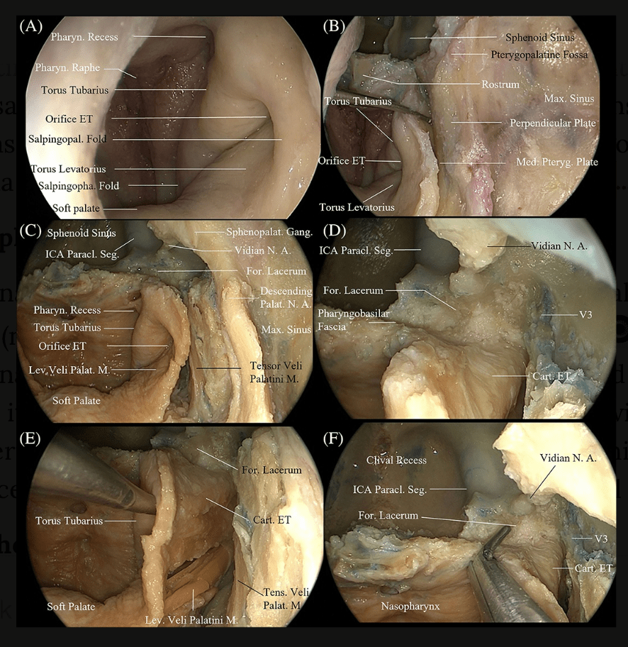
- Organized around the Eustachian tube orifice
- Floor / inferior boundary:
- Open inferiorly, communicating with the oropharynx via the pharyngeal isthmus:
- Upper surface of the soft palate forms the effective floor, and the isthmus closes during swallowing to prevent nasal reflux:
- The nasopharyngeal cavity opens into the cavity of the oropharynx through the pharyngeal isthmus, which lies between the posterior border of the soft palate and the posterior pharyngeal wall:
- This region is marked on the posterior pharyngeal wall by a fold of mucosa formed by the palatopharyngeal sphincter between the palatopharyngeus muscle and the superior pharyngeal constrictor muscle:
- During swallowing, elevation of the soft palate and constriction of the palatopharyngeal sphincter seals off the pharyngeal isthmus, separating the nasopharynx from the oropharynx:
- This prevents the retrograde flow of materials into the nasopharynx and nasal cavity
- During swallowing, elevation of the soft palate and constriction of the palatopharyngeal sphincter seals off the pharyngeal isthmus, separating the nasopharynx from the oropharynx:
- This region is marked on the posterior pharyngeal wall by a fold of mucosa formed by the palatopharyngeal sphincter between the palatopharyngeus muscle and the superior pharyngeal constrictor muscle:
- The nasopharyngeal cavity opens into the cavity of the oropharynx through the pharyngeal isthmus, which lies between the posterior border of the soft palate and the posterior pharyngeal wall:
- Upper surface of the soft palate forms the effective floor, and the isthmus closes during swallowing to prevent nasal reflux:
- Open inferiorly, communicating with the oropharynx via the pharyngeal isthmus:
- Anterior:
- Lateral wall structures:
- Torus tubarius:
- Prominent posterior lip of the tubal orifice:
- Elevated by tubal cartilage
- Prominent posterior lip of the tubal orifice:
- Salpingopalatine fold:
- Anterior lip of the orifice
- Torus levatorius:
- Inferior elevation overlying the levator veli palatini
- Salpingopharyngeal fold:
- Mucosal fold below the torus tubarius overlying salpingopharyngeus
- Fossa of Rosenmüller (pharyngeal recess):
- Deep lateral mucosal depression behind the torus tubarius:
- Its apex points toward the internal carotid artery:
- With the foramen lacerum above:
- Most common site of origin of nasopharyngeal carcinoma and a key surgical landmark
- With the foramen lacerum above:
- Its apex points toward the internal carotid artery:
- Deep lateral mucosal depression behind the torus tubarius:
- Torus tubarius:
- Musculature:
- Tensor veli palatini:
- Opens the Eustachian during swallowing.
- Levator veli palatini:
- Elevates the soft palate:
- Forms the torus levatorius
- Elevates the soft palate:
- Salpingopharyngeus:
- Runs in the salpingopharyngeal fold:
- Mucosal fold below the torus tubarius
- Runs in the salpingopharyngeal fold:
- Superior constrictor:
- Forms the muscular posterolateral wall
- Pharyngobasilar fascia:
- Suspends the pharynx from the skull base.
- Tensor veli palatini:
- Lymphoid tissue
- Prominent components of Waldeyer’s ring:
- Nasopharynx-associated lymphoid tissue
- The nasopharyngeal tonsil (adenoids):
- Lies on the roof / posterior wall:
- Is prominent in children and atrophies with age:
- Hypertrophy can obstruct nasal breathing or the Eustachian tube orifice
- Is prominent in children and atrophies with age:
- Lies on the roof / posterior wall:
- Tubal tonsils sit near the tubal orifices
- Prominent components of Waldeyer’s ring:
- Epithelium
- Largely respiratory (ciliated pseudostratified columnar) epithelium:
- With areas of stratified squamous epithelium at contact / friction sites
- Largely respiratory (ciliated pseudostratified columnar) epithelium:
- Neurovascular and clinical relations
- Sensory innervation:
- Trigeminal (V2, pharyngeal branch) and glossopharyngeal nerves
- Deep lateral-wall structures:
- Parapharyngeal internal carotid artery (lateral to the fossa of Rosenmüller)
- Foramen lacerum and clivus posteriorly
- Foramen ovale (V3) and pterygopalatine fossa contents (vidian nerve, sphenopalatine ganglion
- V2 laterally / anteriorly:
- These explain pathways of local spread and perineural invasion in nasopharyngeal carcinoma
- Sensory innervation:
- References
- Endoscopic endonasal transpterygoid nasopharyngectomy: Anatomical considerations and technical note. Liu J, Zhao J, Wang Y, et al. Head & Neck. 2024;46(2):306-320. doi:10.1002/hed.27581.
- Anatomy and assessment of the pediatric airway. Adewale L. Paediatric Anaesthesia. 2009;19 Suppl 1:1-8. doi:10.1111/j.1460-9592.2009.03012.x.