Nodal criteria use a 6-cm size cutoff and the caudal border of the cricoid cartilage as a landmark
T0 defined by EBV-positive cervical nodes without identifiable primary
Workup culminates in classification into M0 vs M1 pathway
Key Practical Points
Endoscopy plus biopsy is the diagnostic gold standard:
Most tumors arise in the fossa of Rosenmüller:
Targeted / blind biopsies appropriate:
When no tumor is visible but suspicion is high
MRI is the preferred modality for local staging:
Reported 100% sensitivity, 84% specificity in one series
FDG-PET / CT is the most sensitive test for nodal and distant metastasis
Plasma EBV DNA is more sensitive / specific than serum IgA / VCA titers:
Correlates with stage, and normalizes with successful treatment:
Useful for baseline risk stratification and post-treatment monitoring
WHO histology:
Keratinizing SCC
Nonkeratinizing carcinoma:
Differentiated
Undifferentiated:
Lymphoepithelioma-like carcinoma is a variant of the undifferentiated type
Basaloid SCC
In non-endemic regions:
A larger fraction of NPC is EBV-negative:
More often keratinizing / HPV-associated):
Lowering EBV DNA diagnostic yield
References
Head and Neck Cancers. National Comprehensive Cancer Network. Updated 2026-05-12.
ACR Appropriateness criteria® for nasopharyngeal carcinoma. Saba NF, Salama JK, Beitler JJ, et al. Head & Neck. 2016;38(7):979-86. doi:10.1002/hed.24423.
The Role of Cross-Sectional Imaging in Suspected Nasopharyngeal Carcinoma. Shayah A, Wickstone L, Kershaw E, Agada F. Annals of the Royal College of Surgeons of England. 2019;101(5):325-327. doi:10.1308/rcsann.2019.0025.
Nasopharyngeal Carcinoma. Chen YP, Chan ATC, Le QT, et al. Lancet (London, England). 2019;394(10192):64-80. doi:10.1016/S0140-6736(19)30956-0.
Comparison of Plasma Epstein-Barr Virus (EBV) DNA Levels and Serum EBV Immunoglobulin a/Virus Capsid Antigen Antibody Titers in Patients With Nasopharyngeal Carcinoma. Shao JY, Li YH, Gao HY, et al. Cancer. 2004;100(6):1162-70. doi:10.1002/cncr.20099.
Epstein-Barr Virus DNA in Nasopharyngeal Carcinoma: A Brief Review. Xue F, He X. Methods in Molecular Biology (Clifton, N.J.). 2020;2204:99-107. doi:10.1007/978-1-0716-0904-0_9.
Circulating Tumor DNA in Head and Neck Cancer. Kansara S, Contrera K, Roof S, et al. JAMA Otolaryngology– Head & Neck Surgery. 2026;:2851926. doi:10.1001/jamaoto.2026.2045.
Is a bilateral, deep fascial compartment of the larynx:
Filled with fat and loose connective tissue
Definition:
The paraglottic space (PGS) is a fat- and muscle-containing connective tissue compartment of the larynx:
Whose greatest clinical importance is:
As a preferential pathway for the spread of laryngeal cancers:
Particularly transglottic tumors
As a prognostic and surgical planning determinant
It surrounds the laryngeal ventricle:
Lies deep to the true and false cords:
Between the mucosa and inner surface of the thyroid cartilage:
It communicates directly with the preepiglottic space:
Allowing tumor to move rapidly between compartments
It spans the supraglottic, glottic, and subglottic regions:
Playing a critical role in clinical oncology:
Because it acts as a primary, hidden pathway for the “transglottic” spread of laryngeal cancers
Anatomical Boundaries
It was first described by:
Tucker and Smith in 1962
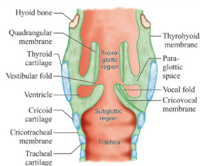
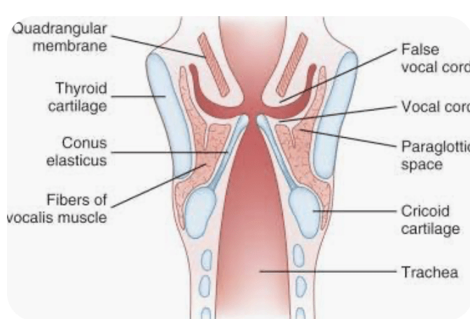
The paraglottic space is a tetrahedral-shaped compartment bounded by several cartilaginous and fibroelastic structures:
Anterolaterally:
The inner surface of the thyroid cartilage
Superomedially:
The quadrangular membrane:
Which separates it from the false vocal folds
The space is continuous with (or, in some specimens, separated by a collagenous septum from) the preepiglottic space
Inferomedially:
The conus elasticus:
Which separates it from the subglottis
Posteriorly / Posterolaterally / Dorsally:
The mucosal lining of the piriform sinus
Anteriorly:
It communicates directly with the pre-epiglottic space
Content of the Space:
The paraglottic space houses several vital neurovascular and muscular structures embedded within its protective layer of loose areolar and adipose tissue:
Adipose tissue:
Abundant fat:
That makes the space highly visible on CT and MRI scans
Intrinsic laryngeal muscles:
Including the thyroarytenoid, lateral cricoarytenoid, and posterior cricoarytenoid muscles
Blood vessels and nerves:
Glands, blood vessels, and branches of the superior and inferior laryngeal nerves
Clinical Significance:
The primary clinical importance of the paraglottic space:
Lies in laryngeal oncology and tumor staging:
Transglottic Spread:
Because the PGS crosses the level of the laryngeal ventricle without physical barriers:
A squamous cell carcinoma originating in the true vocal cords can easily migrate vertically through this space into the false vocal cords (or vice versa):
This is known as transglottic spread
Cancer Staging:
Invasion of the inferior paraglottic space can limit vocal fold mobility
In the TNM staging system, tumor invasion into the paraglottic space:
Automatically elevates a glottic or supraglottic laryngeal cancer to a T3 stage:
Altering the treatment trajectory from localized therapy to systemic options or extensive surgery
Surgical Planning:
Knowledge of the paraglottic space borders is vital for performing voice-sparing, partial laryngectomies
If a tumor completely infiltrates this space:
A total laryngectomy may be required to ensure clean margins
References:
Joo YH, Park JO, Cho KJ, Kim MS. Relationship between paraglottic space invasion and cervical lymph node metastasis in patients undergoing supracricoid partial laryngectomy. Head Neck. 2012.
Tamaki A, Miles BA, Lango M, Kowalski L, Zender CA. AHNS Series: Do you know your guidelines? Review of current knowledge on laryngeal cancer. Head Neck. 2018.
Reidenbach MM. The Paraglottic Space and Transglottic Cancer: Anatomical Considerations. Clin Anat. 1996.
Reidenbach MM. Borders and Topographic Relationships of the Paraglottic Space. Eur Arch Otorhinolaryngol. 1997.
Joo YH, Park JO, Cho KJ, Kim MS. Relationship between paraglottic space invasion and cervical lymph node metastasis in patients undergoing supracricoid partial laryngectomy. Head Neck. 2012.
Ferrari M, Schreiber A, Mattavelli D, et al. Surgical anatomy of the parapharyngeal space: Multiperspective, quantification-based study. Head Neck. 2019.
Wang XR, Wang H, Gao Y, et al. Study on the Ultrasonic Characteristics of the Paraglottic Space at the Glottic Level in Normal Adults. Head Neck. 2026.
Virós Porcuna D, Pollán Guisasola CM, Viña Soria C, et al. Transoral robotic parapharyngeal space dissection. Head Neck. 2024.
Mohamed A, Paleri V, George A. A cadaveric study quantifying the anatomical landmarks of the facial artery and its parapharyngeal branches for safe transoral surgery. Head Neck. 2019.
Hearn MW, Vogel CT, Laughlin RM, et al. Review of Spaces. Atlas of Operative Oral and Maxillofacial Surgery. 2022.
Dammann F, Wartenberg J. Diseases of the Head and Neck. Radiology-Nuclear Medicine Diagnostic Imaging. 2023.
Primary squamous cell carcinoma of the parotid gland (pSCCP):
Is a rare, aggressive salivary gland malignancy diagnosed only by exclusion:
After metastatic / secondary spread from a cutaneous or mucosal head and neck SCC, high-grade mucoepidermoid carcinoma, salivary duct carcinoma with squamous differentiation, and direct extension have all been ruled out
It is a contested entity:
Contemporary genomic data show that nearly all “parotid SCCs” carry a UV-induced mutational signature indicating cutaneous origin:
So the great majority of cases coded as primary are in fact metastatic (mSCCP)
Definition and the diagnosis-of-exclusion problem
True pSCCP is thought to arise from squamous metaplasia of excretory duct epithelium:
By convention requires no evidence of any other primary squamous source
pSCCP and mSCCP are histologically nearly identical:
Every criterion proposed to favor a primary (e.g., ductal dysplasia) can also occur when metastatic tumor grows along preexisting ducts
SCC differentiation can also be seen in genetically defined salivary carcinomas :
SCC accounts for roughly 0.3% to 1.5% of salivary gland tumors in classic series:
Though registry data (which cannot distinguish primary from metastatic) label a much higher share:
Up to ~ 23% of parotid cancers in one German national study, and ~ 2% of parotid neoplasms at a single institution after strict histologic review
Registry-based incidence has risen (~ 0.87 to 2.0 per million in SEER over 42 years):
Paralleling rising cutaneous SCC incidence and an aging population:
Most likely reflecting misclassified metastatic disease
The German registry found:
SCC-type parotid cancer was strongly associated with a prior head and neck SCC (adjusted OR 5.48), median interval 365 days:
Supporting frequent secondary origin
Typical patient:
Elderly (7th decade)
Male predominance:
Roughly 2:1
Presentation is often advanced, with a parotid mass, facial nerve dysfunction, and / or cervical nodes:
The majority present at stage IV
Pathohistology and immunohistochemistry:
Keratinizing to nonkeratinizing infiltrative squamous nests with intercellular bridges / keratinization:
Keratinization is seen in ~1 in 6 metastatic SCC cases and is rare in the main mimics
Squamous markers p40, p63, CK5/6 are positive:
Mucicarmine should be negative:
Positive mucin points to mucoepidermoid carcinoma
Androgen receptor (AR) and / or HER2 positivity:
Should raise salivary duct carcinoma with squamous differentiation rather than pSCCP:
AR / CK7 expression does not exclude SDC
FNA differential includes:
Warthin tumor (well-differentiated cases)
High-grade mucoepidermoid carcinoma
Salivary duct carcinoma
UV-signature mutational analysis:
Is now the most useful ancillary tool to establish cutaneous (metastatic) origin
Work-up:
The NCCN Head and Neck Cancers guidelines outline evaluation of any parotid mass:
Complete H&P with full head and neck exam (including mirror / fiberoptic exam), FNA biopsy, and, as clinically indicated, contrast CT / MRI from skull base to clavicle, chest CT, dental / nutrition / speech evaluation, and multidisciplinary consultation
Because SCC histology mandates excluding a metastatic source:
Workup should additionally include a thorough cutaneous exam of the face / scalp / ear (cutaneous SCC is the most common tumor to metastasize to the parotid), and an occult-primary / mucosal SCC evaluation when no skin primary is found:
EUA, direct laryngoscopy, tonsillectomy, chest / abdomen / pelvis CT or FDG-PET/CT, with p16 / HPV and EBV testing
FNA:
Reliably distinguishes benign from malignant (sensitivity ~ 80%, specificity ~ 97%) and is highly accurate for high-grade cancers
Staging:
Historically staged with the AJCC 8th edition (2017) TNM for major salivary glands
T category is size / extraparenchymal-extension based:
T1 ≤ 2 cm
T2 > 2 to 4 cm
T3 > 4 cm and / or extraparenchymal extension
T4a skin / mandible / ear canal / facial nerve
T4b skull base / pterygoid plates / carotid encasement)
An important caveat:
The newly published AJCC / UICC 9th edition (TNM9) creates a unified salivary-gland-specific system but explicitly excludes squamous cell carcinoma (along with neuroendocrine and basosquamous carcinoma) from salivary gland staging:
Practically, parotid SCC is therefore staged using the head and neck cutaneous / mucosal SCC or cervical-node / unknown-primary frameworks rather than the salivary-gland schema
Management:
No prospective trials exist; management is extrapolated from salivary gland and cutaneous / metastatic SCC guidelines and retrospective series
The consistent message across data sets is that surgery is essential:
Outcomes without resection are poor
The core approach for resectable disease is:
Total (at least superficial) parotidectomy + neck dissection + adjuvant radiotherapy
Per NCCN for major salivary gland cancers:
Primary treatment:
Complete surgical resection, with neck dissection for high-grade and / or T3 to 4 tumors (or clinically N+ disease):
The facial nerve is preserved when feasible
Adjuvant therapy:
Adjuvant RT is preferred for adverse features:
High grade, close / positive margins, perineural / neural invasion, nodal metastases, lymphovascular invasion, or high-grade T3 to T4a tumors (SCC qualifies as high grade)
–Unresectable / gross residual disease:
Definitive RT, or concurrent systemic therapy / RT (category 2B)
ASCO guidelines:
Recommend that for advanced (T3 to T4) or high-grade parotid cancers, at least a superficial parotidectomy with removal of additional at-risk parotid tissue be performed (given planned adjuvant RT):
Balancing oncologic clearance against facial nerve risk:
There are no data that more aggressive parotidectomy improves survival or locoregional control when adjuvant RT is used
Prognosis:
Uniformly aggressive regardless of primary-vs-metastatic classification
Reported median survival is roughly 13 to 24 months, and 5-year overall survival does not exceed ~ 50%
Larger tumor size, extraparenchymal extension, nodal metastases, distant metastases, and advanced age independently predict worse survival
Recurrent disease carries a particularly poor prognosis (median ~14.5 months)
Secondary (metastatic) parotid SCC has worse survival than true primary salivary cancers and higher rates of facial nerve sacrifice
References:
Primary Squamous Cell Carcinoma of the Parotid Gland: Study and Review of the Literature. Horáková Z, Stárek I, Kalfert D, et al. In Vivo (Athens, Greece). 2024 Jan-Feb;38(1):358-364. doi:10.21873/invivo.13446.
Metastatic Cutaneous Squamous Cell Carcinoma Accounts for Nearly All Squamous Cell Carcinomas of the Parotid Gland. Bradley PJ, Stenman G, Thompson LDR, et al. Virchows Archiv : An International Journal of Pathology. 2024;485(1):3-11. doi:10.1007/s00428-024-03798-5.
Squamous Carcinoma in a Major Salivary Gland: A Review of the Diagnostic Considerations. Taxy JB. Archives of Pathology & Laboratory Medicine. 2001;125(6):740-5. doi:10.5858/2001-125-0740-SCIAMS.
Histogenesis of Salivary Gland Neoplasms. Regezi JA, Batsakis JG. Otolaryngologic Clinics of North America. 1977;10(2):297-307.
Salivary duct carcinoma with squamous differentiation: histomorphological and immunophenotypical analysis of six cases. Dababneh MN, Griffith CC, Magliocca KR, Stojanov IJ. Histopathology. 2024;85(4):590-597. doi:10.1111/his.15217.
Squamous cell carcinoma of the parotid gland. Ying YL, Johnson JT, Myers EN. Head & Neck. 2006;28(7):626-32. doi:10.1002/hed.20360.
Squamous Cell Carcinoma of the Parotid Gland: A Population-Based Registry Study of Primary Versus Metastatic Disease. Oesterling F, Möller L, Kajüter H, et al. Cancer Epidemiology. 2026;102:103085. doi:10.1016/j.canep.2026.103085.
Primary Squamous Cell Carcinoma of the Parotid Gland: The Importance of Correct Histological Diagnosis. Flynn MB, Maguire S, Martinez S, Tesmer T. Annals of Surgical Oncology. 1999;6(8):768-70. doi:10.1007/s10434-999-0768-y.
Incidence and survival trends of parotid malignancies over 42 years. Gupta A, Koochakzadeh S, Neskey DM, Nguyen SA, Lentsch EJ. Head & Neck. 2020;42(9):2308-2315. doi:10.1002/hed.26172.
Prognostic factors for squamous cell cancer of the parotid gland: An analysis of 2104 patients. Chen MM, Roman SA, Sosa JA, Judson BL. Head & Neck. 2015;37(1):1-7. doi:10.1002/hed.23566.
The key radiologic and cytomorphologic features of oncocytic and oncocytoid lesions of the salivary gland. Lubin D, Song S, Zafar HM, Baloch Z. Diagnostic Cytopathology. 2019;47(6):617-636. doi:10.1002/dc.24175.
Head and Neck Cancers. National Comprehensive Cancer Network. Updated 2026-05-12.
Cutaneous squamous cell carcinoma of the head and neck metastasizing to the parotid gland—A review of current recommendations. O’Hara J, Ferlito A, Takes RP, et al. Head & Neck. 2011;33(12):1789-95. doi:10.1002/hed.21583.
Occult Primary (Cancer of Unknown Primary [CUP]). National Comprehensive Cancer Network. Updated 2026-05-04.
Management of Salivary Gland Malignancy: ASCO Guideline. Geiger JL, Ismaila N, Beadle B, et al. Journal of Clinical Oncology : Official Journal of the American Society of Clinical Oncology. 2021;39(17):1909-1941. doi:10.1200/JCO.21.00449.
Key Updates on the Version 9 AJCC/UICC Staging System for Salivary Gland Carcinoma. Huang SH, Seethala RR, Patel SG, et al. Annals of Surgical Oncology. 2026;33(6):4958-4963. doi:10.1245/s10434-026-19350-5.
Proposed Version Nine of the AJCC and UICC TNM Classification for Salivary Gland Carcinoma. Huang SH, Cotler J, Palis B, et al. JAMA Otolaryngology– Head & Neck Surgery. 2026;152(4):366-375. doi:10.1001/jamaoto.2025.5396.
Primary Squamous Cell Carcinoma of the Parotid Gland: Clinicopathological Characteristics, Treatment, and Prognosis. Xiao M, Liu J, You Y, Yang X, Wang Y. International Journal of Oral and Maxillofacial Surgery. 2021;50(2):151-157. doi:10.1016/j.ijom.2020.06.010.
Primary and Secondary Tumors of the Parotid Gland: Clinical Features and Prognosis. Pecorari G, Pizzo C, Briguglio M, Cravero E, Riva G. Cancers. 2023;15(4):1293. doi:10.3390/cancers15041293.
The third (terminal) part of the maxillary artery and its accompanying branches
Nerve of the pterygoid canal (Vidian nerve):
Carrying sympathetic (via the deep petrosal nerve) and parasympathetic fibers (via the greater petrosal nerve) into the fossa
Communications and Openings:
The fossa communicates with surrounding anatomic regions via several key foramina and fissures:
Middle cranial fossa:
Via the foramen rotundum
Orbit:
Via the inferior orbital fissure
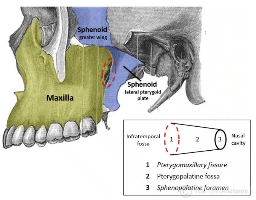
Nasal cavity:
Via the sphenopalatine foramen
Oral cavity / Palate:
Via the greater and lesser palatine canals
Infratemporal fossa:
Via the pterygomaxillary fissure
Pharynx / Nasopharynx:
Via the pharyngeal / palatovaginal canal
Clinical Significance:
Because of its numerous pathways and high density of nerves and blood vessels:
The pterygopalatine fossa can act as a conduit for the spread of infections and malignant tumors from the face or nasal cavity into the cranial vault
Surgeons also target this area for nerve blocks or approaches to control severe bleeding (epistaxis)
Left infratemoporal fossa demonstrating the opening of the pterygopalatine fossa (circled in red). Note: the zygomatic arch has been removed in this image.Screenshot
Remains one of the most powerful and actionable prognostic factors in oral tongue / oral cavity SCC
Classically, Scholl and colleagues:
Reviewed 268 patients with squamous carcinoma of the oral tongue:
They found that 54 (20.1%) had microscopic “cut-through” at the intraoperative frozen section margin:
An initially positive margin
Even when additional resection converted these to final negative margins:
Local control remained significantly worse:
Than in patients whose margins were clear on the first pass
They also reported that margin involvement patternsdiffered by T stage:
T1 to T2 tumors:
More often had positive mucosal margins
T3 to T4 tumors:
Commonly failed at the deep / soft-tissue margins
Similar observations were made in classic series evaluating “positive” epidermoid carcinoma margins in the head and neck:
Looser, Shah, and Strong:
Demonstrated that patients with involved margins:
Had substantially higher local recurrence than those with negative margins:
With early reports quoting local recurrence in roughly two-thirds to three-quarters of patients with positive margins versus about one-third with negative margins
Loree and Strong:
Subsequently examined 398 oral cavity SCCs:
Showing that positive or “close” margins (tumor at or within 0.5 mm of the inked edge, or significant premalignant change / in situ carcinoma at the margin):
Were associated with a doubling of local recurrence (36% vs 18%) and inferior 5-year survival compared with negative margins (52% vs 60%)
The reliability and utility of intraoperative frozen section (FS) margin assessment have also been extensively studied:
Spiro et al:
Reported an overall intraoperative FS diagnostic accuracy of:
Roughly 89% for oral tongue cancer:
Importantly found that accuracy was similar whether sections were taken directly from the patient’s tumor bed or from the oriented surgical specimen
Positive or “close” margins:
Defined in their series as tumor present at the ink or within roughly one high-power field of the resection edge:
Were associated with a significantly increased risk of local recurrence (p < 0.003)
Subsequent work by Byers and others:
Confirmed the prognostic and therapeutic value of frozen section (FS):
Guided re-resection in HNSCC:
But also highlighted that FS cannot fully compensate for suboptimal initial resection planes
Definitions of margin status and distance:
There is now better consensus on margin nomenclature
Most contemporary series and guidelines define:
Positive margin:
Invasive carcinoma or severe / high-grade dysplasia:
At the inked edge, or < 1 mm from the inked edge
Close margin:
Invasive carcinoma typically 1 to 4 or 1 to 5 mm from the inked edge:
Cut-off values vary:
But a 5 mm microscopic threshold is most commonly used in oral cavity SCC
Clear margin:
≥ / > 5 mm from invasive tumor to the inked edge after formalin fixation
Tasche et al., in a large JAMA Otolaryngology analysis:
Proposed that a distance < 1 mm behaves biologically like an involved margin:
With similarly high local recurrence risk
Whereas 1 to 5 mm margins had intermediate risk and ≥ 5 mm margins were associated with the lowest recurrence
More recent multicenter work emphasizes the importance of deep margin distance in particular:
With data suggesting that deep margins ≤ 3 mm carry a significantly higher risk of local failure compared with > 3 mm:
Even when the mucosal margin is wide
Impact of positive and close margins on outcomes:
Multiple retrospective series and meta-analyses now support and refine the early observations of Scholl, Looser, Loree, and Strong:
Positive final margins are consistently associated with:
~ 2-fold higher risk of local recurrence, increased regional / distant failure in some series, and significantly worse disease-specific and overall survival
Binahmed et al. and McMahon et al:
Both showed that patients with involved margins:
Had roughly double the local recurrence and significantly poorer survival compared with those with clear margins, and that close margins behaved intermediately between clearly negative and frankly positive margins
Liao et al:
Identified margin status, together with T stage, DOI, and perineural invasion:
As major predictors of local tumor control in oral cavity SCC
A 2019 systematic review and meta-analysis by Gorphe:
Concluded that positive margins carry an approximately two-fold increased risk of death and local failure across head and neck sites, independent of other factors
More granular contemporary analyses, including Buchakjian et al. and Szewczyk et al., have shown that:
Close margins (1 to 4.9 mm) often do not independently worsen outcomes if other adverse factors (lymphovascular invasion, perineural invasion, ENE, nodal disease, advanced T stage):
Are absent and if appropriate adjuvant therapy is given when indicated
The prognostic effect of close margins is modulated by:
Depth of invasion (DOI), pattern of invasion, and composite histologic risk models (e.g., Brandwein-Gensler)
Subsite-specific studies have further refined this:
Tongue and floor-of-mouth tumors are particularly prone to failure at the deep margin:
In several series, deep margin positivity or ≤ 2 to 3 mm clearance:
Has been more predictive of local recurrence than mucosal margin distance
Microscopic cut-through and “revised” margins:
Building on Scholl’s original work, the concept of microscopic tumor cut-through (MTCT):
A positive FS margin that is revised to negative on final pathology, has been extensively studied
Patel et al. (Head & Neck 2010) showed that MTCT:
Was associated with significantly worse local control and disease-specific survival compared with margins that were negative from the outset:
Particularly in patients with nodal disease
Guillemaud et al. similarly reported that intraoperative cut-through, even if revised to R0:
Predicted higher local recurrence and worse outcomes in oral cavity SCC
A meta-analysis by Bulbul et al. concluded that clearance of a positive margin improves outcomes relative to leaving it unrevised:
But patients with MTCT still fare worse than those whose margins were always negative:
Suggesting MTCT is a marker of more aggressive biology and / or challenging local anatomy
More recently, Agne et al. evaluated T3 to T4 OCSCC and confirmed that MTCT:
Remained an independent predictor of local recurrence on multivariable analysis (HR ~1.8–2.2 for local failure):
Although its effect on disease-specific survival attenuated when controlling for nodal stage and other high-risk features
These data support considering MTCT as a high-risk feature warranting discussion of treatment intensification:
For example (e.g., adjuvant chemoradiotherapy) in a multidisciplinary tumor board, even when final margins are technically negative
Kwok et al. addressed the related question of “clear versus revised margins” in 417 patients with oral and pharyngeal carcinoma:
Patients who required immediate re-resection for a positive FS margin but ended with R0 status:
Had survival similar to those with primary R0 resection, and both groups did substantially better than patients left with residual microscopic or macroscopic disease
This suggests that while MTCT carries biologic risk:
An aggressive intraoperative strategy to convert to R0 is still beneficial and should remain standard practice
Intraoperative margin assessment:
Specimen vs tumor bed:
There is growing recognition that how margins are sampled:
Is almost as important as the final measurement
Meier et al.’s AHNS survey and several subsequent series have documented wide variation in intraoperative margin practices (tumor bed vs specimen mapping, number of samples, definition of “adequate” clearance), and a substantial rate of FS–permanent section discrepancy
Key contemporary points include:
Specimen-based mapping (oriented and inked, with communication between surgeon and pathologist):
Tends to provide more reliable correlation between FS and final margins than random tumor-bed biopsies
FS accuracy remains high (often ~ 85% to 95%):
But false-negatives and false-positives still occur:
Particularly at the deep margin, in previously irradiated fields, and in specimens with significant shrinkage
In some series, “complete FS margins” with a measurable 1 to 5 mm histologic buffer were associated with improved local control compared with conventional limited sampling
Recent reviews and consensus statements (e.g., Kubik et al., Kain et al., Chen et al. 2024) now recommend:
A planned 1 to 1.5 cm gross resection margin in vivo for oral tongue SCC, anticipating ~30% to 50% shrinkage with formalin fixation and tissue relaxation
Routine use of oriented, inked specimens with targeted FS from high-risk areas (deep margin, close relationship to muscle bundles or neurovascular structures)
Consideration of advanced adjuncts—near-infrared fluorescence mapping, specimen 3D-mapping, and emerging augmented-reality registration—for difficult tongue and floor-of-mouth resections
Integration with histologic risk models:
Finally, margin status must be interpreted in the context of overall histologic risk
The Brandwein-Gensler model:
Worst pattern of invasion, perineural invasion, lymphocytic host response and later refinements:
Have shown that high-risk tumors have markedly increased recurrence and disease-specific mortality even when margins are clear
Conversely, some low-risk early-stage tumors with close (but not involved) margins may do well without aggressive adjuvant therapy
This supports a nuanced, risk-adapted approach in which:
Positive margins or MTCT → strong indication for adjuvant chemoradiotherapy in most patients
Close margins (1 to 4 mm) → individualized decision based on DOI, nodal status, PNI/LVI, pattern of invasion, and patient-specific factors
Clear margins (≥ 5 mm) → lowest risk group, managed according to other adverse features
Reviewed:
Scholl P, Byers RM, Batsakis JG, Wolf P, Santini H. Microscopic cut-through of cancer in the surgical treatment of squamous carcinoma of the tongue: prognostic and therapeutic implications. Am J Surg. 1986;152:354-360.
Looser KG, Shah JP, Strong EW. The significance of “positive” margins in surgically resected epidermoid carcinomas. Head Neck Surg. 1978;1:107-111.
Loree TR, Strong EW. Significance of positive margins in oral cavity squamous carcinoma. Am J Surg. 1990;160:410-414.
Spiro RH, Guillamondegui O, Paulino AF, et al. Pattern of invasion and margin assessment in patients with oral tongue cancer. Head Neck. 1999;21:408-413.
Chen TY, Emrich LJ, Driscoll DL. The clinical significance of pathological findings in surgically resected margins of the primary tumor in head and neck carcinoma. Int J Radiat Oncol Biol Phys. 1987;13:833-837.
McMahon J, O’Brien CJ, Pathak I, et al. Influence of condition of surgical margins on local recurrence and disease-specific survival in oral and oropharyngeal cancer. Br J Oral Maxillofac Surg. 2003;41:224-231.
Binahmed A, Nason RW, Abdoh AA. The clinical significance of the positive surgical margin in oral cancer. Oral Oncol. 2007;43:780-784.
Liao CT, Chang JTC, Wang HM, et al. Analysis of risk factors of predictive local tumor control in oral cavity cancer. Ann Surg Oncol. 2008;15:915-922.
Patel RS, Goldstein DP, Guillemaud J, et al. Impact of positive frozen section microscopic tumor cut-through revised to negative on oral carcinoma control and survival rates. Head Neck. 2010;32:1444-1451.
Guillemaud J, Patel RS, Goldstein DP, et al. Prognostic impact of intraoperative microscopic cut-through on frozen section in oral cavity squamous cell carcinoma. J Otolaryngol Head Neck Surg. 2010;39:370-377.
Kwok P, Gleich O, Hübner G, Strutz J. Prognostic importance of “clear versus revised margins” in oral and pharyngeal cancer. Head Neck. 2010;32:1479-1484.
Gorphe P. A systematic review and meta-analysis of margins in head and neck cancer. Oral Oncol. 2019;95:93-101.
Tasche KK, Buchakjian MR, Pagedar NA, Sperry SM. Definition of “close margin” in oral cancer surgery and association of margin distance with local recurrence rate. JAMA Otolaryngol Head Neck Surg. 2017;143:1166-1172.
Buchakjian MR, Tasche KK, Robinson RA, et al. Association of main specimen and tumor bed margin status with local recurrence and survival in oral cancer surgery. JAMA Otolaryngol Head Neck Surg. 2016;142:1191-1198.
Kain JJ, Birkeland AC, Udayakumar N, et al. Surgical margins in oral cavity squamous cell carcinoma: current practices and future directions. Laryngoscope. 2020;130:128-138. Szewczyk M, et al. A matter of margins in oral cancer—how close is enough? Cancers (Basel). 2024;16(8):1488.
Agne GR, et al. Oncologic outcomes of microscopic tumor cut-through in locally advanced oral squamous cell carcinoma. Arch Head Neck Surg. 2022;51:e20220013. Chen Y, et al. Surgical margins in head and neck squamous cell carcinoma. Int J Surg. 2024;109:54-66.
Brandwein-Gensler M, et al. Oral squamous cell carcinoma: histologic risk assessment, but not margin status, is strongly predictive of local disease-free and overall survival. Am J Surg Pathol. 2005;29:167-178.
Does adding concurrent chemotherapy to definitive RT improve outcomes vs RT alone in stage III to IV oropharyngeal cancer? PubMed
Who was enrolled?
n=226 adults with stage III to IV oropharynx SCC:
No planned primary surgery
Arms well balanced by age, sex, stage, PS, site PubMed
Treatment and dosing (the “GORTEC” CRT)
RT (both arms):
70 Gy in 35 fractions
CRT arm chemotherapy:
Carboplatin 70 mg/m²/day (IV bolus) + 5-FU 600 mg/m²/day (continuous infusion), days 1 to 4, given during weeks 1, 4, and 7 of RT (total 3 cycles) PubMed+1
Endpoints:
Primary intent:
Improve DFS:
Reported OS, DFS, and locoregional control (LRC) at 3 and 5 years; plus acute and late toxicity PubMed+1
Perioperative Immunotherapy – KEYNOTE-689 Continues to Shape Practice:
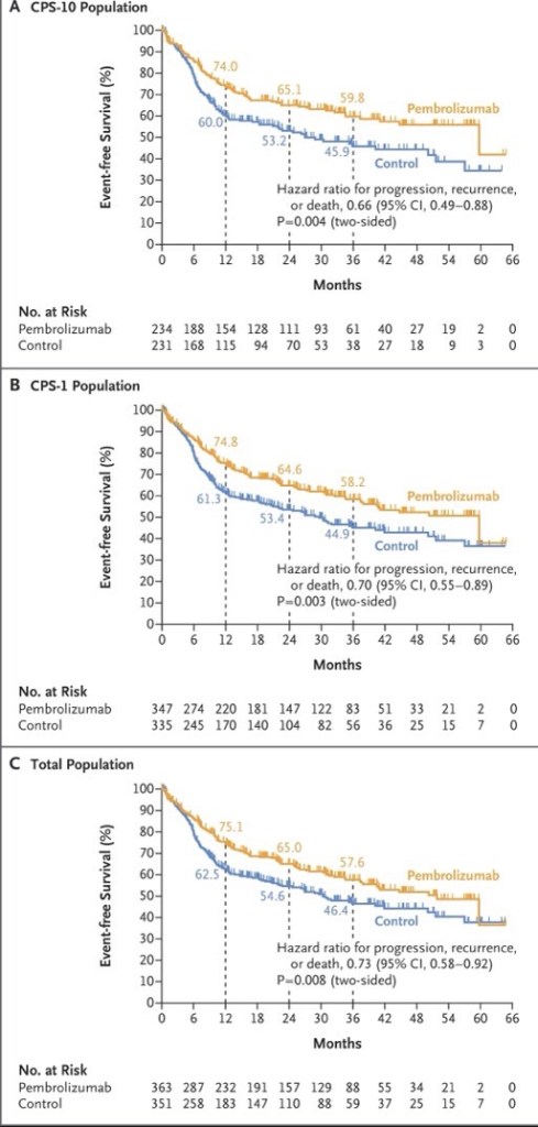
The KEYNOTE-689 phase 3 trial:
Which led to the FDA approval of perioperative pembrolizumab (June 2025):
For resectable locally advanced HNSCC with PD-L1 CPS ≥ 1:
Remains a central focus
New ASCO 2026 exploratory analyses demonstrate that the EFS benefit of neoadjuvant / adjuvant pembrolizumab:
Persists across surgical outcome subgroups, including:
Patients with and without extranodal extension or positive margins
Notably, fewer patients in the pembrolizumab arm had ENE or positive margins post-surgery:
Suggesting neoadjuvant pembrolizumab contributes to pathologic downstaging
A pooled meta-analysis of three phase 3 trials (KEYNOTE-689, NIVOPOSTOP, and IMvoke010; n=1,786) presented at ASCO 2026:
Confirmed a significant improvement in event-free / disease-free survival with perioperative or adjuvant PD-1 / PD-L1 blockade (pooled HR 0.79, 95% CI 0.68–0.91):
With no meaningful increase in treatment-related deaths
The following figure from the KEYNOTE-689 trial illustrates the event-free survival benefit across PD-L1 subgroups:
Neoadjuvant Bispecific Antibody Combinations: Ivonescimab Leads the Way:
Several ASCO 2026 abstracts highlight the emerging role of bispecific antibodies in the neoadjuvant setting:
All patients with CPS >30 achieved pCR, and 100% laryngeal / pharyngeal preservation was achieved
A randomized phase II trial comparing ivonescimab (PD-1 / VEGF), cadonilimab (PD-1 /CTLA-4), and penpulimab (PD-1 alone), each combined with chemotherapy:
Showed the highest pCR rate with ivonescimab at 60%, compared to 42.1% with cadonilimab and 40% with single-agent PD-1
An 87.5% ORR and a 95.8% larynx preservation rate in resectable LA-HNSCC:
With all p16-positive and CPS ≥ 20 patients responding
Novel Agents in Recurrent / Metastatic HNSCC:
Bispecific antibodies are generating significant excitement in the recurrent / metastatic setting:
Ficerafusp alfa (EGFR × TGF-β) + pembrolizumab:
Two-year follow-up data showed a confirmed ORR of 54% in HPV-negative R/M HNSCC (21% CR), with a median DOR of 21.7 months and median OS of 21.3 months
The phase 2 / 3 FORTIFI-HN01 trial is now actively enrolling
Petosemtamab (EGFR × LGR5) + pembrolizumab:
Updated phase 2 data showed a 60% ORR (including 5 CRs) in first-line PD-L1+ R/M HNSCC, with median DOR of 11 months
Two phase 3 trials (LiGeR-HN1 and LiGeR-HN2) are recruiting
CRB-701 (Nectin-4 ADC):
A phase 1 / 2 study in heavily pretreated R/M HNSCC (85% refractory to immunotherapy and platinum):
Showed a confirmed ORR of 33.3% at both 2.7 and 3.6 mg/kg doses, regardless of HPV status
Becotatug vedotin (EGFR ADC):
A new randomized phase II trial is evaluating neoadjuvant becotatug vedotin alone or combined with immune checkpoint inhibitors in resectable LA-HNSCC
The deep response rate was 87.9%, with 86% of patients receiving de-escalated definitive therapy
At 23 months median follow-up, 2-year PFS was 86% and OS was 100%
Circulating tumor HPV-DNA was significantly associated with recurrence
Evolving First-Line R / M HNSCC Pipeline:
A landscape analysis presented at ASCO 2026 identified 145 active regimens (111 unique assets) in the first-line R / M HNSCC pipeline
Key trends include a shift toward chemotherapy-free combinations alongside PD-1 blockade:
With bispecific antibodies (10.8%), ADCs (10.8%), and cancer vaccines (9.0%) representing the most common novel modalities
Eight key phase 3 trials are underway:
All incorporating PD-1 as a target, with pembrolizumab as the backbone in 7 of 8
Overall, ASCO 2026 highlights a transformative period in head and neck oncology:
With perioperative immunotherapy now established as a new standard, bispecific antibodies and ADCs showing compelling early efficacy, and biomarker-driven strategies (PD-L1 CPS, HPV status, ctHPV-DNA, MRD) increasingly guiding treatment selection
References
Neoadjuvant and Adjuvant Pembrolizumab in Locally Advanced Head and Neck Cancer. Uppaluri R, Haddad RI, Tao Y, et al. The New England Journal of Medicine. 2025;393(1):37-50. doi:10.1056/NEJMoa2415434.
FDA approves neoadjuvant and adjuvant pembrolizumab for resectable locally advanced head and neck squamous cell carcinoma | FDA. Food and Drug Administration. 2025-06-13.
Neoadjuvant and adjuvant pembrolizumab (pembro) plus standard of care (SOC) for resectable locally advanced head and neck squamous cell carcinoma (LA HNSCC): Efficacy by surgical outcomes in the phase 3 KEYNOTE-689 trial.. Adkins D, Haddad R, Tao Y, et al. Journal of Clinical Oncology. 2026;44(Suppl 16):6057. doi:10.1200/JCO.2026.44.16_suppl.6057.
Perioperative or adjuvant PD-1/PD-L1 blockade with curative-intent multimodality therapy for locally advanced head and neck squamous cell carcinoma: A systematic review and meta-analysis of randomized trials.. Daher S, Daher H, Altal H, et al. Journal of Clinical Oncology. 2026;44(Suppl 16):e18074. doi:10.1200/JCO.2026.44.16_suppl.e18074.
Neoadjuvant ivonescimab (AK112, a PD-1/VEGF bispecific antibody) combined with nab-paclitaxel and cisplatin (AP) for resectable locally advanced head and neck squamous cell carcinoma (LA-HNSCC): An exploratory phase II study.. Kunyu Yang, Xiaomeng Zhang and Lu Wen. Journal of Clinical Oncology. 2026;44(Suppl 16):6014. doi:10.1200/JCO.2026.44.16_suppl.6014.
Neoadjuvant immunotherapy in combination with chemotherapy in resectable locally advanced head and neck squamous cell carcinoma: Updated efficacy and safety data from a randomized phase II trial.. Liu L, Chen F, Li Y, et al. Journal of Clinical Oncology. 2026;44(Suppl 16):6091. doi:10.1200/JCO.2026.44.16_suppl.6091.
Neoadjuvant adebrelimab plus chemotherapy in untreated locally advanced head and neck squamous cell carcinoma: Efficacy and biomarker insights from a single-arm phase 2 trial.. Fang R, Lei W, Huang B, et al. Journal of Clinical Oncology. 2026;44(Suppl 16):6106. doi:10.1200/JCO.2026.44.16_suppl.6106.
Ficerafusp Alfa (BCA101) With Pembrolizumab for Recurrent or Metastatic Head and Neck Squamous Cell Carcinoma: Two-Year Results of an Expansion Cohort of a Phase I/Ib Trial. Hanna GJ, Zandberg DP, Wong DJ, et al. Journal of Clinical Oncology : Official Journal of the American Society of Clinical Oncology. 2026;:JCO2502027. doi:10.1200/JCO-25-02027.
A multicenter, randomized, double-blind, phase 2/3 study of ficerafusp alfa (BCA101) or placebo in combination with pembrolizumab for first-line treatment of HPV-negative, PD-L1–positive, recurrent or metastatic (R/M) head and neck squamous cell carcinoma (HNSCC): FORTIFI-HN01.. Ferrarotto R, Kaczmar J, Spigel D, et al. Journal of Clinical Oncology. 2026;44(Suppl 16):TPS6129. doi:10.1200/JCO.2026.44.16_suppl.TPS6129.
Petosemtamab (MCLA-158) with pembrolizumab as first-line (1L) treatment of PD-L1+ recurrent/metastatic (r/m) head and neck squamous cell carcinoma (HNSCC): Phase 2 trial. Herpen C, Daste A, Arrazubi V, et al. Journal of Clinical Oncology. 2025;43(Suppl 16):6024. doi:10.1200/JCO.2025.43.16_suppl.6024.
LiGeR-HN Phase III Trials of Petosemtamab + Pembrolizumab and Petosemtamab Monotherapy in Recurrent or Metastatic HNSCC. Machiels JP, Fayette J, Haddad R, et al. Future Oncology (London, England). 2025;21(16):2007-2016. doi:10.1080/14796694.2025.2511470.
A phase 1/2 study of the next-generation nectin-4–targeting antibody-drug conjugate CRB-701 (SYS6002) in patients with recurrent or metastatic head and neck squamous cell carcinoma.. Mantia C, Hanna G, Loriot Y, et al. Journal of Clinical Oncology. 2026;44(Suppl 16):6062. doi:10.1200/JCO.2026.44.16_suppl.6062.
A randomized, non-comparative, multicenter phase II trial of neoadjuvant becotatug vedotin alone or combined with immune checkpoint inhibitors (penpulimab/ivonescimab) in resectable locally advanced head and neck squamous cell carcinoma.. Wei X, Xiang Z, Zeng Y, et al. Journal of Clinical Oncology. 2026;44(Suppl 16):TPS6135. doi:10.1200/JCO.2026.44.16_suppl.TPS6135.
Neoadjuvant HPV16-specific viral immunotherapy (HB200) plus chemotherapy with response-adapted de-escalation in HPV16+ oropharyngeal squamous cell carcinoma: TARGET-HPV trial.. Rosenberg A, Juloori A, Cursio J, et al. Journal of Clinical Oncology. 2026;44(Suppl 16):6097. doi:10.1200/JCO.2026.44.16_suppl.6097.
Emerging trends in research strategies in the first-line recurrent or metastatic head and neck cancer (R/M SCCHN) landscape: A top-level analysis by Oncofocus.. Shukla A, Keeshara V, Chamaria M, et al. Journal of Clinical Oncology. 2026;44(Suppl 16):e18029. doi:10.1200/JCO.2026.44.16_suppl.e18029.
Recent Highlights and Breakthroughs in Immunotherapy for Head and Neck Cancers. Vuille JA, Szturz P. Current Opinion in Oncology. 2026;38(3):201-211. doi:10.1097/CCO.0000000000001211.
Immunotherapy in Locally Advanced Head and Neck Squamous Cell Carcinoma: The Current Status and Future Outlook. Köylü B, Selçukbiricik F, Aksoy S, Güven DC. Critical Reviews in Oncology/Hematology. 2026;:105145. doi:10.1016/j.critrevonc.2026.105145.
The Groupe d’Oncologie Radiotherapie Tete et Cou trial evaluated the concomitant approach in patients with oropharynx cancer only
A total of 226 patients were randomly assigned to either:
Radiation therapy alone (70 Gy) or
Radiation therapy (70 Gy) with concurrent carboplatin and infusion 5-FU
Significant benefits in 5-year overall survival (22% versus 16%, p = .05) and locoregional control (48% versus 25%, p = .002) were noted in the combined treatment arm
Complete responses were observed in a significant number of patients:
Thus avoiding the sequelae and short-term morbidity of surgical resection
Other Phase II trials also support the feasibility of administering other chemotherapy regimens concurrently with radiation therapy for patients with locoregionally advanced head and neck cancer, including but not limited to:
Chang SL, Wu TC, Yiu CY. Tornwaldt’s Cyst Formation After Concurrent Chemoradiotherapy for Nasopharyngeal Carcinoma. J Laryngol Otol. 2006.
Sekiya K, Watanabe M, Nadgir RN, et al. Nasopharyngeal Cystic Lesions: Tornwaldt and Mucous Retention Cysts of the Nasopharynx: Findings on MR Imaging. J Comput Assist Tomogr. 2014.
Ikushima I, Korogi Y, Makita O, et al. MR Imaging of Tornwaldt’s Cysts. AJR Am J Roentgenol. 1999.
Turan S, Gürbüz MK, Kaya E, et al. Is Transnasal Endoscopic Marsupialization Sufficient in Thornwaldt Cysts? J Craniofac Surg. 2020.