Blog

Evaluation of a Breast Mass on Ultrasound

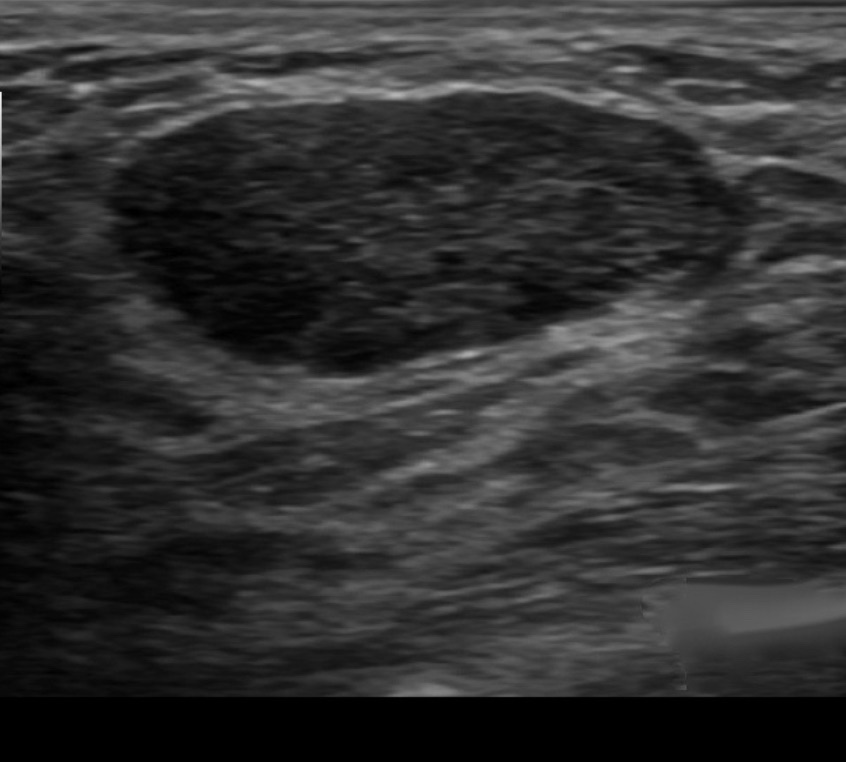
  • You first evaluate the lesion for any of the 10 malignant signs:
    • Shadowing
    • Hypoechoic echotexture
    • Spiculation
    • Angular margins
    • Thick echogenic halo
    • Microlobulation
    • Taller than wide
    • Duct extension
    • Branching pattern
    • Calcifications
  • Finding none, you move on to the second step in the evaluation process:
    • Specifically look for one of the 3 strictly defined benign signs:
      • If any of them are found, the lesion can be considered BIRADS 3
    • The 3 benign findings defined by Stavros are:
      • A purely hyperechoic lesion with no hypoechoic area larger than a normal duct or lobule
      • Elliptical, wider than tall, well-circumscribed and thin echogenic capsule
      • Gently lobulated, wider than tall, well-circumscribed and thin echogenic capsule
    • Combining the elliptical or gently lobulated shapes with the presence of a complete, thin echogenic capsule:
      • Is necessary because many circumscribed carcinomas and most ductal carcinoma in situ are encompassed in a thin, echogenic capsule:
        • However, the shape of circumscribed invasive carcinoma or pure ductal carcinoma in situ is rarely elliptical or gently lobulated
  • BIRADS 3:
    • A 6-month follow-up ultrasound would be appropriate unless the anxiety of the patient makes core biopsy a better option
  • References
    • Madjar H, Mendelson EB. The Practice of Breast Ultrasound. 2nd ed. Thieme; 2008;141-144.
    • Stavros AT. Breast Ultrasound. Philadelphia, PA: Lippincott Williams & Wilkins; 2004.
#Arrangoiz #Doctor #Surgeon #CancerSurgeon #SurgicalOncologist #BreastSurgeon #MountSinaiMedicalCenter #MSMC #Miami #Mexico #BreastCancer

Ultrasound Appearance of Axillary Lymph Nodes

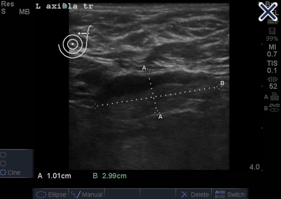
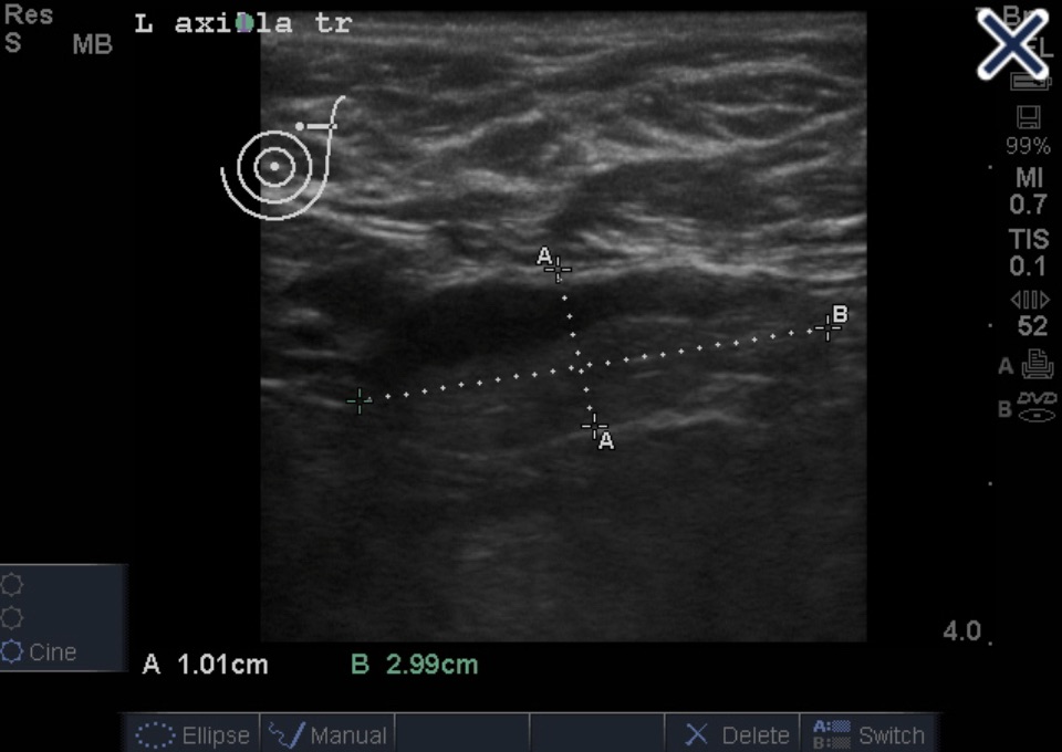
Abnormal lymph node with eccentric cortical thickening
  • This axilla ultrasound shows a case of severe eccentric compression and displacement of the hilum to the edge of the node:
    • Favoring metastatic disease
  • Typical lymph node ultrasound appearances include:
    • Uniformly mildly thickened cortex
      • Typical of inflamed or reactive lymph node
    • Eccentric cortical thickening:
      • Favors metastatic disease (Image)
    • Convex indentations of the hilum:
      • Favors metastatic disease
    • Severe compression of the hilum to slit-like configuration:
      • Can occur in metastatic or severely inflamed lymph node
    • Severe eccentric compression and displacement of the hilum to the edge of the node:
      • Favoring metastatic disease
    • Complete obliteration of the hilum and rounding of the node:
      • Favoring metastatic disease:
        • Can also occur in severe necrotizing lymphadenitis
    • Perinodal invasion by metastasis:
      • The outer thin echogenic capsule cannot be identified:
        • Angular margins
Severe eccentric compression and displacement of the hilum to the edge of the node.
  • References
    • Stavros AT. Evaluation of regional lymph nodes in breast cancer patients. In: Stavros AT. Breast Ultrasound. Philadelphia, PA: Lippincott Williams & Wilkins; 2004:858-859.
    • AIUM curriculum for fundamentals of ultrasound physics and instrumentation. JUltrasound Med 2019;38(8):1933-1935. https://onlinelibrary.wiley.com/doi/epdf/10.1002/jum.15088. Accessed July 16, 2020.
#Arrangoiz #Doctor #Surgeon #CancerSurgeon #SurgicalOncologist #Breast Surgeon #MountSinaiMedicalCenter #MSMC #Miami #Mexico #BreastCancer

Types of Calcifications 1

  • Ductal calcifications:
    • Have a wide variety of presentations depending upon the underlying process that created them
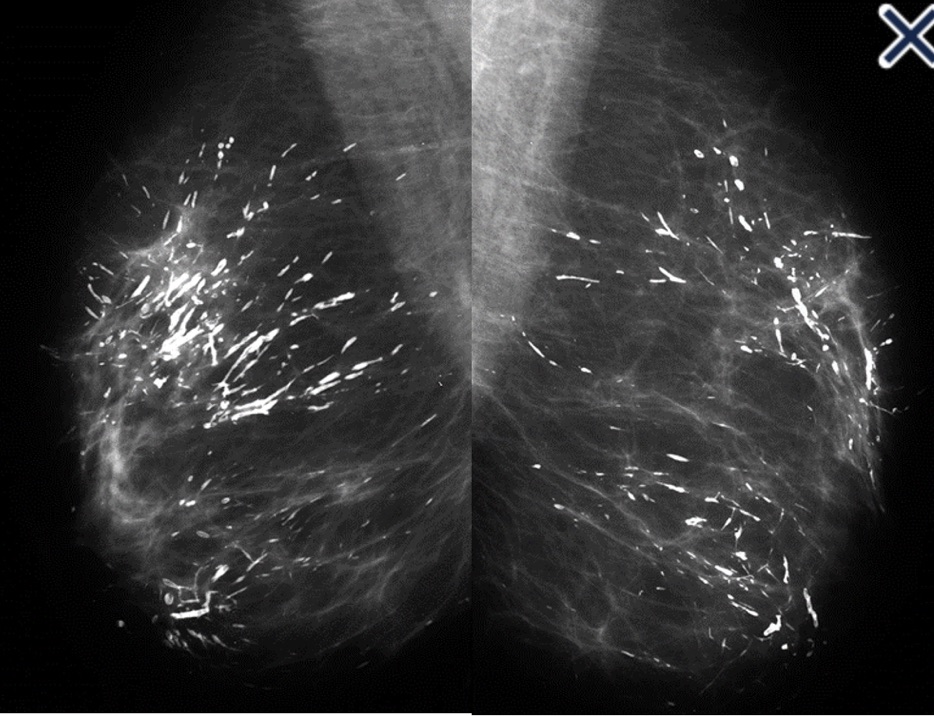
  • When coarse rod-like ductal calcifications are diffuse, bilateral, and not confined to a single lobe:
    • They can be confidently assumed to result from plasma cell mastitis, and do not require further evaluation or biopsy (Image)
  • The process is called secretory disease:
    • Because there is a stagnant, viscous fluid that eventually petrifies and results in the smooth contoured calcifications
    • Some of them are branching and look like malignant casting type calcifications:
      • But the key distinguishing feature is the diffuse, multilobe, bilateral nature of the process:
        • Calcifications become much more worrisome when they are confined to a single lobe
  • The most frequent malignant, ductal “casting type” calcifications are:
    • Fragmented, linear, and branching, and they are the most reliable mammographic sign of malignancy (Image)
Casting type calcification.
  • The presence of fragmented and / or dotted casting type calcifications on the mammogram restricted to one lobe:
    • Is a pathognomonic sign of a diffuse, grade 3 breast cancer subtype that originates in the major ducts and usually has a solid or micropapillary pattern:
      • Traditionally, this subtype has been called “comedo carcinoma”
    • The cancer cells either produce a viscous, proteinaceous fluid, which gradually concentrates and eventually calcifies, or they undergo necrosis (apoptosis) followed by calcification:
      • In both instances, the intraluminal pressure increases, distending the ducts considerably
  • Dotted casting type calcifications:
    • Have been referred to as “snakeskin-like calcifications” and they accumulate in the fluid produced by either micropapillary or solid cancer cell growth patterns (Image)
    • The tips of the micropapillary growths may become detached and eventually calcify, contributing to the intraluminal calcifications (Image)
Snakeskin type calcifications.
The micropapillary growths break off and calcify in the lumen, resulting in the individual dots of calcification (the dark, almost black stained structures).
  • Occasionally, malignant ductal calcifications present in a manner that can be easily mistaken for a benign process:
    • It occurs when fluid production, rather than necrosis, dominates the picture
    • The intraductal carcinoma can be grades 1, 2, or 3 and a micropapillary and/or cribriform architecture is present

Dense Breast Screening

  • Debate on adjunct screening in women with dense breasts:
    • Has resulted from legislation mandating that women be informed if their mammograms show dense breast tissue, including informing them that other screening modalities are available
  • In addition to MRI and molecular imaging:
    • Both tomosynthesis and breast ultrasound are additional techniques for enhanced screening in patients with dense breasts
  • Multiple studies show significant benefits from the addition of tomosynthesis to conventional digital mammography alone in screening programs:
    • Ciatto et al:
      • Found an increase in detection rate of invasive breast cancer:
        • From 5.3/1000 to 8.1/1000
      • While also decreasing the recall rate by 17%
    • Skaane et al:
      • Found a 40% increase in the detection of invasive cancers with a 15% reduction in false negatives
    • Rose et al.3 and Haas et al:
      • Showed statistically significant relative reductions in recall rates of 37% and 30%, respectively
    • A recent retrospective review of 454,850 examinations in 13 screening centers in the United States:
      • Demonstrated a 41% increase in invasive cancer detection, a 15% reduction in call backs, and a 49% increase in the positive predictive value for recall
  • Implementation of tomosynthesis:
    • Did not lead to a significant reduction in biopsy rates as compared to digital mammography screening
  • As yet, there are no data that show a reduction in mortality with enhanced screening in dense breasts
  • A prospective multicenter study compared tomosynthesis with bilateral physician hand-held ultrasound screening in 3,231 asymptomatic patients with mammography-negative dense breasts:
    • In all, 24 additional cancers were detected, 23 of which were invasive
    • Tomosynthesis detected 13 cancers, and ultrasound detected 23
    • These data suggest that even though tomosysthesis significantly increases the number of cancers found in dense breasts, in the hands of a skilled breast radiologist, ultrasound may be even better
  • References
    • Ciatto S, Houssami N, Bernardi D, Caumo F, Pellegrino M, Brunelli S, et al. Integration of 3D digital mammography with tomosynthesis for population breast-cancer screening (STORM): a prospective comparison study. Lancet Oncol. 2013;14(7):583-589.
    • Skaane P, Bandos AI, Gullien R, Eben EB, Ekseth U, Haakenaasen U, et al. Comparison of digital mammography alone and digital mammography plus tomosynthesis in a population-based screening program. Radiology. 2013;267(1):47-56.
    • Haas BM, Kalra V, Geisel J, Raghu M, Durand M, Philpotts LE. Comparison of tomosynthesis plus digital mammography and digital mammography alone for breast cancer screening. Radiology. 2013;269(3):694-700.
    • Rose SL, Tidwell AL, Bujnoch LJ, Kushwaha AC, Nordmann AS, Sexton R Jr. Implementation of breast tomosynthesis in a routine screening practice: an observational study. AJR Am J Roentgenol. 2013;200(6):1401-1408.
    • Friedewald SM, Rafferty EA, Rose SL, Durand MA, Plecha DM, Greenberg JS, et al. Breast cancer screening using tomosynthesis in combination with digital mammography. JAMA. 2014;311(24):2499-2507.
    • Tagliafico AS, Calabrese M, Mariscotti G, Durando M, Tosto S, Monetti F, et al. Adjunct screening with tomosynthesis or ultrasound in women with mammography-negative dense breasts: interim report of a prospective comparative trial. J Clin Oncol. 2016;34(16):1882-1888.
#Arrangoiz #Doctor #Surgeon #CancerSurgeon #SurgicalOncologist #Breast Surgeon #MountSinaiMedicalCenter #MSMC #Miami #Mexico #BreastCancer

Tissue Harmonic Imaging in Breast Cancer

  • Tissue harmonic imaging:
    • Creates images derived solely from higher frequencies
  • The ultrasound beam:
    • Is transmitted centered at 1 frequency, e.g., 6 MHz
    • Received centered at a multiple of the transmitted frequency, e.g., 12 MHz
  • Different techniques can be used to process the received signals so that only the returning high-frequency harmonic signal is used to produce the image:
    • Whereas echoes from the fundamental / lower frequencies are rejected
  • THI increases signal-to-noise ratio:
    • Resulting in better tissue contrast
  • THI reduces reverberation, clutter, and speckle artifacts:
    • Improving contrast resolution
    • It accentuates real echoes in addition to suppressing artifactual echoes
    • The suppression of speckle artifact by coded harmonics makes solid nodules more hypoechoic and conspicuous than they are with fundamental imaging (Images a and b)
A. Infiltrating ductal carcinoma with fundamental imaging.
B. The same infiltrating ductal carcinoma as 2a with coded harmonic imaging.
  • It makes the thin, echogenic capsule that surrounds most benign lesions appear to be thinner, more echogenic and more complete than with fundamental imaging
  • THI cannot be combined with simultaneous color Doppler imaging because the resulting frame rate would be unacceptable:
    • When Doppler is required, the image that is interlaced with Doppler must be constructed at fundamental rather than harmonic frequencies
  • THI is of limited value in differentiating benign from malignant lesions.
  • Another method of reducing artifactual echoes, improving contrast, and making the thin, echogenic capsule more conspicuous is real time spatial compounding of images:
    • In conventional imaging, each frame is created by a single sweep of the beam at a 90 degree angle to the long axis of the transducer
    • In compound imaging, there are multiple sweeps of the beam from different angles, creating a spatially and temporally compounded image from multiple angles over time
    • Among other things, the lateral borders of lesions can be seen better with compound imagin
  • References
    • Mesurolle B, Helou T, El-Khoury M, Edwardes M, Sutton EJ, Kao E. Tissue harmonic imaging, frequency compound imaging, and conventional imaging use and benefit in breast sonography. J Ultrasound Med. 2007;26(8):1041-1051.
    • Cha JH, Moon WK, Cho N, Kim SM, Park SH, Han BK, et al. Characterization of benign and malignant solid breast masses: comparison of conventional US and tissue harmonic imaging. Radiology. 2007;242(1):63-69.
    • Stavros AT. Breast ultrasound equipment requirements. In: Stavros AT. Breast Ultrasound. Philadelphia, PA: Lippincott Williams & Wilkins; 2004:16-41.
#Arrangoiz #BreastSurgeon

Calcifications Identified on Mammogram

Straight lateral magnification view. “Milk of Calcium” Calcifications.
  • The images clearly show benign “milk of calcium” type calcifications:
    • Which do not warrant biopsy or interval follow-up regardless of how many are present
  • In fibrocystic change:
    • An apocrine metaplastic cell layer lines the cystically dilated acini:
      • Which are filled with fluid and contain numerous psammoma body-like calcifications
    • The appearance of the calcifications on the mammogram:
      • Will depend on the shape of the summation of the calcified particles in the cystically dilated acini
    • When the shape happens to be “teacup-like” (i.e., crescent-shaped on the mediolateral projection and low density, circular/oval on the craniocaudad projection):
      • The diagnosis of fibrocystic change can be made with confidence
Images a and b show a galactogram performed on a woman with greenish cloudy nipple discharge. The ducts are distended by fluid (duct ectasia) and the acini of a single terminal ductal lobular unit are cystically distended. The contrast media shows a teacup-like appearance, seen from the side (Image a) and seen from above (Image b).
Images a-c show 3D histology images of the aggregate of the psammoma body-like calcification corresponding to the mammogram. The impression is that the teacup-like calcification is a single calcification, but Image c shows that it is the summation / aggregate of many tiny psammoma body-like calcifications.
  • Benign and malignant type calcifications:
    • Can increase or decrease in number and density, or even remain unchanged for years:
      • So changes in the appearance on follow-up examination do not constitute a reliable way to exclude malignancy
  • In general, calcifications should be determined to be benign by:
    • Their appearance and distribution or they should have a large bore core needle biopsy
  • Six month follow-up mammography is not a good way to determine if calcifications are benign
  • In fibrocystic changes:
    • The most frequently occurring calcifications are the psammoma body-like calcifications that float in fluid in microcysts:
      • They should cause little diagnostic confusion
    • When the calcifications are imaged in a craniocaudad projection:
      • The calcifications are spread out over the entire microcyst yielding a smudgy image of the calcifications
    • In a straight lateral image:
      • The microcysts are imaged in a vertically oriented direction, and gravity causes the calcium rich fluid to settle to the bottom of the cysts yielding the teacup-like appearance of the calcifications
        • When these images are present, the diagnosis of benign milk of calcium is secure.
  • Low-grade in situ carcinoma:
    • Would cause powdery calcifications
  • Intermediate grade in situ carcinoma:
    • Would cause crushed stone-type calcifications on the mammogram
  • References
    • Monda LA. Differentiation of breast calcifications. Radiol Technol. 2001;72(6):532-544.
    • Baldwin P. Breast calcification imaging. Radiol Technol. 2013;84(4):383M-404M.

How to Evaluate a Breast Nodule on Ultrasound of the Breast?

  • You first evaluate the lesion for:
    • Any of the 10 malignant signs:
      • Shadowing
      • Hypoechoic echotexture
      • Spiculation
      • Angular margins
      • Thick echogenic halo
      • Microlobulation
      • Taller than wide
      • Duct extension
      • Branching pattern
    • Calcifications
  • Finding none:
    • You move on to the second step in the evaluation process:
      • Specifically look for one of the 3 strictly defined benign signs:
        • If any of them are found:
          • The lesion can be considered BIRADS 3:
            • A 6-month follow-up ultrasound would be appropriate unless the anxiety of the patient makes core biopsy a better option
    • The 3 benign findings defined by Stavros are:
      • A purely hyperechoic lesion:
        • With no hypoechoic area larger than a normal duct or lobule
      • Elliptical, wider than tall, well-circumscribed and thin echogenic capsule
      • Gently lobulated, wider than tall, well-circumscribed and thin echogenic capsule
      • Combining the elliptical or gently lobulated shapes:
        • With the presence of a complete, thin echogenic capsule:
          • Is necessary because many circumscribed carcinomas and most ductal carcinoma in situ are encompassed in a thin, echogenic capsule:
            • However, the shape of circumscribed invasive carcinoma or pure ductal carcinoma in situ:
              • Is rarely elliptical or gently lobulated:
  • References:
    • Madjar H, Mendelson EB. The Practice of Breast Ultrasound. 2nd ed. Thieme; 2008;141-144.
    • Stavros AT. Breast Ultrasound. Philadelphia, PA: Lippincott Williams & Wilkins; 2004.
#Arrangoiz #BreastSurgeon #CancerSurgeon #SurgicalOncologist #Surgeon #Doctor #MountSinaiMedicalCenter #MSMC #Miami #Mexico

Breast Calcifications on Mammogram

  • Screening mammography:
    • Is important in identifying breast cancer at an early stage
  • Calcifications have many forms:
    • The characteristics of the calcifications help identify whether they are associated with a benign or malignant process:
      • Smooth, round, large and layering calcium are generally associated with benign findings
      • Fine, irregular, punctate, linear and branching, and pleomorphic calcifications are characteristics generally associated with malignant findings
  • Magnification views of the calcifications:
    • Are essential in helping to evaluate the calcifications to determine the appearance and to be able to accurately interpret the findings
  • References
    • Brant W, Helms C. Fundamentals of Diagnostic Radiology, 5th Edition. Lippincott, Williams & Wilkins; 2019
    • Baldwin P. Breast calcification imaging. Radiol Technol. 2013;84(4):383M-404M.
#Arrangoiz #Doctor #Surgeon #CancerSurgeon #SurgicalOncologist #BreastSurgeon #BreastCancer #MountSinaiMedicalCenter #MSMC #Miami Mexico

TP53 Mutation in Breast Cancer

  • The TP53 gene:
    • Is a key tumor-suppressor gene that acts as a checkpoint control for DNA damage
    • Due to its critical role in controlling cellular damage:
      • A TP53 germline mutation predisposes patients to multiple malignancies, including breast cancer and soft tissue sarcomas:
        • The associated familial syndrome was first observed in 1969 and is known as the Li-Fraumeni syndrome
    • The penetrance of breast cancer related to TP53 mutations:
      • Is higher than seen in the more common BRCA1 or BRCA2 mutations:
        • With a cumulative incidence reported for TP53 of 85% by age 60
      • TP53-associated breast cancers present at an early age (median age of diagnosis is 34) and the majority are hormone receptor positive and / or HER-2 positive.
    • Due to this high penetrance and associated increased risk for a secondary breast cancer:
      • Bilateral prophylactic mastectomy is recommended for management of an early-stage breast cancer in patients with a mutation in TP53
      • This is especially true in younger women as contralateral breast cancer risk inversely correlates with the patient’s age
    • The recommendation for mastectomy is further supported by the concern regarding radiation use in this patient population already at increased risk for soft tissue sarcomas:
      • Radiation should be used with extreme caution and careful consideration of the risk / benefit
  • For patients presenting with a known TP53 germline mutation and without a diagnosis of breast cancer:
    • NCCN guidelines recommend:
      • Annual breast MRI at 20 to 29 years and annual MRI and mammography at 30 to 75 years for high-risk breast cancer screening
    • Consideration of prophylactic risk-reducing mastectomy should be made in context of the age of presentation:
      • As breast cancer risk increases significantly after the second decade of life in these patients:
        • Bilateral mastectomy should be considered starting at age 20
      • The risk of breast cancer peaks at age 40 to 45 and then decreases, and therefore bilateral mastectomy offers significantly less benefit in women over 60 years of age
  • References
    • Mai PL, Best AF, Peters JA, DeCastro RM, Khincha PP, Loud JT, Bremer RC, Rosenberg PS, Savage SA Risks of first and subsequent cancers among TP53 mutation carriers in the National Cancer Institute Li-Fraumeni syndrome cohort. Cancer. 2016 Dec 1; 122(23):3673-3681.
    • Masciari S, Dillon DA et al. Breast cancer phenotype in women with TP53 germline mutations: a Li Fraumeni syndrome consortium effort. Breast Cancer Res Treat. 2012;133(3):1125–1130.
    • Schon, K, Tischkowitz, M. Clinical implications of germline mutations in breast cancer: TP53 Breast Cancer Res Treat. 2018; 167(2): 417–423.
    • National Comprehensive Cancer Network (2014) Genetic/familial high risk assessment: breast and ovarian. Li Fraumeni syndrome management. NCCN Clinical Practice Guidelines in Oncology. Version 1.2023.
#Arrangoiz #Doctor #Surgeon #CancerSurgeon #SurgicalOncologist #BreastSurgeon #MountSiniaMedicalCenter #MSMC #Miami #Mexico #BreastCancer

Hormone Replacement Therapy After Risk Reducing Bilateral Salpingo Ooforectomy (BSO) in Young Women

  • BRCA mutation carriers:
    • Are recommended to undergo prophylactic BSO to decrease risk of developing ovarian cancer
  • Risk-reducing surgery:
    • Should be performed after completion of childbearing and is recommended at:
      • Ages of 35 to 40 years for BRCA1 mutation carriers
      • Ages of 40 to 45 years for BRCA2 mutation carriers
  • Women who undergo BSO at a young age:
    • Are at increased risk of premature menopausal symptoms including:
      • Vasomotor symptoms
      • Sexual dysfunction
      • Vulvo-vaginal atrophy
    • In addition, premature menopause:
      • May be associated with decreased bone density and cardiovascular disease
  • Short-term hormone replacement therapy (HRT):
    • Can be beneficial to alleviate the symptoms of estrogen deprivation:
      • A literature review on the use of HRT among BRCA mutation carriers following risk-reducing BSO:
        • Summarized data supporting the use of HRT for improvement in menopausal symptoms, quality of life, and sexual function, and suggests an improvement in bone health, cardiovascular health, and cognitive function
  • When assessing breast cancer risk with HRT in this population:
    • The summary of available literature does not show an increase in breast cancer risk with short-term HRT use following risk-reducing BSO:
      • However, there is notable concern regarding increased breast cancer risk with the use of combination estrogen and progesterone HRT compared to estrogen alone:
        • Kotsopoulos and colleagues reported a prospective, longitudinal cohort study of BRCA1 mutation carriers who underwent risk-reducing BSO from 80 centers in 17 countries 1995-2017:
          • BRCA1 carriers with a follow-up of 7.6 years following BSO were included
          • Of the population, 43% of women used some form of HRT following BSO for a mean duration of 3.9 years
          • The authors did not find an associated increase in breast cancer risk with the use of HRT in this population of BRCA1 carriers post-BSO (HR 0.97, p=.89 for any HRT use vs. none)
          • In this cohort, most women took estrogen alone (69%) while 18% took combination estrogen plus progesterone and 32% used another formulation of hormone therapy
          • While there was no significant difference in the 10-year actuarial risk for any HRT regimen compared to none (p=0.72), there was a difference in women who used estrogen alone, with a reported breast cancer incidence of 12% compared with 22% in those who used estrogen plus progesterone (p=0.04)
          • In general, estrogen-alone HRT is recommended for women who have undergone a hysterectomy, while combination estrogen plus progesterone is recommended for women with an intact uterus to reduce the risk of endometrial hyperplasia and cancer (a risk with unopposed estrogen)
          • Given these differences in risk, a thoughtful discussion is necessary to balance the risks and benefits of HRT with appropriate gynecologic surgery.
  • While premature menopause symptoms are a common concern among young women undergoing risk reducing BSO, few BRCA mutation carriers are currently recommended to consider HRT for symptom management:
    • A study reporting on BRCA mutation carriers from the ‘Facing Our Risk of Cancer Empowered’ group found that 81% of the postmenopausal population became menopausal prematurely secondary to surgery or medications
    • Of this group, the majority reported concerns of libido /sexuality (78%), cardiovascular disease (78%), and osteoporosis (65%), but HRT use was reported in only 13% of women with no prior cancer history with only 26% of women reporting that this was favored by their healthcare provider
    • Continued provider and patient education is needed to educate patients that short-term hormone replacement therapy is safe in BRCA1 mutation carriers following risk reducing bilateral salpingo-oophorectomy
  • References
    • Gordhandas S, Norquist BM, Pennington KP, Yung RL, Laya MB, Swisher EM. Hormone replacement therapy after risk reducing salpingo-oophorectomy in patients with BRCA1 or BRCA2 mutations; a systematic review of risks and benefits. Gynecol Oncol. 2019;153(1):192-200.
    • Kotsopoulos J, Gronwald J, Karlan BY, Huzarski T, Tung N5, Moller P, et al. Hormone replacement therapy after oophorectomy and breast cancer risk among BRCA1 mutation carriers. JAMA Oncol. 2018;4(8):1059-1065.
    • Birrer N, Chinchilla C, Del Carmen M, Dizon DS. Is Hormone Replacement Therapy Safe in Women With a BRCA Mutation?: A Systematic Review of the Contemporary Literature. Am J Clin Oncol. 2018 Mar;41(3):313-315.
#Arrangoiz #Doctor #Surgeon #CancerSurgeon #SurgicalOncologist #BreastSurgeon #MountSiniaMedicalCenter #MSMC #Miami #Mexico #BreastCancer