Blog

Atypical Lobular Hyperplasia (ALH) of the Breast

  • Atypical lobular hyperplasia (ALH):
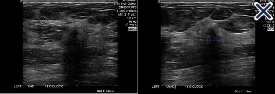
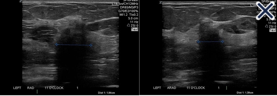
    • Is generally an incidental finding on core needle biopsy without specific defining characteristics on mammography, ultrasound, or MRI
  • A palpable breast mass which yields ALH at core needle biopsy:
    • Is discordant and should prompt further diagnostic work-up with a second biopsy, either core or excisional
  • ALH should only be considered for observation:
    • When there is radiologic, pathologic, and clinical concordance:
      • As the risk of upstaging to carcinoma in this scenario:
        • Is less than 5%
  • ALH alone confers a 4 to 5-fold increased risk of future breast cancer
  • Relevant indications for genetic testing include:
    • A personal history of breast cancer ≤ age 45
    • Triple negative breast cancer ≤ age 60
    • A first-degree relative with breast cancer ≤ age 50
    • Two or more first- or second-degree relatives with breast cancer at any age
    • Patient or relative with bilateral breast cancer
    • Male breast cancer in a relative at any age
  • Risk-reducing mastectomy can be considered in patients with very high lifetime breast cancer risk:
    • Usually reserved for women with high-penetrance gene mutations, such as BRCA 1 or 2
  • References
    • Morrow M, Schnitt SJ, Norton L. Current management of lesions associated with an increased risk of breast cancer. Nat Rev Clin Oncol. 2015;12(4):227–238.
    • Smart CE, Furnival CM, Lakhani SR. Chapter 17. High-Risk Lesions: ALH/LCIS/ADH. In: Kuerer HM ed. Kuerer’s Breast Surgical Oncology. New York, NY: McGraw-Hill, 2010.
    • Murray MP, Luedtke C, Liberman L, Nehhozina T, Akram M, Brogi E. Classic lobular carcinoma in situ and atypical lobular hyperplasia at percutaneous breast core biopsy: outcomes of prospective excision. Cancer. 2013;119(5):1073-1079.
    • The American Society of Breast Surgeons (2016). Consensus Guideline on Concordance Assessment of Image-Guided Breast Biopsies and Management of Borderline or High-Risk Lesions https://www.breastsurgeons.org/docs/statements/Consensus-Guideline-on-Concordance-Assessment-of-Image-Guided-Breast-Biopsies.pdf. Accessed February 23, 2020.
#Arrangoiz #Doctor #Surgeon #CancerSurgeon #SurgicalOncologist #BreastSurgeon #MountSiniaMedicalCenter #MSMC #Miami #Mexico #BreastCancer

Breast Cancer Risk in Identical Twins

  • Lifetime breast cancer risk is 13% for identical twins of breast cancer patients compared to 9% for dizygotic twins
  • Twin studies are important because they help us understand the contribution of genetics to risk stratification in various populations
  • Other twin studies have estimated that:
    • 12% to 30% of breast cancer is primarily genetic in origin
  • In a recent update on cancer risk in the population of twins in Nordic countries:
    • The familial breast cancer risk was 28% for monozygotic twins and 20% for dizygotic twins at a median follow-up of 32 years
  • Thus, a minority of breast cancers are directly attributed to germline genetics and only 5% to 10% are thought to be due to inheritance of mutations in major autosomal dominant breast cancer predisposition genes
  • References
    • Baker SG, Lichtenstein P, Kaprio J, Holm N. Genetic susceptibility to prostate, breast, and colorectal cancer among Nordic twins. Biometrics. 2005;61(1):55-63.
    • Lichtenstein P, Holm NV, Verkasalo PK, Iliadou A, Kaprio J, Koskenvuo M, et al. Environmental and heritable factors in the causation of cancer–analyses of cohorts of twins from Sweden, Denmark, and Finland. N Engl J Med. 2000;343(2):78-85.
    • Locatelli I, Lichtenstein P, Yashin AI. The heritability of breast cancer: a Bayesian correlated frailty model applied to Swedish twins data. Twin Res. 2004;7(2):182-191.
    • Mucci LA, Hjelmborg JB, Harris JR, Czene K, Havelick DJ, Scheike T, et al. Nordic Twin Study of Cancer (NorTwinCan) collaboration. familial risk and heritability of cancer among twins in Nordic countries. JAMA. 2016;315(1):68-76.
#Arrangoiz #BreastSurgeon

Phyllodes Tumors

  • Introduction
    • Phyllodes tumors (PTs) of the breast are considered a rare fibroepithelial neoplasms of the breast and are considered a challenging for both pathologists and surgeons
    • The World Health Organization (WHO) has classified PTs histologically as:
      • Benign
      • Borderline
      • Malignant
    • PTs can be detected in all ages:
      • However, the median age of presentation is 45 years
    • PTs can mimic fibroadenoma in clinical presentations
    • Breast imaging is also similar to fibroadenomas
    • Cytological diagnosis of PTs by biopsy is usually unreliable:
      • However, a core needle biopsy is superior to fine-needle aspiration
    • Surgery is considered the mainstay treatment for PTs of the breast:
      • With a goal of achieving negative margins
    • Adjuvant chemotherapy and radiation therapy use for malignant PTs are controversial

Screening for Breast Cancer in Women with A History of Mantle Radiation Prior to the Age of 30

  • Women who receive thoracic (i.e., mantle) radiation prior to age 30:
    • Are at increased risk of breast cancer:
      • Although standardized incidence ratios vary from 13 to 55 based on patient, disease, and treatment factors
  • In the Late Effects Study Group trial:
    • The relative risk of breast cancer varied by follow-up interval and was greatest at 15 to 19 years after radiation exposure
  • Screening guidelines for those under age 25 include:
    • An annual clinical exam beginning 10 years after the radiation exposure
  • Screening guidelines for those over age 25:
    • Include an annual clinical exam beginning 8 to 10 years after the radiation exposure, with the addition of annual screening mammogram for patients ≥ age 30
    • Annual MRI is recommended for patients ≥ age 25
  • Recent studies reporting the persistence of gadolinium deposits in the brain following serial contrast MRI scans have led to a related FDA safety alert:
    • However, deposition is associated with only some gadolinium based contrast agents, and there is no clinical data that this results in detrimental long-term cognitive effects
  • There is currently no evidence that biannual MRI is more valuable than annual MRI for screening.
  • References
    • Henderson TO, Amsterdam A, Bhatia S, Hudson MM, Meadows AT, Neglia JP, et al. Systematic review: surveillance for breast cancer in women treated with chest radiation for childhood, adolescent, or young adult cancer. Ann Intern Med.2010;152(7):444-454.
    • van Leeuwen FE, Klokman WJ, Stovall M, Dahler EC, van’t Veer MB, Noordijk EM, et al. Roles of radiation dose, chemotherapy, and hormonal factors in breast cancer following Hodgkin’s disease. J Natl Cancer Inst. 2003;95(13):971-980.
    • National Comprehensive Cancer Network. Breast Cancer Screening and Diagnosis, Version 1.2019. https://www.nccn.org/professionals/physician_gls/pdf/breast-screening.pdf. Accessed February 23, 2020.
    • Ramalho J, Ramalho M, Jay M, Burke LM, Semelka RC. Gadolinium toxicity and treatment. Magn Reson Imaging. 2016;34(10):1394-1398.
    • Stojanov D, Aracki-Trenkic A, Benedeto-Stojanov D. Gadolinium deposition within the dentate nucleus and globus pallidus after repeated administrations of gadolinium-based contrast agents-current status. Neuroradiology. 2016;58(5):433-441.
    • Olchowy C, Cebulski K, Lasecki M, et al. The presence of the gadolinium-based contrast agent depositions in the brain and symptoms of gadolinium neurotoxicity – A systematic review. PLoS One. 2017;12(2):e0171704.
#Arrangoiz #Doctor #Surgeon #CancerSurgeon #BreastSurgeon #SurgicalOncologist #BreastCancer #Miami #Mexico #MountSinaiMedicalCenter #MSMC

Hormone Replacement Therapy in Postmenopausal Patients with a History of Breast Cancer – Is it Safe?

  • Two randomized clinical trials:
    • Were initiated and terminated early due to an increase in breast cancer events in patients in the hormone replacement therapy (HRT) arm with a personal history of breast cancer
      • The HABITS (Hormonal Replacement Therapy after Breast Cancer- Is It Safe?) trial:
        • Randomized 442 patients with a personal history of breast cancer to:
          • HRT with estradiol with or without progesterone (based on the presence of a uterus) vs. nonhormonal symptom management
        • More women in the HRT arm than the non-HRT arm had hormone receptor-positive breast cancer (62.3% vs 54.5%), and approximately half of patients in each group had taken HRT before their diagnosis of breast cancer
        • At median follow-up of 4 years, new breast cancer events occurred twice as frequently in the HRT group (hazard ratio [HR] 2.4) compared to the nonhormonal symptom management group
    • A similar trial in Stockholm found no difference in breast cancer recurrence in patients randomized to HRT vs. no HRT at 11 years of follow-up:
      • But reported an increased risk of contralateral breast cancer in those receiving HRT (HR 3.6, p=0.013)
  • Estrogen alone:
    • Can be used to reduce symptoms for postmenopausal women without a personal history of breast cancer who have had a hysterectomy, and was associated with a nonsignificant lower risk of breast cancer in the Women’s Health Initiative trial at 13 years of follow-up (HR 0.79)
    • However, the WHI cohort did not include women with a personal history of breast cancer, and therefore these data cannot be extrapolated to this population
  • Expert guidelines:
    • Recommend the use of topical low-dose vaginal estrogen therapy for women with bothersome genitourinary symptoms and contraindications to the use of systemic HRT:
      • This is a reasonable strategy in breast cancer survivors due to the local nature of vaginal estrogen and its low systemic absorption
  • References
    • Holmberg L, Iversen OE, Rudenstam CM, Hammar M, Kumpulainen E, Jaskiewicz J, et al. Increased risk of recurrence after hormone replacement therapy in breast cancer survivors. J Natl Cancer Inst. 2008;100(7):475-482.
    • Fahlén M, Fornander T, Johansson H, Johansson U, Rutqvist LE, Wilking N, et al. Hormone replacement therapy after breast cancer: 10 year follow up of the Stockholm randomised trial. Eur J Cancer. 2013;49(1):52-59.
    • Manson JE, Chlebowski RT, Stefanick ML, Aragaki AK, Rossouw JE, Prentice RL, et al. Menopausal hormone therapy and health outcomes during the intervention and extended post-stopping phases of the Women’s Health Initiative randomized trials. JAMA. 2013;310(13):1353-1368.
    • The NAMS 2017 Hormone Therapy Position Statement Advisory Panel. The 2017 hormone therapy position statement of The North American Menopause Society. Menopause. 2017;24(7):728-753.
#Arrangoiz #BreastSurgeon #CancerSurgeon #MountSinaiMedicalCenter #MSMC #Miami #Mexico

Breast Cancer Risk Models

  • The Tyrer-Cuzick model:
    • Estimates breast cancer risk based largely on family history, and it includes breast density as part of the risk caclulation
  • The Gail model:
    • Which calculates breast cancer risk based on age, race/ethnicity, age at menarche, age at first live birth, number of prior breast biopsies, personal history of atypical hyperplasia, and family history of breast cancer in first-degree relatives, does not include breast density or genetic testing results
  • BRCAPRO and BOADICEA:
    • Are both Mendelian models that estimate breast cancer risk based on the probability that the individual carries a mutation in a major breast cancer susceptibility gene, such as BRCA1 or BRCA2
    • These models do not incorporate nulliparity or breast density into the calculation
  • The Claus model:
    • Is based only on family history of breast cancer in first- and second-degree relatives, is not thoroughly validated in independent cohorts, and does not include factors such as nulliparity
  • References
    • Amir E, Freedman OC, Seruga B, Evans DG. Assessing women at high risk of breast cancer: a review of risk assessment models. J Natl Cancer Inst. 2010;102(10):680-691.
    • Tyrer J, Duffy SW, Cuzick J. A breast cancer prediction model incorporating familial and personal risk factors. Stat Med. 2004;23(7):1111-1130. [See comment in Stat Med. 2005;24:1610-161; erratum appears in Stat Med. 2005;24:156].
    • Gail MH, Brinton LA, Byar DP, Corle DK, Green SB, Schairer C, et al. Projecting individualized probabilities of developing breast cancer for white females who are being examined annually. J Natl Cancer Inst. 1989;81(24):1879-1886.
    • Claus EB, Risch N, Thompson WD. Autosomal dominant inheritance of early-onset breast cancer implications for risk prediction. Cancer. 1994;73(3):643-651.
#Arrangoiz #BreastSurgeon

Lobular Carcinoma In Situ

  • The rate of cancer development in setting of LCIS:
    • Has been reported to be approximately 2% per year:
      • Translating into a cumulative long-term rate of 26% at 15 years
  • While a minority of patients elect to proceed with bilateral prophylactic mastectomy for LCIS:
    • The estimated breast cancer incidence is generally not considered high enough to justify such extensive surgery
    • Furthermore, women with a history of LCIS:
      • Typically develop low- or intermediate-grade malignancies clinically detected at an early stage
  • Multiple studies support a low upgrade rate on excision (1% to 5% for patients diagnosed with LCIS on core biopsy and rad-path concordant) with a 1% upgrade rate reported in a prospective study with central pathology review:
    • Therefore surgical excision is not routinely indicated
  • The risk conferred by LCIS:
    • Is independent of family history
  • Chemoprevention:
    • Reduces breast cancer risk by 40% to 65% in women at elevated risk:
      • Data from the NSABP P-1 trial suggest that women with LCIS derive even greater benefit than this estimate
  • References:
    • King TA, Pilewskie M, Muhsen S, Patil S, Mautner SK, Park A, et al. Lobular carcinoma in situ: a 29-year longitudinal experience evaluating clinicopathologic features and breast cancer risk. J Clin Oncol. 2015;33(33):3945–3952.
    • Wong SM, King T, Boileau JF, Barry WT, Golshan M. Population-based analysis of breast cancer incidence and survival outcomes in women diagnosed with lobular carcinoma in situ. Ann Surg Oncol. 2017;24(9):2509-2517.
    • Nakhlis F, Gilmore L, Gelman R, et al. Incidence of Adjacent Synchronous Invasive Carcinoma and/or Ductal Carcinoma In-situ in Patients with Lobular Neoplasia on Core Biopsy: Results from a Prospective Multi-Institutional Registry (TBCRC 020). Ann Surg Oncol. 2016;23(3):722-728.
    • King MC, Wieand S, Hale K, et al. Tamoxifen and breast cancer incidence among women with inherited mutations in BRCA1 and BRCA2: National Surgical Adjuvant Breast and Bowel Project (NSABP-P1) Breast Cancer Prevention Trial. JAMA. 2001;286(18):2251-2256.
#Arrangoiz #Surgeon #Doctor #CancerSurgeon #BreastSurgeon #SurgicalOncologist #LCIS #LobularCarcinomaInSitu #MountSinaiMedicalCenter #MSMC #Miami #Mexico

Lobular Carcinoma In Situ

  • The rate of cancer development in setting of LCIS:
    • Has been reported to be approximately 2% per year:
      • Translating into a cumulative long-term rate of 26% at 15 years
  • While a minority of patients elect to proceed with bilateral prophylactic mastectomy for LCIS:
    • The estimated breast cancer incidence is generally not considered high enough to justify such extensive surgery
    • Furthermore, women with a history of LCIS:
      • Typically develop low- or intermediate-grade malignancies clinically detected at an early stage
  • Multiple studies support a low upgrade rate on excision (1% to 5% for patients diagnosed with LCIS on core biopsy and rad-path concordant) with a 1% upgrade rate reported in a prospective study with central pathology review:
    • Therefore surgical excision is not routinely indicated
  • The risk conferred by LCIS:
    • Is independent of family history
  • Chemoprevention:
    • Reduces breast cancer risk by 40% to 65% in women at elevated risk:
      • Data from the NSABP P-1 trial suggest that women with LCIS derive even greater benefit than this estimate
  • References:
    • King TA, Pilewskie M, Muhsen S, Patil S, Mautner SK, Park A, et al. Lobular carcinoma in situ: a 29-year longitudinal experience evaluating clinicopathologic features and breast cancer risk. J Clin Oncol. 2015;33(33):3945–3952.
    • Wong SM, King T, Boileau JF, Barry WT, Golshan M. Population-based analysis of breast cancer incidence and survival outcomes in women diagnosed with lobular carcinoma in situ. Ann Surg Oncol. 2017;24(9):2509-2517.
    • Nakhlis F, Gilmore L, Gelman R, et al. Incidence of Adjacent Synchronous Invasive Carcinoma and/or Ductal Carcinoma In-situ in Patients with Lobular Neoplasia on Core Biopsy: Results from a Prospective Multi-Institutional Registry (TBCRC 020). Ann Surg Oncol. 2016;23(3):722-728.
    • King MC, Wieand S, Hale K, et al. Tamoxifen and breast cancer incidence among women with inherited mutations in BRCA1 and BRCA2: National Surgical Adjuvant Breast and Bowel Project (NSABP-P1) Breast Cancer Prevention Trial. JAMA. 2001;286(18):2251-2256.
#Arrangoiz #Surgeon #Doctor #CancerSurgeon #BreastSurgeon #SurgicalOncologist #LCIS #LobularCarcinomaInSitu #MountSinaiMedicalCenter #MSMC #Miami #Mexico

BRCA 1 and BRCA 2 Hereditary Breast Cancer

  • BRCA1 and BRCA2:
    • Are genes that produce tumor suppressor proteins:
      • Which help repair damaged DNA
    • They are the most common gene alterations seen in the hereditary breast cancer population
      • They are associated with an increased risk of breast cancer:
        • Estimated to be 55% to 70% for BRCA 1 carriers and 45% to 70% in BRCA2 carriers by age 70
    • While both BRCA1 and BRCA2 mutations are associated with an increased risk of breast cancer:
      • BRCA1 breast cancers more commonly occur in younger, premenopausal women and are more likely to be triple negative
    • BRCA1 is associated with a higher risk of ovarian cancer compared to BRCA2:
      • With a lifetime risk of 40% to 45% in BRCA1 carriers compared to 15% to 20% in BRCA2 carriers
    • BRCA2 breast cancers:
      • More closely resemble the sporadic breast cancer pattern, with a predominance of hormone receptor positive cancers in women greater than 50 years
  • CHEK2 and PALB2:
    • Are moderate penetrance genes that are less common than BRCA mutations
    • Similar to BRCA 2 deleterious mutations:
      • CHEK2 and PALB2 mutations are associated with hormone receptor positive postmenopausal breast cancer
  • References
#Arrangoiz #Doctor #Surgeon #CancerSurgeon #SurgicalOncologist #BreastSurgeon #MountSinaiMedicalCenter #MSMC #Miami #Mexico #BreastCancer #HormoneReplacementSurgery

Indications for Risk Reducing Bilateral Salpingectomy-Oophorectomy (BSO)

  • Women with a pathogenic variant in BRCA1:
    • Have a lifetime cumulative ovarian cancer risk of 44% (95% CI 36% to 53%)
  • The NCCN guidelines recommend:
    • Risk-reducing bilateral salpingectomy-oophorectomy (BSO), for women who have completed childbearing:
      • Be performed by age 35 to 40 in BRCA1 patients
    • Of note, hysterectomy at the time of BSO is not routinely recommended
  • There is a lack of data to support survival benefit to ovarian cancer screening
  • Pelvic MRI serves no role in screening for ovarian cancers
  • For high-risk patients who decline or defer BSO:
    • Screening is typically offered and includes transvaginal ultrasound and cancer antigen (CA) 125 every six months beginning at age 30 or 5 to 10 years before the earliest age of first diagnosis of ovarian cancer in the family:
      • This screening strategy appears most effective with use of longitudinal algorithms as opposed to a single cutoff for CA 125
  • There is no current effective chemoprevention option for risk-reduction in ovarian cancer, though research is underway
  • References
    • Kuchenbaecker KB, Hopper JL, Barnes DR, et al. Risks of breast, ovarian, and contralateral breast cancer for BRCA1 and BRCA2 mutation carriers. JAMA. 2017;317(23):2402-2416.
    • National Comprehensive Cancer Network. Genetic/familial high risk assessment: breast and ovarian, Version 1.2020 https://www.nccn.org/professionals/physician_gls/pdf/genetics_bop.pdf Accessed February 23, 2020.
    • Menon U, Karpinskyj C, Gentry-Maharaj A. Ovarian cancer prevention and screening. Obstet Gynecol. 2018;131(5):909-927.
    • Kathawala RJ, Kudelka A, Rigas B. The chemoprevention of ovarian cancer: the need and the options. Curr Pharmacol Rep. 2018;4(3):250-260.
#Arrangoiz #Doctor #Surgeon #CancerSurgeon #SurgicalOncologist #BreastSurgeon #MountSinaiMedicalCenter #MSMC #Miami #Mexico #BreastCancer #HormoneReplacementSurgery