Blog

Simple Breast Cyst

  • Although most of these lesions are benign:
    • The presence of a mass can cause significant anxiety because of the concern that the mass represents a cancer
  • A mass can be solid or cystic and is not always seen on a mammogram
  • An ultrasound of the breast:
    • Can determine whether a breast mass is cystic or solid
  • Cysts are most frequently seen in women who are 40 to 49 years old:
    • But they may be present at any age
  • Cysts are most often hormonally regulated and may occur in the perimenopausal period
  • Cysts on ultrasound:
    • Can be either simple or complex
  • Ultrasound is 90% to 100% accurate for characterization of benign cysts when strict criteria are used
  • Simple cysts:
    • Have a negligible risk of cancer, and current evidence indicates that a simple cyst does not need to aspirated:
      • Unless it is symptomatic, and no other work-up is necessary
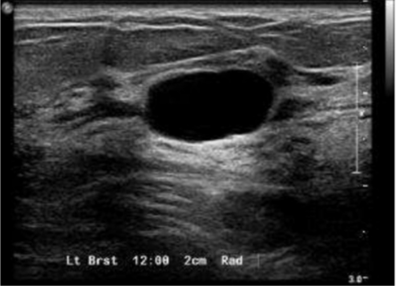
  • Simple cysts on ultrasound are:
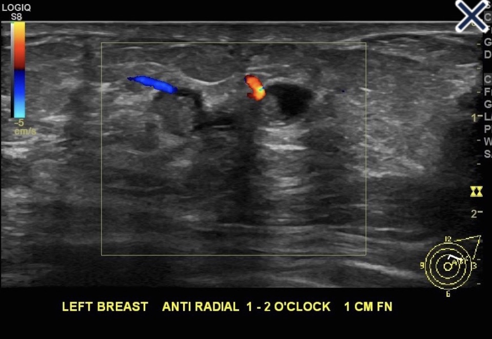
    • Anechoic, well circumscribed, with smooth walls and posterior enhancement, as depicted in the image
  • Complex cysts:
    • Have a very low malignancy rate (0.3%) and a 6-month follow-up ultrasound is reasonable
    • However, a complex cystic lesion with a significant solid component:
      • May have an associated cancer risk of up to 23%
    • Therefore, if the mass has a solid component:
      • A biopsy is indicated to rule out an associated malignancy
  • References:
    • Berg WA, Campassi CI, Ioffe OB. Cystic lesions of the breast: sonographic-pathologic correlation. Radiology. 2003;227:183-191.
    • Morrow M, Wong S, Vetna L. The evaluation of breast masses in women younger than forty years of age. Surgery. 1998;124:643-640.
    • Osborne MP, Boolbol S, Asad J. Benign conditions of the breast. In Kuerer H (ed.): Breast Surgical Oncology. New York, NY: McGraw-Hill; 2010:165-179.
    • Venta LA, Kim JP, Pelloski CE, et al. Management of complex breast cysts. Am J Roentgenol. 1999;173:1331-1336.
#Arrangoiz #BreastSurgeon #CancerSurgeon #SurgicalOncologist #BreastCancer #BreastCysts #MSMC #MountSinaiMedicalCenter #Miami #Mexico

Axillary Ultrasound in the Evaluation of Breast Cancer Patients

Normal Axillary Lymph Node
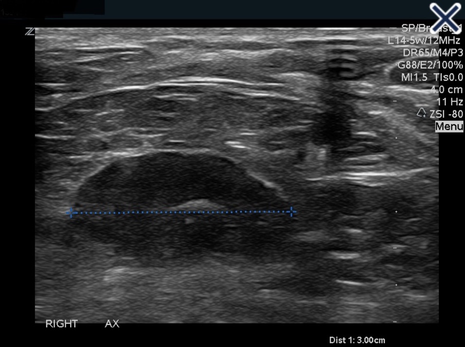
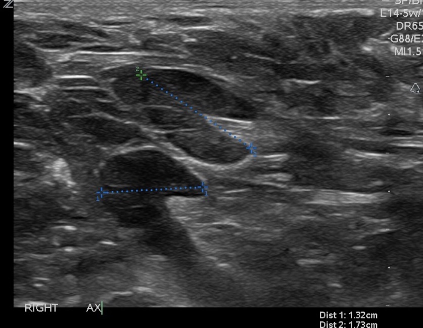
  • A normal axillary lymph node (Image):
    • Is elliptical in shape (or bean shaped), and has a narrow, symmetrical, hypoechoic cortex surrounding an isoechoic to hyperechoic fatty hilum (mediastinum)
    • The cortex of a normal node is composed largely of lymphatic tissue and fluid-filled cortical sinuses:
      • Thus the hypoechoic echogenicity
    • The hilum contains alternating medullary cords and sinusoids that have innumerable acoustic interfaces:
      • Thus the higher degree of echogenicity
Metastatic Axillary Lymph Node. Another presentation of a metastatic node is an asymmetric cortex where the tumor can be seen invading the hilum with convex indentations that look like “rat bites”
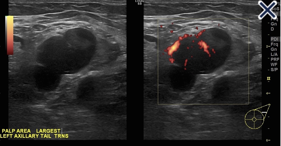
Lymph node completely replaced with metastatic carcinoma obliterating the fatty hilum. Doppler shows more than a single blood vessel supplying the node.
  • When a lymph node is completely replaced with metastatic cancer:
    • It will be rounded, hypoechoic, and the hilum will be completely obliterated (Image)
    • Before the node reaches the stage of complete replacement:
      • It can have an asymmetric, thickened cortex, with an eccentric hilum (Image)
  • Another presentation of a metastatic node:
    • Is an asymmetric cortex where the tumor can be seen invading the hilum with convex indentations that look like “rat bites” (First Image)
  • A metastatic node can also have severe compression of a central hilum resulting in a slit-like central hyperechoic band (Image)
Severe compression of the hilum by metastatic carcinoma resulting in a “slit-like” central hilum
Lymph nodes with eccentric hila and asymmetric, thickened cortices.
  • Reactive nodes can be difficult to distinguish from metastatic nodes (Image)
Reactive node.
  • In general, benign causes of nodal enlargement:
    • Tend to thicken the cortex diffusely
  • In addition, it is not uncommon to have a metastatic node adjacent to a normal node:
  • But all nodes in a region tend to be reactive when the cause is benign
  • Finally, a reactive node has blood supply on Doppler examination through a single hilum, whereas metastatic nodes tend to have multiple transcapsular vessels
  • References
  • Rahbar H, Partridge SC, Javid SH, Lehman CD. Imaging axillary lymph nodes in patients with newly diagnosed breast cancer. Curr Probl Diagn Radiol. 2012;41(5):149-158.
  • Stavros AT. Breast Ultrasound. Philadelphia, PA: Lippincott Williams & Wilkins; 2004.

Ultrasound Evaluation of a Solid Breast Nodule

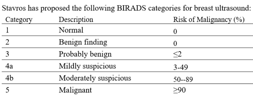
  • Stavros has proposed the following BIRADS categories for breast ultrasound (see Table 1)
Proposed BIRADS categories for breast ultrasound
  • The American College of Radiology classification subdivides category 4 into:
    • BIRADS 4a:
      • Which has a 2% to 10 % risk of malignancy
    • BIRADS 4b:
      • Which has a 10% to 50 % risk
    • BIRADS 4c:
      • Which has a 50% to 95% risk
  • BIRADS 5 has 95% or greater chance of malignancy
  • In evaluating a solid sonographic nodule:
    • One should first look for any of the 10 signs of malignancy:
      • If even one of them is present:
        • The lesion cannot be considered BIRADS 3
    • The 10 signs of malignancy include:
      • Shadowing
      • Hypoechoic echotexture
      • Spiculation
      • Angular margins
      • Thick echogenic halo
      • Microlobulation
      • Taller than wide
      • Duct extension
      • Branching pattern
      • Calcifications
  • Note that Stavros compares the echogenicity of lesions to that of breast fat, not breast parenchyma:
    • Therefore, a lesion with hypoechoic echotexture would be very hypoechoic if breast parenchyma is used as the reference
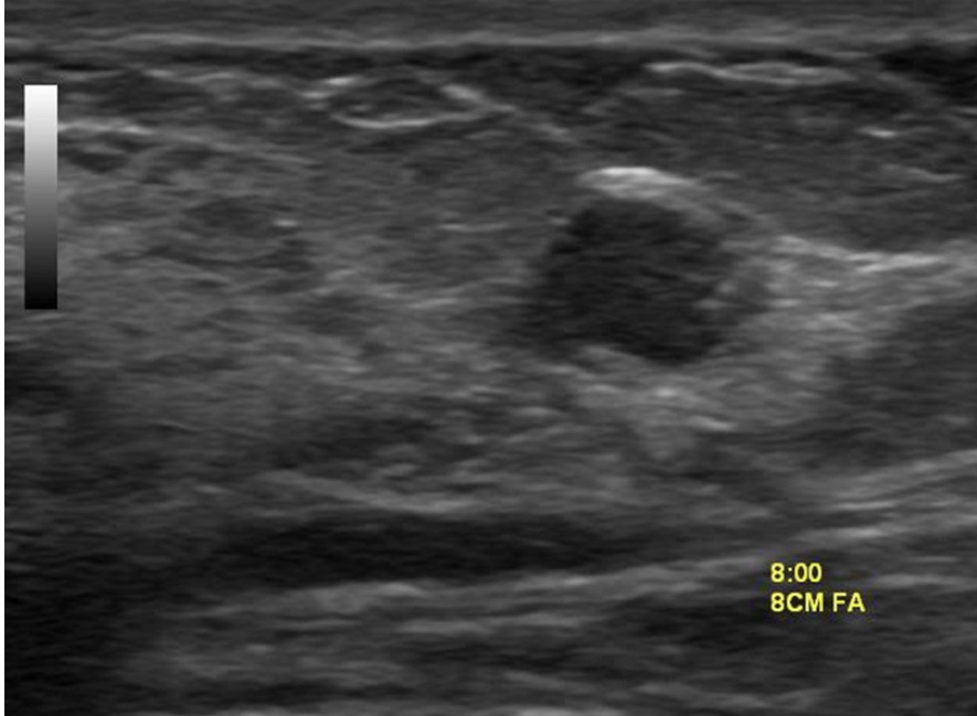
  • The hypoechoic lesion in the image does not have smooth margins but appears microlobulated
  • Regardless of whether the classification of Stavros or the American College of Radiology is used:
    • The risk of the lesion in this patient is not low enough to be considered BIRADS 3 nor high enough risk to warrant BIRADS 5:
      • Thus, it falls somewhere in the BIRADS 4 range:
        • Biopsy is required
  • References
    • D’Orsi CJ, Sickles EA, Mendelson EB, Morris EA. ACR BI-RADS® Atlas, Breast Imaging Reporting and Data System. 5th ed. Reston, VA: American College of Radiology; 2013
    • Jales RM, Sarian LO, Torresan R, Marussi EF, Alvares BR, Derchain S. Simple rules for ultrasonographic subcategorization of BI-RADS®-US 4 breast masses. Eur J Radiol. 2013;82(8):1231-1235.
    • Stavros AT. Breast Ultrasound. Philadelphia, PA: Lippincott Williams & Wilkins; 2004.

Breast Density on Imaging Studies

  • Women over age 40 with heterogeneously dense breasts or extremely dense breasts:
    • At average risk for developing breast cancer:
      • 12% to 13% lifetime risk:
        • Require only annual mammography
  • The decision to pursue additional imaging in patients with elevated risk should supplement but never entirely replace mammography
  • Use of screening ultrasound or MRI of the breast are appropriate for women at increased risk:
    • But the benefit remains to be determined in women of average risk for breast cancer
  • Breast fibroglandular composition:
    • Is defined by one of the following four descriptions:
      • Almost entirely fatty
      • Scattered areas of fibroglandular density
      • Heterogeneously dense
      • Extremely dense
  • The U.S. population distribution of breast density is as follows:
    • 10% almost entirely fatty
    • 40% scattered areas of fibroglandular density
    • 40% heterogeneously dense
    • 10% extremely dense
  • Women with heterogeneously dense or extremely dense breasts:
    • Are considered to have dense breasts:
      • Sensitivity of mammography decreases as breast density increases
      • Increased breast density not only has a masking effect, which may obscure masses:
        • But also serves as an independent risk factor for breast cancer
        • It has been reported that the increased risk:
        • May be as much as 4 to 6-fold
          • Estimates this high are obtained when comparing women with dense breasts to those with fatty replaced breasts
          • Since only 10% of women have fatty replaced breasts:
            • It makes more sense to make the comparison with women of average breast density
          • The relative risk for cancer in women with heterogeneously dense breasts compared with the average woman is approximately 1.2, and the relative risk for cancer in women with extremely dense breasts compared with the average woman is approximately 2.1
  • In general, breast density decreases with increasing age and increasing body mass index:
    • So it is not the absolute density that is a risk factor:
      • But the difference in the observed and expected density
  • Several states have passed legislation requiring women with dense breasts to be specifically informed of their breast density:
    • Such women are informed of the limitations of mammography in dense breasts and are instructed to discuss further management with their physicians
    • An informed decision regarding potential use of supplemental screening options, in addition to mammography, should be discussed, factoring in elements such as overall breast cancer risk as well as the positives and negatives of additional screening, including likelihood of additional benign biopsies
    • Guidelines for enhanced screening have been developed using lifetime risk calculations as calculated by models stressing family history, such as the Tyrer Cuzick Model, and not purely using breast density
  • References
    • 1. D’Orsi CJ, Sickles EA, Mendelson EB, Morris EA. ACR BI-RADS® Atlas: Breast Imaging Reporting and Data System, 5th ed. Reston, VA: American College of Radiology; 2013. 2. 3. Freer PE. Mammographic breast density: impact on breast cancer risk and implications for screening. Radiographics. 2015;35(2):302-315.
    • Brentnall AR, Harkness EF, Astley SM, Donnelly LS, Stavrinos P, Sampson S, et al. Mammographic density adds accuracy to both the Tyrer-Cuzick and Gail breast cancer risk models in a prospective UK screening cohort. Breast Cancer Res. 2015;17(1):147.
    • American College of Radiology. ACR statement on reporting breast density in mammography reports and patient summaries. https://www.acr.org/Advocacy-and-Economics/ACR-Position-Statements/Reporting-Breast-Density. November 26, 2017. Accessed July 16, 2020.
#Arrangoiz #BreastSurgeon #BreastCancer #MountSinaiMedicalCenter

Galactoceles

  • Galactoceles:
    • Are milk retention cysts:
      • That result from a blocked milk duct
  • They present as cystic, sometimes very large masses:
    • During pregnancy, lactation, and after weaning
  • They are often painless unless they become infected
  • Initially, they contain milky fluid:
    • But over time, contents become thicker and more creamy or oily as the fluid is reabsorbed
  • Ultrasound is the primary diagnostic method
    • Typical findings include a:
      • Well-defined lesion with thin echogenic walls
      • The internal appearance consists of either homogeneous contents or heterogeneous contents with fluid clefts and anechoic rims
  • Management consists of:
    • Needle aspiration demonstrating milky contents:
      • Which both confirms the diagnosis and excludes malignancy
  • Surgical resection is reserved for:
    • Cases refractory to conservative management
  • References
    • Sawhney S. Petkovska L, Ramadan S, Al-Muhtaseb S, Jain R, Sheikh M.Sonographic appearance of galactoceles. J Clin Ultrasound. 2002;30(1):18-22.
    • Sabate JM, Clotet M, Torrubia S, Gomez A, Guerrero R, de las Heras P. Radiologic evaluation of breast disorders related to pregnancy and lactation. Radiographics.2007;27(Suppl 1):S101-S124.

#Arrangoiz #BreastSurgeon #BreastCancer #Galactocele #CancerSurgeon

Less Frequent Mammography for Breast Cancer Survivors Has No Impact on Survival

  • Certain breast cancer survivors can safely reduce the frequency with which they undergo surveillance mammography 3 years after surgery, according to findings presented at the 2023 San Antonio Breast Cancer Symposium
  • The results are important, because currently in the US, breast cancer survivors are told to undergo an annual mammogram every year for an indefinite period of time after their surgery:
    • In the UK, survivors routinely undergo an annual mammogram for the first 5 years after surgery then de-escalate to once every 3 years afterward for those aged 50 and older
  • These findings presented at SABCS suggest that survivors can get screened less frequently than current guidelines allow.
  • In the phase 3 Mammo-50 trial:
    • Researchers randomly assigned women who underwent curative surgery for breast cancer to one of two groups.
    • One was assigned to undergo a mammogram every year, and another received mammograms less frequently: once every 2 years for those who had breast-conservation surgery and once every 3 years for those who had mastectomies
    • After a 5-year follow-up period, the groups had similar rates of both overall survival (OS) and breast-cancer specific survival
    • The annual group had a breast-cancer specific survival rate of 98.1% and an OS of 94.7%, whereas the less-frequent group had a breast-cancer specific survival rate of 98.3% and an OS of 94.5%
    • The two groups also had similar recurrence rates:
      • 5.9% for the annual group and 5.5% for those assigned to less frequent screening
    • Although the researchers acknowledged that the de-escalation arm had a lower compliance rate than the annual arm:
      • At 69% compared with 83%, they added that a sensitivity analysis revealed this difference in adherence did not impact their final results
  • In a concurrent substudy evaluating the participants’ quality of life using four different validated questionnaires, researchers reported no differences between the two groups in the domains of distress, mental well-being, and other areas
  • Researchers acknowledged that the study was limited by the fact that it focused only on women older than 50 who were already cancer-free for at least 3 years
  • De-escalation of mammographic surveillance reduces the burden on the health care system, decreases the inconvenience for women having to undergo these mammograms, and reduces the associated stress of waiting for results:
    • The trial demonstrated that the outcomes from undergoing less frequent mammograms were no worse than undergoing annual mammograms for this group of women

Younger Postmenopausal Patients May Skip Adjuvant Radiotherapy After Early Breast Cancer Surgery

  • Younger postmenopausal patients with low-risk, stage I HR-positive breast cancer and certain genetic markers may be able to safely forgo adjuvant radiotherapy:
    • According to study findings presented at the 2023 San Antonio Breast Cancer Symposium in Texas
  • In the IDEA (Individualized Decisions for Endocrine therapy Alone) trial:
    • 200 postmenopausal patients with stage I HR-positive, HER2-negative breast cancer:
      • All of whom had scores of 18 or less on the Oncotype DX recurrence assay:
        • Elected to skip adjuvant radiotherapy:
          • Provided they were willing to continue with endocrine therapy for 5 years
    • All participants were aged between 50 and 69 years and required to have a margin of 2 mm or more after breast-conserving surgery
    • The primary outcome:
      • Was the rate of disease recurrence at 5 years after surgery
    • Patients had a mean recurrence score of 11.2
    • Eighty-five patients had grade 1 tumors, whereas 109 had grade 2 tumors and 6 had grade 3 tumors
    • The tumors were a mean of 10 mm
    • The median follow-up was 5.21 years
    • Both the overall survival rate and breast-cancer survival rate were 100% at 5 years:
      • The researchers reported, with a 5-year freedom-from-recurrence rate of 99% (95% CI, 96-100):
        • However, 2 patients died after the 5-year follow-up period had expired
    • There were two breast cancer related events during the follow-up period:
      • One event, which occurred at the 21-month mark:
        • Was an isolated axillary recurrence:
          • This was treated with axillary dissection and breast irradiation as well as local irradiation
      • The other was an ipsilateral breast event:
        • Which occurred at 49 months and was treated with another breast-conserving surgery
    • Another 6 patients experienced recurrence after the 5-year follow-up period, the researchers reported:
      • Five of these were ipsilateral breast events, and one was an ipsilateral breast event with regional occurrence
    • For the entire follow-up period:
      • The crude rate of ipsilateral breast events was:
        • 3.3% (n = 2 of 60) for patients who were 50 to 59 years old
        • 3.6% (n = 5 of 140) for those who were 60 to 69 years old
      • The crude overall relapse rates in these age groups were 5% (n = 3) and 3.6%, respectively
  • These findings indicate that younger postmenopausal patients with stage I breast cancer who skip radiotherapy after breast-conserving surgery:
    • Have a very low risk of disease recurrence within 5 years:
      • However, 5 years is an early time point for this population, and longer-term follow-up of this study and others will be essential to determine whether this option can be safely offered to women in this age group
  • Reference:
    • Jagsi, R. Five-year outcomes of the IDEA trial of endocrine therapy without radiotherapy after breast-conserving surgery for postmenopausal patients age 50-69 with genomically-selected favorable stage I breast cancer. Abstract GS02-08. SABCS 2023.
#Arrangoiz @Doctor #Surgeon #CancerSurgeon #SurgicalOncologist #BreastSurgeon #BreastCancer #MSMC #MountSiniaMedicalCenter #Miami #Mexico

Radiation Changes in the Breast Seen on Mammogram

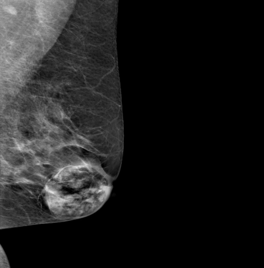
Right craniocaudad image.
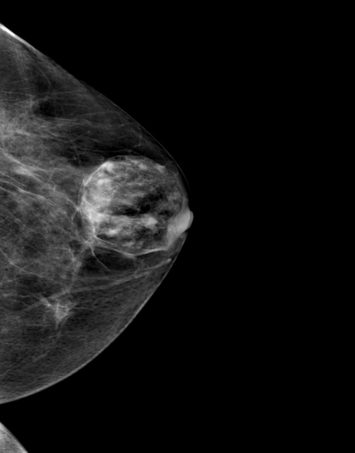
Right mediolateral oblique image.
  • The mammogram:
    • Shows trabecular thickening and an increased thickness of the skin
  • Trabecular thickening:
    • Is an increased prominence of linear structures in the breast:
      • Primarily corresponding to Cooper’s ligaments
    • It is almost always seen in conjunction with skin thickening:
      • It indicates significant breast edema
    • It has a number of different causes, including:
      • Congestive heart failure
      • Renal failure
      • Venous or lymphatic obstruction
      • Diffusely infiltrating carcinoma
      • Radiation
    • When the cause is lymphatic obstruction:
      • The obstruction can be the result of surgery, radiation, or lymphatic invasion by cancer
  • Unilateral breast edema with increased trabecular and skin thickening:
    • Sometimes occurs with congestive heart failure
  • The pedal edema that develops during the daytime:
    • Decreases at night and the fluid accumulates in the dependent breast in patients who sleep on their side
  • References
    • Berg, WA, Birdwell RL, Kennedy A. Diagnostic Imaging: Breast. Salt Lake City, UT: Amirsys; 2006.
    • Verbelen H, Gebruers N, Beyers T, De Monie AC, Tjalma W. Breast edema in breast cancer patients following breast-conserving surgery and radiotherapy: a systematic review. Breast Cancer Res Treat. 2014;147(3):463-471.
    • Wratten CR, O’Brien PC, Hamilton CS, Bill D, Kilmurray J, Denham JW. Breast edema in patients undergoing breast-conserving treatment for breast cancer: assessment via high frequency ultrasound. Breast J. 2007;13(3):266-273.
    • Menta A, Fouad TM, Lucci A, Le-Petross H, Stauder MC, Woodward WA, et al. Inflammatory breast cancer: what to know about this unique, aggressive breast cancer. Surg Clin North Am. 2018;98(4):787-800.
#Arrangoiz #Doctor #Surgeon #CancerSurgeon #SurgicalOncologist #BreastSurgeon #MountSinaiMedicalCenter #MSMC #Miami Mexico #BreastCancer

Sonographic Appearance of Lymph Nodes

  • The sonographic appearance of a normal lymph node:
    • Is elliptical with a thin, hypoechoic cortex and an isoechoic to hyperechoic fatty hilum (Image)
Normal axillary lymph node
  • Metastatic carcinoma in a lymph node:
    • Would usually have an asymmetric thick cortex or have near-total or total obliteration of the hilum:
    • Resulting in a rounded, hypoechoic mass (Image)
Node with metastatic carcinoma
  • Axillary adenopathy can occur in association with rheumatoid arthritis:
    • But the sonographic findings would usually be a symmetrical, mild thickening of the cortex:
      • Usually with preservation of the hilum
  • Silicone granulomas:
    • Classically create a snowstorm appearance:
      • Which allows a definitive diagnosis by ultrasound alone
Silicone Granulomatous
  • The mass in the image has a rounded anterior border and “dirtyincoherent shadowing that obscures the posterior border of the lesion:
    • Nothing other than silicone can cause this sonographic appearance:
      • But it is difficult to distinguish free silicone that has migrated to the axilla from a node that has been replaced with silicone
  • In different stages of development, silicone extravasation can also mimic complex cysts or fibrotic nodules, depending on the amount of silicone extravasated and the amount of time from extravasation:
    • If a large amount of silicone is released into the tissue, the ultrasound pattern is that of a complex cyst
  • Silicone’s presence in tissues can cause a local inflammatory reaction:
    • Which may cause tissue fibrosis and a fibrotic nodule:
      • This is a late finding seen following extravasating
  • References:
    • Berg WA, Caskey CI, Hamper UM, Anderson ND, Chang BW, Sheth S, et al. Diagnosing breast implant rupture with MR imaging, US, and mammography. Radiographics. 1993;13(6):1323-1336.
    • Juanpere S, Perez E, Huc O, Motos N, Pont J, Pedraza S. Imaging of breast implants‒a pictorial review. Insights Imaging. 2011;2(6):653-670.

Mammographic Images in Diffusely Invasive Breast Cancer

  • Diffusely invasive carcinoma:
    • Has a mammographic appearance of:
      • Diffuse architectural distortion
    • Usually involving a large area:
      • Often larger than a lobe:
        • With no central tumor mass and no calcifications
    • It sometimes has the appearance of:
      • A “spider’s web” (Image 1)
    • The diffusely infiltrating cancer forms concave contours with the surrounding fat in a manner similar to normal fibroglandular tissue (Images 2 a-b)
Image 1: Mediolateral oblique and craniocaudal projections.
Mastectomy slice radiographs (a) and large format 3D histology image (b) showing concave contours similar to normal breast
  • The imaging findings of diffusely infiltrating breast cancer:
    • Are strikingly different from the imaging findings of breast cancers originating either from the terminal ductal lobular units (TDLUs) or the lactiferous ducts:
      • Suggesting that it may have a different site of origin
  • It has been recently proposed that diffusely infiltrating breast cancers may originate from:
    • Mesenchymal stem cells (progenitors):
      • Through a complex process of both epithelial-mesenchymal transformation and more frequently, mesenchymal-epithelial transformation
    • The clinical presentation is typically a:
      • Recently detected, extensive, firm lesion:
        • Often appearing as an interval cancer following a previous mammogram which was interpreted as normal
    • On clinical breast examination:
      • The cancer does not have a distinct tumor mass or focal skin retraction seen in other cancers:
        • But rather an indistinct “thickening” and eventually a shrinkage of the breast
    • In order to make the diagnosis before the development of a palpable mass and a decrease in size of the breast:
      • The radiologist and breast surgeon must have a high level of suspicion and a thorough knowledge of the underlying pathophysiology
    • The subgross (3D) histopathology images:
      • Show how growth of the mesenchymal tissue distorts the normal, harmonious connective tissue framework by causing nonuniform thickening of the fine sheets of connective tissue (Images 3a -b)
Large format subgross (3D) histology images of a diffusely infiltrating breast cancer
  • The predominance of mesenchyme in the diffusely infiltrating breast malignancy:
    • Allows it to be imaged with greater sensitivity by ultrasound than by mammography
  • The thin sheets or veils of tissue reflect the ultrasound waves, but are relatively easily penetrated by x-rays:
    • The structural / architectural distortion, while difficult to detect mammographically:
      • Is readily detectable on 2-mm thick coronal sections of automated breast ultrasound (Image 3c)
    • The hypoechoic changes can also usually be seen on hand-held ultrasound (Image 4).
3D automated ultrasound images. The 2-mm thick multi-slice series demonstrate the extensive architectural distortion, corresponding to the 3D histology
Hand-held ultrasound of diffusely infiltrating carcinoma
  • The growth pattern and cell type of diffusely invasive breast cancer:
    • Is very similar to that of diffuse gastric carcinoma (linitis plastica):
      • Both of these diseases can be associated with a deleterious mutation in the CDH1 gene:
        • Which is located on chromosome 16q22:
          • It codes for e-cadherin protein (Image 5a, Image 5b)
Large format histology slide of diffusely infiltrating breast cancer similar to growth pattern of linitis plastica
High-power histology of pleomorphic infiltrating breast cancer with cell type similar to linitis plastica.
  • References
    • Hansford S, Kaurah P, Li-Chang H, Woo M, Senz J, Pinheiro H, et al. Hereditary diffuse gastric cancer syndrome: CDH1 mutations and beyond. JAMA Oncol. 2015;1(1):23-32.
    • Tot T. The diffuse type of invasive lobular carcinoma of the breast: morphology and prognosis. Virchows Arch. 2003;443(6):718-724.
    • Tot T. Diffuse invasive breast carcinoma of no special type. Virchows Arch. 2016;468(2):199-206.
#Arrangoiz #Doctor #Surgeon #CancerSurgeon #SurgicalOncologist #Breast Surgeon #MountSinaiMedicalCenter #MSMC #Miami #Mexico #BreastCancer