New Research Alert: ER-Low Breast Cancer & Endocrine Therapy (2026)
Liu et al. Breast Cancer Res Treat. Published Jan 6, 2026.
🔬 Key Focus
This study evaluated outcomes and benefits of endocrine therapy (ET) in early breast cancer patients with low estrogen receptor (ER) expression (1%–10%) compared to ER-high and ER-negative cancers.
📊 Major Findings
🧬 ER-Low Phenotype Is Distinct
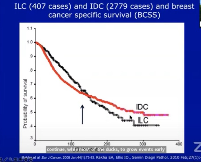
ER-low (~4.4% of cases) behaves more like ER-negative disease in HER2-negative patients. Worse breast cancer-free survival compared to ER-high. Similar risk to triple-negative disease in HER2-negative subgroup.
💊 Endocrine Therapy Benefit Varies by Subtype
📍 HER2-Negative ER-Low:
✔ ET significantly reduced locoregional recurrence & distant metastasis
✔ Improved breast cancer–free survival (BCFS)
📍 HER2-Positive ER-Low:
❌ No clear survival or BCFS benefit from ET observed
🧠 Clinical Implications
✅ Consider ET in HER2-negative, ER-low early breast cancer
⚠️ The benefit of ET in HER2-positive ER-low remains uncertain
🧪 ER-low shouldn’t be treated the same as classical ER-high luminal tumors
📌 Why This Matters
ER-low tumors are being increasingly recognized as a biologically unique subgroup. This research supports more nuanced treatment planning, particularly regarding the value of endocrine therapy based on HER2 status.
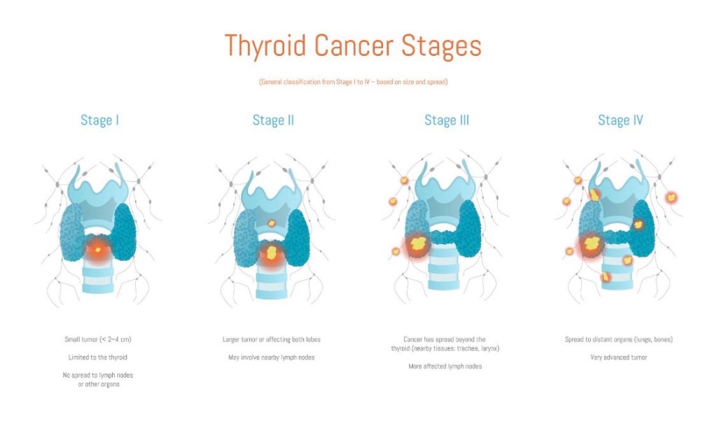
Poorly differentiated thyroid cancer (PDTC) is an uncommon but important subtype that sits between differentiated thyroid cancer (papillary/follicular) and anaplastic thyroid cancer in terms of aggressiveness.
🧠 What defines PDTC? Tumors show loss of typical thyroid differentiation Often described with insular, trabecular, or solid growth patterns More aggressive than papillary or follicular cancer Less responsive to radioactive iodine than well-differentiated cancers
🔍 How is PDTC diagnosed? Definitive diagnosis is made on surgical pathology Based on specific histologic criteria (e.g., high mitotic rate, necrosis, insular pattern) FNA may suggest aggressive disease but is not always definitive
⚖️ How is PDTC treated?
Management requires multidisciplinary, risk-adapted care: Total thyroidectomy is usually recommended Therapeutic lymph node dissection when nodal disease is present Radioactive iodine may be considered, but effectiveness is variable External beam radiation and systemic therapy in selected cases Close, long-term surveillance
📈 Prognosis Worse than papillary or follicular thyroid cancer Better than anaplastic thyroid cancer Outcomes depend on: Tumor stage Completeness of surgical resection Presence of distant metastases
🦋 Early recognition and expert pathology review are critical for optimal outcomes.
👨⚕️ Dr. Rodrigo Arrangoiz, MD Surgical Oncologist – Thyroid, Head & Neck, Breast Mount Sinai Medical Center
📌 Take-home message: Poorly differentiated thyroid cancer is uncommon but serious—timely diagnosis and comprehensive care matter.
📚 References Lloyd RV et al. WHO Classification of Tumours of Endocrine Organs Haugen BR et al. ATA Guidelines for Differentiated Thyroid Cancer. Thyroid Volante M et al. Poorly differentiated thyroid carcinoma. Endocr Relat Cancer
Failure to identify a parathyroid gland during cervical exploration:
Is most commonly explained by:
Ectopic location rather than true absence
Large anatomic and surgical series demonstrate that approximately 15% to 16% of parathyroid glands are ectopic:
With predictable distributions:
Based on embryologic origin (Taterra et al., Surg Radiol Anat, 2019):
Consequently, a structured search strategy:
Guided by embryology and prevalence data is recommended
General intra-operative principles:
Careful inspection of the orthotopic field:
Is mandatory before declaring a gland ectopic
Approximately 80% to 90% of parathyroid glands are located within a few millimeters of the posterior thyroid capsule (Taterra et al., 2019):
Gentle subcapsular dissection along the posterior surface of the thyroid should be completed before expanding the field
Intrathyroidal parathyroid glands:
Account for 2% to 3% of all glands and up to 20% to 22% of ectopic glands, particularly inferior glands (Phitayakorn & McHenry, Am J Surg, 2006):
For this reason, inspection and palpation of the thyroid specimen is considered standard practice in experienced centers (Noussios et al., Exp Clin Endocrinol Diabetes, 2012)
Reoperative series demonstrate that most “missed” glands:
Are found in standard embryologic locations:
Most commonly the tracheoesophagealgroove, thyrothymic ligament, or superior mediastinum:
Emphasizing the importance of a systematic rather than random exploration (Silberfein et al., Arch Surg, 2010)
Superior parathyroid gland – Evidence-Based search pattern:
Typical location:
Superior parathyroid glands:
Fourth pharyngeal pouch origin:
Exhibit limited migration and are therefore relatively constant in position
They are typically located on the posterior aspect of the upper thyroid pole:
Approximately 1 cm above the intersection of the recurrent laryngeal nerve (RLN) and the inferior thyroid artery:
Frequently within the tracheoesophageal groove (Scharpf et al., Surg Oncol Clin N Am, 2016)
Common ectopic locations:
When ectopic:
Superior parathyroid glands are most often displaced posteriorly, rather than inferiorly:
Tracheoesophageal or para-esophageal groove the most common ectopic site for superior glands (Noussios et al., 2012; Taterra et al., 2019)
Retro-esophageal or retro-pharyngeal space, particularly in undescended glands (Scharpf et al., 2016)
Posterior mediastinum, where enlarged glands may descend along the esophagus but remain posterior in relation to the RLN (Phitayakorn & McHenry, 2006)
Stepwise surgical approach:
If a superior gland is not identified in its orthotopic location, the recommended sequence is:
Systematic exploration of the tracheoesophageal groove following the RLN superiorly
Blunt dissection of the para- and retro-esophageal spaces
Evaluation of the high posterior neck for undescended glands
Inspection of the thyroid specimen for an intrathyroidal gland (Noussios et al., 2012; Silberfein et al., 2010)
Descend with the thymus and demonstrate significantly greater variability
Orthotopically, they are most often located near the lower thyroid pole, anterior to the RLN, frequently within or adjacent to the thyrothymic ligament (Scharpf et al., 2016)
Common ectopic locations:
Inferior glands account for the majority of ectopic parathyroids:
Intrathymic or within the cervical thymus:
Approximately 30% of ectopic inferior glands (Phitayakorn & McHenry, 2006)
Anterosuperior mediastinum, often contiguous with thymic tissue (Noussios et al., 2012)
Intrathyroidal:
Accounting for ~ 20% to 22% of ectopic inferior glands (Phitayakorn & McHenry, 2006)
High cervical or carotid sheath locations, representing failed embryologic descent (Noussios et al., 2012)
Stepwise surgical approach:
When an inferior gland is not identified at the lower pole:
The thyrothymic ligament should be followed inferiorly toward the thymus
A limited cervical thymectomy should be performed when clinically appropriate:
Given the high incidence of intrathymic glands
The lower thyroid pole and specimen should be inspected for intrathyroidal tissue
The carotid sheath and high cervical region should be explored in cases suspicious for undescended glands (Phitayakorn & McHenry, 2006; Silberfein et al., 2010)
Lessons from re-operative surgery:
In contemporary re-operative parathyroidectomy series, previously missed glands were most commonly located in the:
Tracheoesophageal groove
Thyrothymic ligament
Superior mediastinum
Confirming that failure is usually related to incomplete exploration of predictable embryologic sites rather than unusual anatomy (Silberfein et al., Arch Surg, 2010)
Key references:
Taterra D, et al. The prevalence and anatomy of parathyroid glands: a meta-analysis. Surg Radiol Anat. 2019.
Phitayakorn R, McHenry CR. Incidence and location of ectopic abnormal parathyroid glands. Am J Surg. 2006;191:418–423.
Noussios G, et al. Ectopic parathyroid glands and their anatomical, clinical and surgical implications. Exp Clin Endocrinol Diabetes. 2012.
Silberfein EJ, et al. Reoperative parathyroidectomy: location of missed glands. Arch Surg. 2010.
Scharpf J, et al. Anatomy and embryology of the parathyroid glands. Surg Oncol Clin N Am. 2016.
Anaplastic thyroid cancer (ATC) is rare (<2%) but represents the most aggressive form of thyroid cancer. It behaves very differently from other thyroid cancers and requires urgent, multidisciplinary care.
🧠 Key characteristics of ATC
Rapidly growing neck mass Often presents with hoarseness, difficulty swallowing, or breathing problems Frequently diagnosed at an advanced stage Can arise from pre-existing differentiated thyroid cancer
🔍 How is ATC diagnosed?
Clinical suspicion due to rapid growth Imaging (CT/MRI) to assess airway and invasion Core needle biopsy or surgical biopsy for confirmation Molecular testing (e.g., BRAF V600E) to guide targeted therapy
⚖️ How is ATC treated?
Management requires a multidisciplinary approach:
Airway protection is often the first priority Surgery when feasible Radiation therapy and systemic therapy Targeted therapy and immunotherapy have significantly improved outcomes in selected patients
📈 Prognosis
Historically poor Modern targeted therapies have changed the landscape, improving survival in carefully selected patients Early referral to specialized centers is critical
🦋 ATC is a medical emergency—time matters.
👨⚕️ Dr. Rodrigo Arrangoiz, MD
Surgical Oncologist – Thyroid, Head & Neck, Breast
Mount Sinai Medical Center
📌 Take-home message:
Anaplastic thyroid cancer is aggressive, but early recognition and modern therapies are improving outcomes.
📚 References
Smallridge RC et al. ATA Guidelines for Anaplastic Thyroid Cancer. Thyroid Bible KC et al. Targeted therapy in ATC. NEJM NCCN Guidelines: Thyroid Carcinoma
ADH and atypical lobular hyperplasia (ALH) are now frequently diagnosed with use of core-needle biopsy
Although atypia can be difficult to distinguish from carcinoma in situ:
Pathologic criteria exist to distinguish the two entities
This distinction is important because, while in situ carcinoma is malignant and may progress to invasive disease:
ADH is a non-obligate cancer precursor and often represents a marker of an elevated future breast cancer risk
ADH is most frequently found by mammography
Atypia alone, with no other risk factors, confers an approximate:
Four-fold to five-fold risk of the development of breast cancer
Although breast MRI is more sensitive to detect intermediate- and high-grade ductal carcinoma in situ (DCIS) as well as invasive cancers:
Breast MRI lacks sufficient diagnostic ability to differentiate ADH versus DCIS or invasive cancers
Excision is indicated for ADH found on core needle biopsy:
As concomitant in situ or invasive cancer will be found in approximately 15% of cases (15% to 30% in some series)
The 10-year risk of developing a breast cancer after a diagnosis of ADH:
Is approximately 17%:
Risk is bilateral:
Breast cancers developing within 5 years of a biopsy of ADH more likely to occur in the ipsilateral breast than those developing more than 5 years (82% ipsilateral in the first 5 years vs 58% ipsilateral after 5 years)
The National Surgical Adjuvant Breast and Bowel Project (NSABP) P-1 trial:
Showed that when atypia is found on a sample obtained with needle biopsy and excision rules out cancer:
Tamoxifen reduces the risk of developing breast cancer by about 86%:
These patients should therefore be referred for discussion about this endocrine prophylaxis
Similarly, the Study of Tamoxifen and Raloxifene (STAR, NSABP P-2) trial:
Which randomized post-menopausal women to tamoxifen or raloxifene, found that raloxifene provided equivalent risk reduction to tamoxifen with less toxicity (e.g., endometrial cancer)
References:
Coopey SB, Mazzola E, Buckley JM, et al. The role of chemoprevention in modifying the risk of breast cancer in women with atypical breast lesions. Breast Cancer Res Treat. 2012;136:627-633.
Heller SL, Moy L. Imaging features and management of high-risk lesions on contrast-enhanced dynamic breast MRI. AJR Am J Roentgenol. 2012;198:249-255.
Krishnamurthy S, Bevers T, Kuerer H, Yang WT. Multidisciplinary considerations in the management of high-risk breast lesions. AJR Am J Roentgenol. 2012;198:W132-140.
Hartmann LC, Radisky DC, Frost MH, et al. Understanding the premalignant potential of atypical hyperplasia through its natural history: a longitudinal cohort study. Cancer Prev Res (Phila). 2014;7:211-217.
Vogel VG, Costantino JP, Wickerham DL, et al; National Surgical Adjuvant Breast and Bowel Project. Update of the National Surgical Adjuvant Breast and Bowel Project Study of Tamoxifen and Raloxifene (STAR) P-2 Trial: Preventing breast cancer. Cancer Prev Res (Phila). 2010;3:696-706.
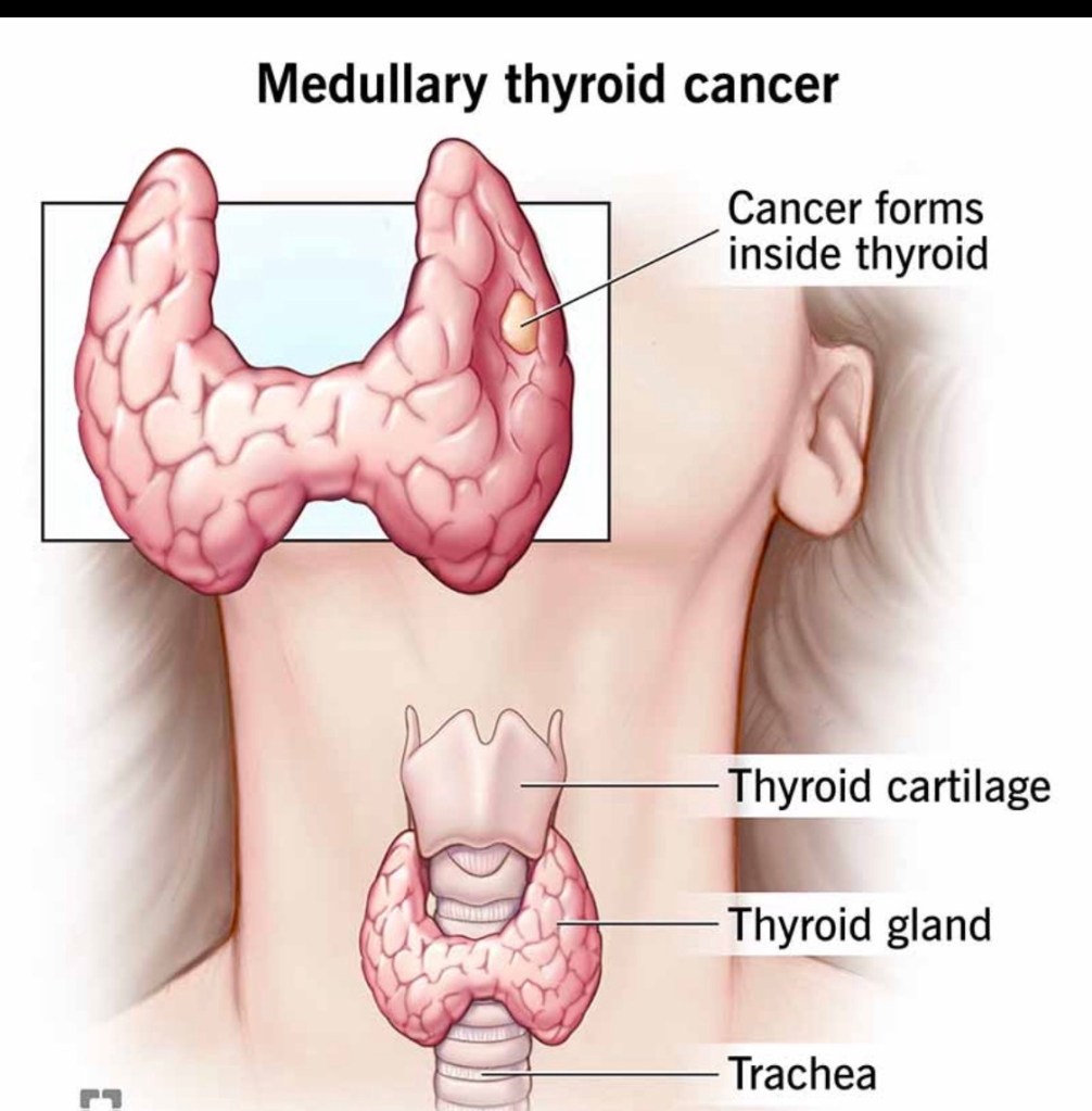
Medullary thyroid cancer (MTC) accounts for ~2–4% of all thyroid cancers and is biologically distinct from papillary and follicular thyroid cancers.
🧠 What makes MTC different?
Arises from parafollicular (C) cells, not follicular cells Produces calcitonin, a key tumor marker Does NOT respond to radioactive iodine Can be sporadic (~75%) or hereditary (~25%)
🧬 The genetic connection
Hereditary MTC is associated with RET mutations Seen in MEN2 syndromes (MEN2A, MEN2B, FMTC) All patients with MTC should undergo genetic testing, regardless of age or family history
🔍 How is MTC diagnosed?
Suspicious thyroid nodule on ultrasound Elevated serum calcitonin (often markedly high) Confirmed by FNA biopsy ± calcitonin washout Imaging to evaluate lymph node involvement
⚖️ How is MTC treated?
The cornerstone of treatment is surgery:
Total thyroidectomy Central neck lymph node dissection Lateral neck dissection when nodes are involved
➡️ Radioactive iodine has no role in MTC.
➡️ Targeted systemic therapies are used in advanced disease.
📈 Prognosis
Highly dependent on stage at diagnosis Early detection → excellent long-term outcomes Lymph node and distant spread worsen prognosis
🦋 Early recognition and expert surgical management are critical.
👨⚕️ Dr. Rodrigo Arrangoiz, MD
Surgical Oncologist – Thyroid, Head & Neck, Breast
Mount Sinai Medical Center
📌 Take-home message:
Medullary thyroid cancer is rare but requires prompt diagnosis, genetic evaluation, and expert surgical care.
📚 References
Wells SA et al. Revised ATA Guidelines for Medullary Thyroid Carcinoma. Thyroid Elisei R et al. Management of Medullary Thyroid Cancer. Lancet NCCN Guidelines: Thyroid Carcinoma
Hürthle cell carcinoma (HCC) is a distinct subtype of differentiated thyroid cancer, accounting for ~3–5% of cases. Although related to follicular tumors, it behaves differently and requires specific management considerations.
🧠 Key characteristics of Hürthle cell carcinoma
Composed of oncocytic (Hürthle) cells rich in mitochondria More common in older patients Less likely to spread to lymph nodes More likely to spread hematogenously (lungs, bone) in higher-risk disease Often less iodine-avid than papillary or follicular thyroid cancer
🔍 How is it diagnosed?
Ultrasound and FNA may suggest a Hürthle cell neoplasm Definitive diagnosis requires surgery, based on: Capsular invasion Vascular invasion
Thyroid lobectomy for small, minimally invasive tumors Total thyroidectomy for larger or invasive disease Radioactive iodine selectively (often less effective than in other subtypes) Close long-term surveillance
📈 Prognosis
Excellent outcomes for minimally invasive disease Prognosis worsens with: Extensive vascular invasion Large tumor size Distant metastases
🦋 Careful pathology review and individualized treatment are essential.
👨⚕️ Dr. Rodrigo Arrangoiz, MD
Surgical Oncologist – Thyroid, Head & Neck, Breast
Mount Sinai Medical Center
📌 Take-home message:
Hürthle cell carcinoma is uncommon but highly treatable when managed by an experienced thyroid team.
📚 References
Haugen BR et al. ATA Guidelines for Differentiated Thyroid Cancer. Thyroid Lloyd RV et al. WHO Classification of Tumours of Endocrine Organs Ganly I et al. Hürthle cell carcinoma outcomes. J Clin Endocrinol Metab