Blog

Diffuse Breast Cancer on Imaging

  • There are two main groups of diffuse breast cancers:
    • That present as large areas of architectural distortion on the mammogram:
      • Neoductgenesis
      • Diffusely infiltrating carcinoma:
        • Which makes up approximately 5% of all breast cancers
  • When the tumor is e-cadherin negative:
    • It is usually called invasive “lobular” carcinoma
  • When it is e-cadherin positive:
    • It is called infiltrating “ductal” carcinoma
  • The designation based on e-cadherin staining is arbitrary:
    • Because the behavior of diffusely invasive carcinoma:
      • Is the same regardless of the staining
  • Lacking calcifications and a central tumor mass:
    • These cancers are notoriously difficult to perceive on mammogram:
      • Even when they are large and palpable or when they occur in fatty involuted breasts:
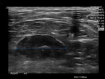
        • However, the associated connective tissue response:
          • Makes this type of cancer quite visible with ultrasound
  • In contrast to diffusely infiltrating cancers:
    • Circular (Image 1) and spiculated (Image 2):
      • Tumors arising in the terminal ductal lobular units (TDLU) have:
        • Bulging, convex contours protruding into the adipose tissue
  • The solid variety of infiltrating lobular carcinoma:
    • Most probably arises within the TDLU:
      • Has a circular / oval shape on breast imaging (Images 3)

Image 1: Lobulated spherical tumor mass

Image 2: Multifocal stellate invasive breast cancer

Image 3: Mammographic, MRI and pathologic images of the solid form of invasive lobular

  • There are two other variants of invasive lobular carcinoma that arise in the TDLUs:
    • The tubulolobular variant:
      •  Is either a unifocal or multifocal spiculated lesion on the mammogram (Image 5)

Image 4: Mammogram and large format histology of a multifocal tubulolobular breast

  • The alveolar type of invasive lobular carcinoma:
    • Is usually mammographically occult;
      • Or it can be seen as a subtle, asymmetric density (Image 5)

Image 5: Mammogram and large format histology alveolar type invasive lobular carcinoma

  • The various forms of invasive lobular carcinoma that develop in the TDLUs and present as localized lesions:
    • Have a significantly better prognosis than the diffusely infiltrating type breast cancer
  • Complex sclerosing lesions:
    • Present mammographically as non-palpable architectural distortion with no central tumor mass and lucent radiating structures, the so called “black star”:
      • As opposed to cancers originating from the TDLU:
        • Which have a dense central tumor mass surrounded by radiopaque spiculation, giving the impression of looking at a “white star
  • Malignant phyllodes tumors:
    • Present as large, high density masses:
      • The borders may be circumscribed or ill defined
  • Fat necrosis:
    • Also presents as a hypoechoic, high-density mass
  • References
  • Tot T. Diffuse invasive breast carcinoma of no special type. Virchows Arch. 2016;468(2):199-206.
  • Tabár L, Dean PB. Teaching Atlas of Mammography. New York, NY: Thieme; 2011.
  • sity mass.

Hot Flashes in Patients Taking Tamoxifen

  • Tamoxifen:
    • Is indicated for premenopausal patients with:
      • Node-negative, hormone receptor–positive, HER2-negative breast cancer with low-risk recurrence scores
    • Is a selective estrogen receptor modulator (SERM):
      • With antiestrogenic activity in breast tissue:
        • Reducing epithelial cell proliferation
  • Hot flashes are one of the most common and bothersome side effects of tamoxifen:
    • Up to 80% of women prescribed tamoxifen complain of hot flashes:
      • About 30% rate them as severe
    • Premenopausal women have a greater increase in hot flashes after starting tamoxifen compared with perimenopausal or postmenopausal women
    • Hot flashes are believed to be due to a central nervous system antiestrogenic effect:
      • Causing thermoregulatory dysfunction
    • Additionally, some data suggest that polymorphisms in drug metabolizing enzymes (cytochrome P450 enzyme, CYP2D6):
      • Decrease the conversion of tamoxifen to its most active metabolite (endoxifen):
        • They may influence the likelihood of tamoxifen-related hot flashes
      • Co administration of drugs that inhibit the activity of CYP2D6:
        • Such as the selective serotonin reuptake inhibitors (SSRIs):
          • Can reduce tamoxifen-related hot flashes
          • Among SSRIs, there is a gradient of potency for inhibition of CYP2D6:
            • For example, paroxetine and fluoxetine are strong CYP2D6 inhibitors, while sertraline and duloxetine are moderate inhibitors.
            • While the strong CYP2D6 inhibitors have the potential to adversely affect drug efficacy, the data to suggest that this issue decreases tamoxifen effect are very weak
            • Venlafaxine is a weak CYP2D6 inhibitor with proven efficacy against hot flashes without risk of significantly interfering with tamoxifen metabolism
  • Black cohosh:
    • Is a substance with purported efficacy treating menopausal symptoms:
      • It is not FDA regulated with reported rare incidence of hepatotoxicity
    • Its use would be contraindicated in a patient on tamoxifen:
      • As it may interfere with CYP2D6 activity
  • References:
  • Aiello Bowles EJ, Boudreau DM, Chubak J, Yu O, Fujii M, Chestnut J, Buist DS. Patient-reported discontinuation of endocrine therapy and related adverse effects among women with early-stage breast cancer. J Oncol Pract. 2012;8(6):e149-e157.
  • Ramaswami R, Villarreal MD, Pitta DM, Carpenter JS, Stebbing J, Kalesan B. Venlafaxine in management of hot flashes in women with breast cancer: a systematic review and meta-analysis. Breast Cancer Res Treat. 2015;152(2):231-237.
  • Johns C, Seav SM, Dominick SA, Gorman JR, Li H, Natarajan L, Mao JJ, et al. Informing hot flash treatment decisions for breast cancer survivors: a systematic review of randomized trials comparing active interventions. Breast Cancer Res Treat. 2016;156(3):415-426.

#Arrangoiz #BreastSurgeon #CancerSurgeon #SurgicalOncologist #BreastCancer #Tamoxifen #Hotflashes #EndocrineTherapy #CASO #CenterforAdvancedSurgicalOncology

De-escalation of Thyroid Cancer

👉Treatment for thyroid cancer used to be a one size fits all

👉New guidelines have allowed physicians to take a more evidence based / personalized approach and include the patient in the decision-making process

👉That’s the way I like to approach this disease process in my patients

👉Classically, thyroid cancer was treated by removing the entire thyroid gland

👉This has significantly changed from that management option to a very evidenced based / individualized management that not only depends on the type of thyroid cancer but also depends on the size of the thyroid cancer and if it has any other aggressive features

👉One option for treating minimally invasive papillary thyroid cancer is active surveillance in very selected cases

👉Another option is surgery, either a thyroid lobectomy va total thyroidectomy

They also said to make sure you get information about all eventual outcomes.

👉70% to 80% of nodules are benign

👉Also the survival rate of most thyroid cancers is in excess of 90%

#Arrangoiz #ThyroidSurgeon #HeadandNeckSurgeon

Pathophysiology and Etiology of Papillary Thyroid Cancer (PTC)

  • Chromosomal rearrangements:
    • Were the first oncogenic events identified in PTC:
      • Encompassing the rearranged during transfection (RET) proto-oncogene:
        • Which arises from a paracentric inversion of chromosome 10
      • In nearly 20% of PTC the RET fusion proteins (the RET / PTC family) seem to have an oncogenic role:
        • With the RET / PTC 1, RET / PTC 2, and RET / PTC 3 representing the vast majority of cases
    • The NTRK 1 and the MET proto-oncogene may be overexpressed and / or amplified
  • Evidence also suggests that some molecules that physiologically regulate the growth of the thyrocytes:
    • Such as interleukin-1 and interleukin-8, or other cytokines (eg, insulin-like growth factor 1, transforming growth factor beta, epidermal growth factor):
      • Could play a role in the pathogenesis of this cancer
  • Mutation in the BRAF gene resulting in the BRAF V600E protein is prominent in PTC:
    • Mathur et al, in a single-institution study reported higher rates of BRAF V600E mutations in PTC from 1991 to 2005:
      • Proposing that this may be contributing to the rise in rates of thyroid cancer
    • The BRAF V600E mutation is related with aggressive clinicopathological characteristics of PTC, including:
      • Lymph node metastasis, extrathyroidal invasion, and loss of radioiodine avidity:
        • Which may lead to failure of radioiodine treatment and disease recurrence
  • The thyroid gland is very sensitive to the effects of ionizing radiation, both accidental and medical exposure to ionizing radiation has been linked to increased risk for thyroid cancer. Around 7% of individuals exposed to the atomic bombs in Japan developed thyroid cancers. The inhabitants, especially children, who lived in Ukraine during the Chernobyl nuclear accident have higher risk of developing PTC [28-30]. It has been documented that PTC in patients who have been exposed to radiation from the Chernobyl accident could be easily distinguished from sporadic PTC in patients with no history of radiation exposure, on the basis of gene expression profiles involving seven genes (ie, SFRP1, MMP1, ESM1, KRTAP2-1, COL13A1, BAALC, PAGE1).

From 1920 to 1960, therapeutic irradiation was used to treat tumors and benign conditions, including acne; excessive facial hair; tuberculosis in the neck; fungus diseases of the scalp; sore throats; chronic coughs; and enlargement of the thymus, tonsils, and adenoids. Approximately 5% to 10% of individuals who were treated with head and neck irradiation for such disorders developed thyroid cancer after a latency period of 30 to 50 years.

The greater exposure to diagnostic radiation, particularly computed tomography, is a potential culprit for the increased incidence of PTC. Individuals who receive radiotherapy for certain types of head and neck cancer, especially during childhood, may have an increased risk of developing thyroid cancer. Factors that heighten the risk for developing PTC after exposure to radiation include female gender, radiation for childhood cancer, and a family history of thyroid cancer.

There is some small evidence that polybrominated diphenyl ethers (PBDEs), that are flame retardants, that may be found in electrical appliances, plastics, televisions, computers, building supplies, foams, carpets, and upholstery, could possibly contribute to the development of thyroid cancer. PBDEs and their metabolites have a structural similarity to thyroxine and can accumulate in tissues. These compounds have been shown to be endocrine disrupters, with thyroid and estrogen effects being the most common. PBDEs have been shown to have increased oncogenic potential in other tissues which has made them a desirable candidate for additional research in thyroid cancer pathogenesis.

Obesity has repeatedly been cited as a possible etiologic factor in the pathogenesis of thyroid cancer and has been postulated to be a possible origin of the increase incidence of this disease worldwide. Undeniably, being overweight and obesity have been associated with an increased risk of developing numerous malignancies, including thyroid, breast, colorectal, kidney, and endometrial cancers. In the United States from 1995 to 2015, one out of every six PTC and two thirds of PTC greater than 4 cm in size have been linked to being overweight or obesity, based on an analysis of data from three large national US databases. Kitahara et al projected that the total relative risk for PTC was 1.26 for persons who are overweight (body mass index [BMI] 25 to 29 kg/m2) and 1.30 for those who are obese (BMI ≥ 30  kg/m2), compared with persons with normal-weight BMI (18.5 to 24.9 kg/m2). The risk in PTCs greater than 4 cm in size was nearly 3-fold higher (hazard ratio [HR] = 2.93, 95% CI 1.25-6.87) with overweight individuals, and more than 5-fold higher (HR = 5.42, 95% CI 2.24-13.1) in obese individuals compared with normal-weight individuals. A study by Leitzmann et al, found that obese adults had a nearly 40% higher risk for developing thyroid cancer when compared with normal-weight individuals. More research is needed to define the exact role of obesity in the development of thyroid cancer, particularly as the incidence of obesity continues to climb throughout the world. 

Most thyroid cancers are sporadic in nature; nonetheless, roughly 5% of non-medullary thyroid cancers are hereditary. These hereditary cases have been divided into two groups: those tumors associated with a familial cancer syndromes, such as Cowden’s disease, familial adenomatous polyposis (FAP), and its variant Gardner’s syndrome, Carney’s complex type 1, and Werner’s syndrome, and those with thyroid neoplasms as the primary feature such as familial non-medullary thyroid cancer (FNMTC). Particularly FAP is associated with an elevated risk of developing a rare variant of PTC called cribriform morula variant of PTC (CMV-PTC). In a study by Uchino et al, of 129 patients with FAP who underwent screening with cervical ultrasound identified 11 cases of PTC, eight of which were CMV-PTC. All the patients with CMV-PTC were women 35 years of age or younger. 

FNMTC is defined by the presence of three or more first-degree relatives with well-differentiated thyroid cancer. When only two family members are affected only 38% will have FNMTC. When three or more family members are affected there is a 96% likelihood of having FNMTC [47]. The pattern of inheritance is autosomal dominant with incomplete penetrance. Individuals with FNMTC will have a more aggressive biology compared to their sporadic counterparts. 

Numerous articles have shown a connection between iodine deficiency and thyroid cancer [49]. Various other ailments have been linked as predisposing factors to PTC, including oral contraceptive use, benign thyroid nodules, late menarche, and late age at first birth. Smoking appears to be associated with a decreased risk of thyroid cancer.

#Arrangoiz #CancerSurgeon #ThyroidSurgeon #HeadandNeckSurgeon #SurgicalOncologist #ThyroidCancer #PapillaryThyroidCancer #CASO #CenterforAdvancedSurgicalOncology #Miami #Hialeah

Epidemiology of Papillary Thyroid Cancer

Epidemiology

  • The American Cancer Society’s most recent estimates for thyroid cancer incidence in the United States for 2021 are 44,280 new cases of thyroid cancer (12,150 in men and 32,130 in women) and approximately 2,200 deaths from thyroid cancer (1,050 men and 1,150 women).
  • The highest incidence of thyroid carcinomas in the world is found among female Chinese residents of Hawaii.
  • The rate of new cases of thyroid cancer was 15.7 per 100,000 men and women per year. The death rate was 0.5 per 100,000 men and women per year. These rates are age-adjusted and based on 2013 to 2017 cases and 2014 to 2018 death.
  • Roughly 1.3% of men and women will be diagnosed with thyroid cancer at some point during their lifetime, based on 2015 to 2017 data.
  • In 2017, there were an estimated 859,838 people living with thyroid cancer in the United States.
  • Thyroid cancer develops in individuals of all ages but is most often seen in persons aged 45 to 54, with a median age at diagnosis is 51 years.
  • In the younger population, PTC tends to occur more often than follicular carcinoma, with a peak in patients aged 30 to 50 years.
  • Approximately 67% of thyroid cancers are localized (confined to the primary site), 28% have spread to regional lymph nodes, and 4% had metastasized to distant sites.
  • The 5-year relative survival for localized thyroid cancer is 99.9%, for patients with regional metastasis is 98.3% and with distant metastasis is 54.9%.

#Arrangoiz #CancerSurgeon #ThyroidSurgeon #HeadandNeckSurgeon #SurgicalOncologist #ThyroidCancer #PapillaryThyroidCancer #PTC #CASO #CenterforAdvancedSurgicalOncology

Fibroadenomas

  1. Fibroadenomas:
    • Are benign tumors composed of stromal and epithelial elements:
      • That are commonly seen in young women
    • Multiple or complex fibroadenomas:
      • May indicate a slightly increased risk for breast cancer:
        • The relative risk of breast cancer in patients with such fibroadenomas:
          • Is approximately twice that of patients of similar age without fibroadenomas
    • A patient’s age determines the preferred imaging method:
      • In general, ultrasonography (US) is preferred:
        • If a palpable mass is found
        • If a patient is younger than 30 years
        • If the patient is pregnant
      • Mammography and US are both useful if the patient has:
        • A palpable mass
        • Is older than 30 years
        • Is not pregnant
      • In patients younger than 30 years:
        • The most appropriate modality is ultrasound:
          • Because the patient is spared radiation exposure and because the likelihood for fibroadenoma is high
      • Mammography is not indicated as the primary imaging study in women younger than 30 years:
        • Unless high-risk factors are present
      • Computed tomography (CT) scanning:
        • Is not initially indicated for assessing a palpable lump in a woman in women younger than 30 years:
          • Because of radiation exposure
          • The inability of CT to demonstrate micro-calcifications
          • The lack of specificity in the findings
      • Magnetic resonance imaging (MRI):
        • Is not initially indicated for assessing a palpable lump in women younger than 30 year:
          • Mainly because of its high cost and the high likelihood of false-positive findings
      • Positron emission tomography:
        • Is expensive and is not universally available
      • On mammograms:
        • Fibroadenomas typically appear as:
          • Circumscribed oval or round masses:
            • Which occasionally have coarse calcifications
      • On ultrasonograms:
        • Fibroadenomas appear as:
          • Circumscribed, homogeneous, oval, hypoechoic masses:
            • That may have gentle lobulations
            • A smooth, thin, echogenic capsule
            • Variable acoustic enhancement; and homogeneity
      • On MRI:
        • Fibroadenomas typically appear as smooth masses with high signal intensity on T2-weighted images and enhancement with the administration of gadolinium-based contrast agent
      • Fibroadenoma:
        • Is a common benign breast lesion:
          • Results from the excess proliferation of connective tissue o
        • Fibroadenomas characteristically contain both:
          • Stromal and epithelial cells
      • Epidemiology:
        • They usually occur in women:
          • Between the ages of 10 and 40 years
          • It is the most common breast mass:
            • In the adolescent and young adult population :
          • Their peak incidence is between:
            • 25 and 40 years
            • The incidence decreases after 40 years
        • Clinical presentation:
          • The typical presentation is in a woman of reproductive age:
            • With a mobile palpable breast lump:
          • Due to their hormonal sensitivity:
            • Fibroadenomas commonly enlarge during pregnancy and involute at menopause:
              • Hence, they rarely present after the age of 40 years
          • The lesions are well defined and well-circumscribed clinically and the overlying skin is normal
          • The lesions are not fixed to the surrounding parenchyma and slip around under the palpating fingers:
            • Hence the colloquial term a breast “mouse”
      • Pathology:
        • A fibroadenoma is a type of adenomatous breast lesion:
          • It contains epithelium:
            • Has minimal malignant potential
        • Multiple fibroadenomas occur in:
          • 10% to 15% of patients:
            • Patients with multiple fibroadenomas:
              • Tend to have a strong family history of these tumors
              • They are assumed to be:
                • Aberrations of normal breast development (ANDI) or the product of hyperplastic processes:
                  • Rather than true neoplasms
        • Fibroadenomas can be stimulated by estrogen and progesterone:
          • Some fibroadenomas also have receptors and respond to:
            • Growth hormone and epidermal growth factor
        • When found in an adolescent girl:
          • The term juvenile fibroadenoma is more appropriate
      • Location:
        • Although they can be located anywhere in the breast:
          • There may be a predilection for the upper outer quadrant
      • Associations:
        • Cyclosporin use o Cowden syndrome
      • Radiographic features:
        • Mammography:
          • Fibroadenomas have a spectrum of features:
            • Well-circumscribed discrete oval mass hypodense or isodense to the breast glandular tissue
            • Mass with macro-lobulation or partially obscured margin
            • Involuting fibroadenomas in older, typically postmenopausal patients may contain:
              • Calcification:
                • Often producing the classic, coarse popcorn calcification appearance
                • In some cases the whole lesion is calcified
                • Calcification may also present as crushed stone-like micro-calcification:
                  • Which makes differentiation from malignancy difficult
        • Breast ultrasound:
          • Typically seen as a well-circumscribed, round to ovoid, or macro-lobulated mass with generally uniform hypoechogenicity
          • Intralesional sonographically detectable calcification:
            • May be seen in approximately 10% of cases
          • Sometimes a thin echogenic rim (pseudo capsule) may be seen sonographically
        • Breast MRI:
          • T1: typically hypo intense or isointense compared with adjacent breast tissue
          • T2: can be hypo- or hyper intense
          • T1 C+ (Gd): can be variable but a majority will show slow initial contrast enhancement followed by a persistent delayed phase (type I enhancement curve); non-enhancing internal septations may be seen
      • Diagnosis:
        • These lesions are easily biopsied under ultrasound guidance
        • When a lesion has the typical features of a fibroadenoma on ultrasound and there are no clinical red flags:
          • They can be safely followed clinically
        • When lesions enlarge or have atypical imaging findings:
          • Ultrasound-guided core biopsy is a minimally invasive outpatient procedure that will give a diagnosis with virtually no complications o
          • There may be a maximum diameter above which a biopsy should be done if no previous imaging is available:
            • The reason for intervention based on size is that a phyllodes tumor may be indistinguishable from a fibroadenoma on ultrasound:
              • A maximum diameter of 2.5 cm may be a useful benchmark for biopsy if you have no previous imaging
          • Interval enlargement is an indication for biopsy
        • Treatment and prognosis:
          • They are benign lesions with minimal or no malignant potential
          • The risk of malignant transformation is extremely low:
            • Has been reported to range around 0.0125% to 0.3%
          • Indications for biopsy include:
            • Enlarging lesion
            • Atypical findings on ultrasound
            • A lesion above 2.5 cm and there are no previous studies for comparison
            • Patient peace of mind:
              • Some patients are simply not happy with a palpable mass in the breast without a histological diagnosis:
                • This is a valid and reasonable indication for biopsy

#Arrangoiz #CancerSurgeon #BreastSurgeon #SurgicalOncologist #CASO #CenterforAdvancedSurgicalOncology #PalmettoGeneralHospital #BreastFibroadenoma #Breast Cancer

CALGB 9343 Trial

  • The Cancer and Leukemia Group B (CALGB) 9343 trial:
    • Enrolled 647 patients:
      • From 1994 until 1999
    • Long-term follow-up data:
      • Were published in 2013:
        • With a median follow-up of:
          • 12.6 years
      • Women age 70 years or older:
        • With clinical stage I:
          • cT1, cN0, cM0
          • ER-positive breast cancer
          • Treated by lumpectomy
      • Were randomly assigned to receive:
        • Tamoxifen plus radiation therapy (TamRT) or Tamoxifen alone (Tam)
        • At 10 years:
          • 98% of women:
            • Receiving TamRT were free from local and regional recurrences
          • Compared to 91% of those receiving Tam
        • The 10-year estimates of overall survival (OS) were:
          • 67% (95% confidence interval [CI], 62–72%) in the TamRT group versus
          • 66% (95% CI, 61%–71%) in the Tam group:
            • But the difference was not statistically significant
      • In addition to concluding that:
        • While RT (in addition to Tam):
          • Reduces locoregional recurrence:
            • The authors noted that “the impact of breast cancer in this select group of older women is much smaller than that of comorbid conditions”:
              • Only 3% of women in study have died as a result of breast cancer whereas 49% have died as a result of other causes
  • References:
    • Hughes KS, Schnaper LA, Bellon JR, et al. Lumpectomy plus tamoxifen with or without irradiation in women age 70 years or older with early breast cancer: long-term follow-up of CALGB 9343. J Clin Oncol. 2013;31:2382-2389

#Arrangoiz #BreastSurgeon #CancerSurgeon #SurgicalOncologist #CASO #CenterforAdvancedSurgicalOncology #BreastCancer

21-Gene Recurrence Score (Oncotype Dx)

  • The 21-gene recurrence score assay:
    • Is a gene-expression assay:
      • That provides prognostic and predictive information:
        • In hormone receptor positive breast cancer
      • The 21-gene recurrence score:
        • Estimates the likelihood of distant recurrence in women with:
          • ER+ breast cancer with up to 3 lymph nodes positive
        • It also predicts who is more likely to benefit from adjuvant chemotherapy:
          • Patients with more than 3 lymph nodes:
            • Usually receive adjuvant chemotherapy and 21-gene signature assay is not routinely ordered
  • The recurrence score based on the 21-gene assay ranges from:
    • 0 to 100:
      • Is predictive of chemotherapy benefit:
        • When it is higher than 25
  • Both tamoxifen and aromatase inhibitors:
    • Have been shown to:
      • Reduce recurrence rates and improve survival in postmenopausal women
    • Although chemotherapy has been shown to be beneficial in many patient subsets:
      • The 21-gene recurrence score was developed:
        • To help ascertain which patients with:
          • ER-positive, node-negative breast cancer:
            • Would be most likely to benefit from chemotherapy in addition to adjuvant tamoxifen
    • In patients with an intermediate recurrence score:
      • The benefit of chemotherapy is unclear
  • The Trial Assigning IndividuaLized Options for Treatment (Rx) [TAILORx] trial:
    • Is a randomized prospective trial:
      • That randomized women with ER-positive breast cancer:
        • With a score of 11 to 25 to:
          • Chemotherapy plus endocrine therapy to endocrine therapy alone
      • This trial is closed to accrual, and the results of this trial for this cohort are pending:
        • However:
          • Outcomes data from a subset of 1626 patients with a recurrence score of less than 10:
            • Were recently published
        • These patients were assigned to:
          • Receive endocrine therapy alone without chemotherapy
        • This study reported a 5-year local recurrence rate of:
          • 0.5% in women with a recurrence score of:
            • Less than 10
        • Furthermore:
          • At 5 years:
            • The invasive disease free survival was:
              • 93.8%
            • The rate of freedom from recurrence of breast cancer at a distant site was:
              • 99.3%
            • At distant or local site was:
              • 98.7%
            • Overall survival rate of:
              • 98%
        • The authors concluded that a favorable gene expression profile:
          • Is associated with a very low rate of recurrence:
            • At 5 years with endocrine therapy alone
  • References:
    • Early Breast Cancer Trialists’ Collaborative Group (EBCTCG), Dowsett M, Forbes JF, et al. Aromatase inhibitors versus tamoxifen in early breast cancer: patient-level meta-analysis of the randomised trials. Lancet. 2015;386(10001):1341-1352.
    • Sparano JA, Gray RJ, Makower DF, et al. Prospective validation of a 21-gene expression assay in breast cancer. N Engl J Med. 2015;373(21):2005-2014.
    • Sparano JA et al, N Engl J Med 2018 Sparano JA, Gray RJ, Makower DF, Pritchard KI, Albain KS, Hayes DF, et al. Adjuvant chemotherapy guided by a 21-gene expression assay in breast cancer. N Engl J Med 2018;379(2):111-121.
    • Paik S, Tang G, Shak S, Kim C, Baker J, Kim W, et al. Gene expression and benefit of chemotherapy in women with node-negative, estrogen receptor-positive breast cancer. J Clin Oncol; 2006;24(23):3726-3734.
    • Dowsett M, Cuzick J, Wale C, Forbes J, Mallon EA, Salter J, et al. Prediction of risk of distant recurrence using the 21-gene recurrence score in node-negative and node-positive postmenopausal patients with breast cancer treated with anastrozole or tamoxifen: a TransATAC study. J Clin Oncol. 2010;29(11):1829-1834.
    • Roberts MC, Miller DP, Shak S, Petkov VI. Breast cancer-specific survival in patients with lymph node-positive hormone receptor-positive invasive breast cancer and Oncotype DX Recurrence Score results in the SEER database. Breast Cancer Res Treat. 2017;163(2):303-310.

#Arrangoiz #CancerSurgeon #BreastSurgeon #BreastCancer #CASO #CenterforAdvancedSurgicalOncology

Thyroid Cancer Epidemiology

  • The American Cancer Society’s most recent estimates for thyroid cancer incidence in the United States for 2021 are:
    • 44,280 new cases of thyroid cancer:
      • 12,150 in men
      • 32,130 in women
    • Approximately 2,200 deaths from thyroid cancer:
      • 1,050
      • 1,150 women
  • The rate of new cases of thyroid cancer was:
    • 15.7 per 100,000 men and women per year
  • The death rate was:
    • 0.5 per 100,000 men and women per year
      • These rates are age-adjusted and based on 2013 to 2017 cases and 2014 to 2018 death
  • Roughly 1.3% of men and women will be diagnosed with thyroid cancer at some point during their lifetime, based on 2015–2017 data
  • In 2017, there were an estimated:
    • 859,838 people living with thyroid cancer in the United States 
  • Approximately 67% of thyroid cancers are localized (confined to the primary site)
  • 28% have spread to regional lymph nodes
  • 4% had metastasized to distant sites
  • The 5-year relative survival for:
    • Localized thyroid cancer is 99.9%
    • For patients with regional metastasis is 98.3%
    • With distant metastasis is 54.9% 

#Arrangoiz #ThyroidSurgeon #CancerSurgeon #ThyroidExpert @CASO #CenterforAdvancedSurgicalOncology

Evolution of HER-2 Targeted Therapies

  • The development of the monoclonal antibody trastuzumab (Herceptin):
    • Has revolutionized the treatment of patients with HER-2-positive breast cancer
  • The exact mechanism of action of trastuzumab remains unclear:
    • It is postulated that its action includes:
      • Antibody-dependent cellular toxicity
      • Inhibition of cell cycle progression
      • Antiangiogenic effects
  • The use of trastuzumab was first approved by the Food and Drug Administration (FDA) in 1998 for the treatment of HER-2-positive metastatic disease:
    • Since that time, drug testing progressed to the adjuvant and neoadjuvant settings
    • Today, trastuzumab is regarded as the standard of care for patients with HER-2-positive breast cancer.
  • Other agents include:
    • The small-molecule tyrosine kinase inhibitor lapatinib
    • Pertuzumab
    • Trastuzumab-emtansine
    • Neratinib
    • Ertumaxomab

#Arrangoiz #CancerSurgeon #BreastSurgeon #SurgicalOncologist #BreastCancer #HERPositiveBreasrtCancer #CASO #CenterforAdvancedSurgicalOncology