Blog

Nipple Discharge Part 1

  • Nipple discharge is a relatively frequent event in females:
    • Being the third most common breast symptom prompting medical care:
      • After breast pain and breast palpable mass
  • Over 80% of females will develop an episode of nipple discharge during their fertile life:
    • Which can be categorized as:
      • Lactational
      • Physiological
      • Pathological 
        • According to the clinical history and the characteristics of the discharge
  • Lactational nipple discharge:
    • Is considered as a normal milk production
    • It is expected during pregnancy and lactation:
      • May persist for up to one-year post-partum or after cessation of breastfeeding
  • When a milky nipple discharge:
    • Occurs in females without recent history of pregnancy or lactation:
      • It is called galactorrhea:
        • Commonly involves bilateral multiple ducts
        • This is the result of an inappropriate increase in prolactin release:
          • Usually supported by a prolactinoma:
            • A prolactin-producing benign tumor of pituitary gland
  • Physiological nipple discharge:
    • Is a benign entity:
      • Usually bilateral
      • White, green, or yellow in color
      • It involves multiple ducts
      • Is associated with nipple squeezing
    • Some causes of physiological nipple discharge are:
      • Hypothyroidism
      • Medication side-effects
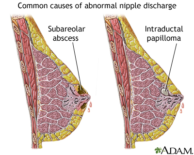
  • Pathologic nipple discharge (PND):
    • Is defined as a clear, serous, or bloody nipple secretion (not green or milky):
      • It is spontaneous
      • Discharging from a single duct and unilateral
    • It is frequently caused by:
      • A benign lesion, such as:
        • Intraductal papilloma(s):
          • 35% to 56% of the cases
        • Ductal ectasia:
          • 6% to 59% of the cases
      • An underlying malignancy can be present in a percentage of cases:
        • Reported to be variable from 5% to 33%
    • Because to differentiate between a benign from a malignant etiology of a PND based on clinical and diagnostic assessment is not easy:
      • Surgical excision has been considered the main way for getting both definitive diagnosis and eliminating the symptom
  • Clinical History and Physical Examination:
    • Clinical history plays an important role:
      • For evaluating the probability of malignancy
    • Predicting factors for malignancy in the presence of PND are:
      • BRCA 1 / mutations
      • History of ipsilateral cancer
      • Previous breast biopsy with diagnosis of atypia
      • Age over 50 years:
        • In a study including 318 patients with nipple discharge (any fluid from the nipple, spontaneous discharge or observed during breast examination):
          • Seltzer has reported a higher incidence of breast cancer:
            • Equal to 9% in females over 50 (95 patients and 9 cancers):
              • While the incidence was of only 1.3% in younger patients (223 patients and 3 cancers)
    • Physical examination:
      • Has the aim of distinguishing between benign and pathological discharge and of verifying the presence of palpable mass or other associated findings
      • It usually includes:
        • complete breast evaluation:
          • With inspection and palpation
          • Followed by a focused inspection of the nipple area:
            • Using a magnifying lamp
      • The physical examination is essential to investigate the:
        • Color of discharge
        • The number of ducts involved
        • The frequency of discharge (persistent or intermittent)
        • If it is unilateral or bilateral
      • spontaneous single-pore bloody and clear discharge:
        • Is suspect for pathological discharge
  • Mammography:
    • Represents the first conventional imaging technique to investigate nipple discharge:
      • At least after 39 years old
    • For patients with PND, aged between 30 and 40 years old with high-family risk:
      • Mammography could be appropriated in order to exclude the presence of microcalcifications
    • As well as for females younger than 30 of age:
      • When initial ultrasound shows suspicious findings
    • The protocol includes:
      • The standard cranio-caudal and mediolateral oblique views
    • Mammography findings that are suspect to be associated to an occult malignancy can range from:
      • Microcalcifications
      • Masses
      • Focal density asymmetry
      • Architectural distortion or ductal ectasia
      • Otherwise no abnormality can be identified
    • Mammography has low sensitivity and limited accuracy:
      • In the detection of retroareolar lesions that are often small, intraductal, and without calcifications
    • Ductal ectasia:
      • May occur as a general increase in density of the retroareolar region and in order to better visualize the area:
        • Spot compression views could be performed
    • In order to improve spatial resolution:
      • Magnification mammography can be performed:
        • To identify microcalcifications and to distinguish between benign or malignant duct disease
    • Microcalcifications with:
      • Branching or linear pattern, variable density, or distributed in a segmental way:
        • Are all highly suspicious of malignancy
      • Whereas round or rod-like calcifications:
        • Suggest for benign disease
    • Bahl et al studied 252 patients with at least one pathological feature of nipple discharge (unilateral, clear or bloody, or spontaneous discharge) who underwent surgical excision or a 2-year follow-up:
      • Of 20 cancers diagnosed:
        • Only three were revealed by mammography:
          • With a 15% (3/20) sensitivity
    • In other studies, the sensitivity of mammography:
      • Ranged from 7% to 26%. 
  • Ultrasound:
    • Offers a better performance than mammography:
      • For detecting intraductal lesions
    • Ductal ectasia:
      • Defined by a duct caliber greater than 3 mm
        • Is one of the most common findings seen on ultrasound:
          • It appears as dilated retroareolar ducts containing anechoid fluid or hypoecoic debris
    • An intraductal papilloma appears as:
      • hypoechoic nodule with a central vascular pedicle on color Doppler:
        • Doppler ultrasound is helpful in differentiating:
          • Intraductal viscous secretion versus intraductal nodule with vascular sign
    • Ultrasound malignant features are:
      • Irregular duct margins
      • Wall thickening
      • Hypoechoic intraductal mass with acoustic shadowing 
    • In a study by Park et al:
      • The detection rate of malignant lesions occult on mammography and ultrasound-detected:
        • Was reported to be 8 of 53 females with PND examined (15%)
    • Yoon et al:
      • Have also reported that adding ultrasound to mammography in the pre-operative setting of PND:
        • Led to the detection of malignancies in 26% of patients (ultrasound detected fivebreast cancers in addition to the 19 breast cancers found by mammography)
    • The role of ultrasound elastography is disputable in predicting malignancy in patients with PND:
      • Guo et al have evaluated the diagnostic accuracy of elastography in patients with PND:
        • Affirming that it is a useful tool for predicting malignancy:
          • With sensitivity for malignancy of 90% and that it could be used as a helpful test before more invasive examination (such as ductoscopy or duct excision):
            • However, it is only a preliminary study and further studies are needed to verify the diagnostic perfor- mance of elastography
  • Nipple discharge cytology:
    • Is performed by squeezing the nipple with a gentle compression of the areola area and spreading the secretion onto a glass slide:
      • After smearing, the slides are immediately fixed by spray fixation or by immersion in 95% ethyl alcohol:
        • Then stained with the Papanicolaou stain
    • It is a simple and fast examination, easy to perform and painless:
      • But strongly limited by a low sensitivity for cancer:
        • With a false negative rate over 50%
      • Moreover, it can be technically impossible when discharge is not present on the moment of the examination
      • According to the American College of Radiology:
        • This examination has not proven to be effective in differentiating benign from malignant lesions:
          • Therefore, discharge cytology is not routinely recommended
    • Nipple discharge smears are classified as abnormal if they contained:
      • Papillary, atypical, suspicious, or malignant cells:
        • Malignant nipple discharge cytology is correlated with higher specificity values

#Arrangoiz #BreastSurgeon #CancerSurgeon #SurgicalOncologist #Teacher #BreastCancer #NippleDischarge #PathologicNippleDischarge #PhysiologicNippleDischarge #CASO #Miami #CenterforAdvancedSurgicalOncology #IntraductalPapilloma #DuctalEctasia

Age as a Prognostic Factor in Thyroid Cancer

👉Thyroid cancer is the only malignancy that has age as a prognostic factor in TNM staging.

👉Due to the favorable prognosis in younger patients the highest possible stage for patients less than 55 is Stage II

AJCC TNM Staging for Thyroid Cancer

#Arrangoiz #ThyroidSurgeon #ThyroidExpert #Teacher #CancerSurgeon #HeadandNeckSurgeon #SurgicalOncologist #CASO #Miami #CenterforAdvancedSurgicalOncology

Papillary Thyroid Cancer Recurrence

👉Up to 20% of patients with papillary thyroid cancer will experience recurrent disease after surgery throughout their lifetime.

👉Certain mutations (e.g. BRAFV600E) are independent risk factors for recurrence.

#Arrangoiz #ThyroidSurgeon #ThyroidExpert #Teacher #CancerSurgeon #HeadandNeckSurgeon #SurgicalOncologist #CASO #Miami #CenterforAdvancedSurgicalOncology

Thyroid Cancer – Lymph Node Recurrences

👉Not all thyroid cancer recurrences require surgery.

👉Small lymph node recurrences in the neck can often be observed, however recurrent nodes greater than 0.8 cm in the central neck and greater than 1 cm in the lateral neck are typically removed.

#Arrangoiz #ThyroidSurgeon #ThyroidExpert #Teacher #CancerSurgeon #HeadandNeckSurgeon #SurgicalOncologist #CASO #Miami #CenterforAdvancedSurgicalOncology

Anatomy of the Nasolacrimal System

  • The purpose of the nasolacrimal system is to:
    • Drain tears from the ocular surface to the lacrimal sac and, ultimately, the nasal cavity
    • Blockage of the nasolacrimal system:
      • Can cause tears to flow over the eyelid and down the cheek:
        • This condition is epiphora
  • Structure and Function:
    • Both the upper eyelid and the lower eyelid have a small opening on the surface of the eyelid margin near the medial canthus:
      • These are called puncta:
        • Each puncta leads to a drainage canal that eventually flows into the lacrimal sac and then the nasal cavity
    • The drainage canal connecting the ocular surface to the nasal cavity consists of multiple parts:
      • Within the lower eyelid:
        • The punctum leads to a 2 mm long ampulla:
          • Which runs perpendicular to the eyelid margin
        • The ampulla turns 90 degrees medially:
          • Becoming the inferior canaliculus and travels 8 to 10 mm before reaching the common canaliculus
        • The upper canaliculus travels 2 mm superiorly in the eyelid before turning 90 degrees medially and moving 8 to 10 mm before connecting to the common canaliculus
      • The common canaliculus:
        • Drains into the lacrimal sac
      • Within the junction between the common canaliculus and the lacrimal sac:
        • Is the valve of Rosenmuller:
          • This apparatus is a one-way valve that prevents reflux from the lacrimal sac to the puncta
    • The lacrimal sac drains:
      • Inferiorly to the nasolacrimal duct:
        • Which is bordered:
          • Medially by:
            • Palatine bone and the inferior turbinate in the nose
          • Laterally by:
            • Maxillary bone
      • The nasolacrimal duct:
        • Opens at the inferior meatus:
          • Located underneath the inferior nasal turbinate
      • The lacrimal sac is:
        • Approximately 10 to 15 mm in axial length and 13 to 20 mm in corneal length
      • The nasolacrimal duct is:
        • 12 to 18 mm long
      • The inferior nasal meatus is partially covered by a mucosal fold:
        • Known as the valve of Hasner
  • Embryology:
    • The nasolacrimal duct:
      • Starts forming around five weeks of gestation
      • It starts out as a linear thickening of ectoderm:
        • Located in a groove between the nasal and maxillary prominences
      • This thickening:
        • Eventually separates into a solid cord and sinks into the surrounding mesenchyme
        • Over time the cord canalizes:
          • Forming the lacrimal sac and the beginning of the nasolacrimal duct
      • The nasolacrimal duct extends:
        • Intranasally until it exits under the inferior turbinate
      • The lacrimal sac extends caudally:
        • To complete the canalicular system
      • The inside of the canal breaks down and forms a lumen:
        • So that the nasolacrimal system is patent:
          • This process is generally complete by the time of birth
  • Blood Supply and Lymphatics:
    • Blood supply to the nasolacrimal area of the face:
      • Is generally from the angular artery:
        • The angular artery is considered a branch of the facial artery:
          • However, some studies have shown that it can originate from the ophthalmic artery in some individuals
        • It terminates in anastomosis with the dorsal nasal branch of the ophthalmic artery
        • The angular artery and vein:
          • Appear alongside the nose near the medial orbit
        • A correlating angular vein drains this region
    • The medial and lateral portions of the eyelids have different lymphatic drainage systems:
      • The medial one-third of the upper eyelid and the medial two-thirds of the lower eyelid:
        • Drain to the submandibular lymph nodes
      • The lateral two-thirds of the upper eyelid and the lateral one-third of the lower eyelid:
        • Drain to the pre-auricular lymph nodes
  • Nerves:
    • Cranial nerve VII:
      • Supplies the motor innervation to the muscles of the face
    • The movement of these muscles:
      • Aid in proper drainage of the tears through the nasolacrimal system:
        • By what is known as the lacrimal pump mechanism
    • Cranial nerve III and cranial nerve VII:
      • Innervate the muscles that control the blinking of the eyelids:
        • This action is the primary driver of the lacrimal pump mechanism
    • Irritation of the ocular surface:
      • Stimulates the ophthalmic branch of cranial nerve five:
        • Which begins the reflex tear arc pathway:
          • The efferent pathway involves cranial nerve VII and parasympathetic fibers
        • The role of the sympathetic nervous system in tear production:
          • Is not well understood
  • Muscles:
    • The action of the orbicularis muscle and surrounding tissues:
      • Helps propel the flow of tears from the canaliculi to the nasolacrimal duct:
        • Via the lacrimal pump mechanism
  • References:
    • Computed tomography dimensions of the lacrimal gland in normal Caucasian orbits., Tamboli DA,Harris MA,Hogg JP,Realini T,Sivak-Callcott JA,, Ophthalmic plastic and reconstructive surgery, 2011 Nov-Dec.
    • An Unusual Case of Nasolacrimal Obstruction Caused by Foodstuffs., Matsumoto H,Matsumoto A,, Case reports in ophthalmology, 2015 Sep-Dec.
    • Lacrimal Gland Volume Changes in Unilateral Primary Acquired Nasolacrimal Obstruction., Yazici A,Bulbul E,Yazici H,Sari E,Tiskaoglu N,Yanik B,Ermis S,, Investigative ophthalmology & visual science, 2015 Jul.
    • Incidence of neoplasia in patients with unilateral epiphora., Bewes T,Sacks R,Sacks PL,Chin D,Mrad N,Wilcsek G,Tumuluri K,Harvey R,, The Journal of laryngology and otology, 2015 Jul.
    • Ducasse A,Arndt C,Brugniart C,Larre I, [Lacrimal traumatology]. Journal francais d’ophtalmologie. 2016 Feb.
    • Modified External Dacryocystorhinostomy in Primary Acquired Nasolacrimal Duct Obstruction., Sharma HR,Sharma AK,Sharma R,, Journal of clinical and diagnostic research : JCDR, 2015 Oct.

#Arrangoiz #CancerSurgeon #HeadandNeckSurgeon #SurgicalOncologist #CASO #Miami #CenterforAdvancedSurgicalOncology

Mucoepidermoid carcinoma (MEC)

  • Mucoepidermoid carcinoma (MEC):
    • Is the most common malignant neoplasm of the:
      • Mayor and minor salivary glands
  • Epidemiology:
    • They encompass between 2.8% to 15.5% of all salivary gland tumors
    • Among 12% to 35% of malignant salivary gland tumors
    • Among 6.5% to 41% of all minor salivary gland tumors:
      • Representing the most common type of malignant minor salivary gland tumor in most series
    • Approximately half the cases occur in the major salivary glands:
      • 65% to 80% of these occur in the parotid
      • 8% to 13% occur in the submandibular gland
      • 2% to 4% involve the sublingual gland
    • MEC of the minor salivary glands:
      • Ordinarily arises on the palate:
        • But a number may also be found in the:
          • Retro molar area
          • Floor of the mouth
          • Buccal mucosa
          • Lip
          • Tongue
    • Its prevalence is highest in:
      • The fourth to fifth decade of life (35 to 65 years of age):
        • With a female preponderance as high as 4:1
  • Grossly:
    • The tumor is poorly circumscribed and measures from 3 to 5 cm
  • Histologically:
    • They are characterized by a mixed population of cells, including:
      • Mucin-producing cells
      • Epidermoid cells with squamoid differentiation
      • Clear cells
      • Intermediate cells:
        • That may predominate in numbers
        • Are believed to be the progenitor of the other types of cells
      • No myoepithelial cells are present
  • The clinical behavior of MEC has proved to be difficult to predict:
    • But correlations to tumor grade and stage have been reported
    • The histologic features that are most useful in predicting the aggressive nature of these tumors are:
      • A minor cystic component (less than 20%)
      • Tumor necrosis
      • Neural invasion
      • Cellular anaplasia
      • Brisk mitotic activity
    • Based on the presence or absence of these features and the clinical behavior, MEC are classified as:
      • Low grade
      • Intermediate grade
      • High grade
    • Low-grade MEC are:
      • Well circumscribed, with pushing margins and dilated cystic areas containing mucin
      • Mucin producing, intermediate, or epidermoid cells make up the lining of these cystic structures
    • Intermediate-grade MEC:
      • As the grade worsens:
        • The tumors become more infiltrative, poorly circumscribed
        • Cystic formations are lost
        • Nests of tumor become more solid and irregular with intermediate or epidermoid cells dominating
    • High-grade MEC are characterized by:
      • The invasion of adjacent structures
      • Atypical mitoses
      • Necrosis
      • Perineural invasion
      • Lymph node metastasis:
        • 40% to 50%
      • Distant metastases
    • Differential diagnosis of these high-grade lesions are:
      • Primary of metastatic squamous cell carcinoma:
        • MEC is differentiated from metastatic SCC by:
          • The presence of intracellular mucin
      • Sebaceous carcinomas
      • Clear cell carcinomas
  • Histologic grade and tumor stage:
    • Appear to have profound effects on survival
    • Aro et al:
      • Found a statistically significant difference in disease free survival (DFS) by grade:
        • Between low-grade MEC and intermediate / high-grade MEC (P = 0.001)

#Arrangoiz #CancerSurgeon #HeadandNeckSurgeon #SurgicalOncologist #SalivaryGlandCancer #MucoepidermoidCarcinoma #ParotidTumors #MEC #CASO #CenterforAdvancedSurgicalOncology #Miami

Cost of Contralateral Prophylactic Mastectomy (CPM) Versus Surveillance

  • There is robust literature to support the use of CPM as a cost-effective strategy:
    • In patients with hereditary breast cancer syndromes
  • Anderson et al. demonstrated that the most cost-effective strategy:
    • With and without quality adjustment:
      • For women with BRCA1 or BRCA2 mutations was:
        • Prophylactic bilateral salpingo-oophorectomy with bilateral mastectomy
  • Simulation models analyzing costs for CPM versus surveillance in patients with sporadic breast cancer reveal disparate findings:
    • An initial Markov model study found that CPM was cost effective compared with surveillance for:
      • Patients younger than 70 years:
        • But this finding was highly dependent on the quality of life assumptions
    • A second study that included operative complications and breast reconstruction costs used a decision-tree model and concluded that:
      • Although CPM resulted in a cost savings over surveillance for women younger than 50 years:
        • It also reduced quality of life years
      • When MRI was inserted in the model as the primary method of screening:
        • The cost-effectiveness of CPM increased
      • Loss of quality of life years was largely attributed to complications from reconstructive procedures
    • The two models differ in the assumptions regarding quality of life:
      • If we assume an improvement in quality of life after CPM:
        • Then CPM could be cost effective
      • Alternatively, if quality of life is decreased,:
        • CPM would not be a cost-effective strategy
  • The available data on cost effectiveness for CPM is limited
  • Summary:
    • CPM is a cost-effective strategy for women with BRCA mutations
    • At this time, there is insufficient evidence to support the concept of superior cost effectiveness for CPM in women with sporadic breast cancer and the cost effectiveness is highly dependent on the quality of life assumptions
  • Reference:
    • Contralateral Prophylactic Mastectomy Consensus Statement from the American Society of Breast Surgeons: Additional Considerations and a Framework for Shared Decision Making. Ann Surg Oncol (2016) 23:3106–3111

#Arrangoiz #BreastSurgeon #BreastCancer #CancerSurgeon #SurgicalOncologist #ContralateralProphylacticMastectomy #Cost-effectiveness #Miami #CASO #CenterforAdvancedSurgicalOncology

  • Laboratory Evaluation:
    • Diagnosis is always established by the measurement of:
      • Sensitive TSH and thyroid hormone levels (free T3 and free T4)
    • Thyrotoxicosis caused by TNG or Graves’ disease is usually characterized by:
      • suppressed TSH level with either:
        • Normal (subclinical) or elevated (overt) free thyroid hormone levels
      • It is insufficient to rely on the measurement of TSH or free thyroid hormones:
        • Alone to diagnose TNG or Graves’ disease:
          • Because suppression of TSH or elevation of thyroid hormones can be associated with clinical conditions other than TNG and Graves’ disease
Low TSHHigh TSH
Secondary hypothyroidismTSH-secreting pituitary tumor
Non-thyroidal illnessThyroid hormone resistance
Glucocorticoid therapy
Amiodarone use
Excessive thyroid hormone therapy
  • Other serologic findings:
    • Such as antithyroid antibodiesantithyroid peroxidase and antithyroglobulin:
      • That support the diagnosis of autoimmune thyroid disease:
        • May be detected in patients with Graves’ disease:
          • However, serum TSHR-Ab:
            • Is occasionally helpful in the diagnosis of Graves’ disease:
              • Though there is no consensus regarding its routine measurement in Graves’ disease
  • Imaging:
    • Radionuclide Imaging:
      • Radionuclide scanning and radioactive iodine uptake (RAIU):
        • Are useful tests to elucidate the cause of hyperthyroidism
      • In toxic nodular goiter (TNG):
        • The radioactive iodine (RAIconcentration is in the nodule(s), and uptake is inhibited in the surrounding tissue:
          • Giving the appearance of “patchy uptake”
        • Consequently, the total RAIU may be either:
        • Slightly raised or at the upper limit of normal
      • In Graves’ disease:
        • Because of the diffuse thyroid involvement:
          • The RAIU is always intense and increased
      • Thyroid radionuclide imaging may not be necessary in every case:
        • When the diagnosis is obvious:
          • But it is helpful in the differentiation of other clinical conditions associated with hyperthyroidism but with low RAIU
Radionuclide scan of toxic nodular goiter demonstrating intense focal uptake of several hot nodules with different degrees of suppression of adjacent thyroid tissue (A) compared with the scan from a patient with non-toxic multi-nodular goiter, showing less intense patchy radioactive iodine uptake (B).
Radionuclide scan in Graves’ hyperthyroidism demonstrating the diffuse and homogeneous nature of increased uptake in both lobes of the thyroid.

Clinical Conditions Associated with Low Radioactive Iodine Uptake and Hyperthyroidism

Thyroiditis
Iodine-induced thyrotoxicosis
Exogenous thyrotoxicosis (factitia)
Ectopic functional thyroid tissue
  • Computed tomography scan:
    • In any patient with compressive or obstructive symptoms and an MNG:
      • Chest radiography and chest computed tomography (CT) are often informative
    • Chest CT is particularly valuable to define the size and extent of the goiter:
      • Especially into the mediastinum
    • Care to avoid iodinated contrast:
      • Until the patient’s thyroid functional status must be taken into account or the significant iodine load CT contrast agent may acutely induce or worsen hyperthyroidism
A, Chest radiography showing a huge solid goiter (horizontal arrow) displacing the trachea without compression (vertical arrow). B, Neck computed tomography of the same goiter (arrows). Thyroidectomy revealed a 290-g benign thyroid gland.
  • Associated metabolic abnormalities:
    • Altered glucose metabolism:
      • Reversible hyperglycemia
      • Elevated C-peptide
      • Elevated intact proinsulin
      • Insulin resistance
    • Increased bone turnover:
      • Elevated markers of bone formation and resorption
        • Are hallmarks of untreated hyperthyroidism
    • Untreated hyperthyroidism is associated with an elevated chromogranin A level:
      • That changes in parallel with thyroid status

Metabolic Abnormalities Associated with Hyperthyroidism

Mild hypercalcemia
Myopathy
Hypokalemic periodic paralysis
Pulmonary hypertension
Cholestatic jaundice

#Arrangoiz #CancerSurgeon #ThyroidSurgeon #ParathyroidSurgeon #HeadandNeckSurgeon #ThyroidExpert #SurgicalOncologist #EndocrineSurgery #CASO #Miami #ThyroidNodule #ToxicNodularGoiter #TNG #MultinodularGoiter #GravesDisease #Hyperthyroidism #Goiter #CenterforAdvancedSurgicalOncology

Sentinel Lymph Node Surgery for Contralateral Prophylactic Mastectomy (CPM) – American Society of Breast Surgeons (ASBrS) Evidence-Based Recommendations

  • The benefit of performing sentinel lymph node (SLN) surgery at the time of CPM is:
    • That the lymph nodes have been evaluated in the event that an occult malignancy is found
  • The downside is:
    • Increased surgical morbidity such as lymphedema
  • By meta-analysis:
    • The risk of lymphedema after SLN alone is:
      • 5.6 % (95 % CI 6.1–7.9 %) and increases with longer follow-up
    • The chance of finding occult invasive disease in a prophylactic mastectomy is:
      • 1.8 %
    • An additional small percent of CPM specimens harbor noninvasive disease:
      • That would not require nodal evaluation
    • The rate of nodal positivity in patients with occult malignancy in CPM is:
      • Only 1.3 %
    • Considering these data:
      • Routine SLN surgery at time of CPM:
        • Places more patients at risk of lymphedema:
          • Than would be expected from the 1% to 2 % of patients with occult disease undergoing axillary dissection
        • Therefore the consensus group:
          • Does not recommend routine SLN for CPM
  • Patients at higher risk of contralateral occult malignancy are:
    • Postmenopausal patients
    • Those with triple-negative
    • Locally advanced
    • Inflammatory breast cancer
    • Invasive lobular disease
  • MRI at the time of breast cancer diagnosis:
    • Identifies occult contralateral disease:
      • 2% to 4 % of the time
    • Suspicious lesions in the contralateral breast should be biopsied:
      • But if a biopsy is not done:
        • SLN surgery should be considered for highly suspicious lesions
  • Summary:
    • Sentinel lymph node surgery on the CPM side should not be routinely performed
  • References:
    • Contralateral Prophylactic Mastectomy Consensus Statement from the American Society of Breast Surgeons: Additional Considerations and a Framework for Shared Decision Making. Ann Surg Oncol (2016) 23:3106–3111

#Arrangoiz #BreastSurgeon #CancerSurgeon #Teacher #SurgicalOncology #CASO #Miami #CenterforAdvancedSurgicalOncology #BreastCancer #ProphylacticMastectomy #CPM #ContralateralProphylacticMastectomy

Lymph Node Staging in Breast Cancer in Patients Greater Than 70 Years of Age

  • Triple-negative breast cancer:
    • Is more responsive to preoperative chemotherapy compared to ER/PR+, HER2neu negative breast cancer:
      • Pathologic complete response is seen in approximately 30% to 40% of patients undergoing treatment with a third-generation regimen:
        • A pathologic complete response:
          • Is highly prognostic in this subset
  • While ER negative breast cancers:
    • Have a lower propensity for regional nodal metastasis compared to ER+ tumors:
      • The difference is relatively small (2% to 5%):
        • Therefore, nodal staging is still a standard practice recommendation
  • The Choosing Wisely guideline:
    • For omission of routine use of sentinel node biopsy in clinically node-negative women ≥ 70 years of age applies to hormone receptor positive breast cancer
  • Sentinel node biopsy may be successfully performed after neoadjuvant chemotherapy and should be performed patients with a clinically negative axilla
  • References
    • Cortazar P, Zhang L, Untch M, et al. Pathologic complete response and long term clinical benefit in breast cancer: the CTNeoBC pooled analysis. Lancet. 2014;384(9938):164-172.
    • Symmans WF, Peintinger F, Hatzis C, et al. Measurement of residual breast cancer burden to predict survival after neoadjuvant chemotherapy. J Clin Oncol. 2007;25(28):4414-4422.
    • von Minckwitz G, Untch M, Blohmer JU, et al. Definition and impact of pathologic complete response on prognosis after neoadjuvant chemotherapy in various intrinsic breast cancer subtypes. J Clin Oncol. 2012;30(15):1796-1804.
    • Viale G, Zurrida S, Maiorano E, et al. Predicting the status of axillary sentinel lymph nodes in 4351 patients with invasive breast carcinoma treated in a single institution. Cancer. 2005;103(3):492-500.
    • Hughes KS, Schnaper LA, Bellon JR, et al. Lumpectomy plus tamoxifen with or without irradiation in women age 70 years or older with early breast cancer: long-term follow-up of CALGB 9343. J Clin Oncol. 2013;31(19):2382-2387.