Wound infections, usually caused by Staphylococcus or Streptococcus species are considered to be rare events in thyroid surgery:
Occurring in 0.3% to 0.8% of cases
Antibiotic prophylaxis:
Is recommended only in immunocompromised patients or in those with valvular cardiac disorders
While mild neck cellulitis frequently regresses under conservative treatment:
Abscesses require rapid incision and evacuation:
Delay of invasive treatment can result in devastating mediastinitis
Clinically evident seromas:
Respond well to percutaneous aspiration
References:
Sosa JA, et al (1998) The importance of surgeon experience for clinical and economic outcomes from thyroidectomy. Ann Surg 228:320–330
Rosato L, et al (2004) Complications of thyroid surgery: analysis of a multicentric study on 14,934 patients oper- ated on in Italy over 5 years. World J Surg 28:271–276
Factors associated with increased contralateral breast cancer risks include:
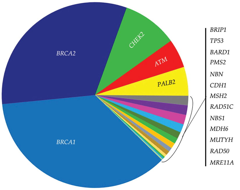
Genetic mutations:
Such as:
BRCA1 mutations
BRCA 2 mutations
Chek2 mutations
Atm mutations
Palb2 mutations
History of mantle radiation
First-degree relative with breast cancer
Young age at diagnosis
Hormone negative disease
To consider contralateral prophylactic mastectomy (CPM):
The disease burden of the primary tumor needs to be lower than the risk of contralateral breast cancer
For an average risk patient:
Contralateral cancer risk ranges from 1% to 5%
Markov models for stage 1 and 2 breast cancer patients:
Showed overall survival benefit of less than 1% in patients who had CPM
CPM may offer a survival advantage for:
Women less than 50 years of age who are diagnosed with triple negative breast cancer
References
Deshmukh AA, Cantor SB, Crosby MA, Dong W, Shen Y, Bedrosian I, et al. Cost of contralateral prophylactic mastectomy. Ann Surg Oncol. 2014;21(9):2823-2830.
Roberts A, Habibi M, Frick KD. Cost-effectiveness of contralateral prophylactic mastectomy for prevention of contralateral breast cancer. Ann Surg Oncol. 2014;21(7):2209-2217.
Frost MH, Hoskin TL, Hartmann LC, Degnim AC, Johnson JL, Boughey JC. Contralateral prophylactic mastectomy: long-term consistency of satisfaction and adverse effects and the significance of informed decision-making, quality of life, and personality traits. Ann Surg Oncol. 2011;18(11):3110-3116.
Barton MB, West CN, Liu IL, Harris EL, Rolnick SJ, Elmore JG, et al. Complications following bilateral prophylactic mastectomy. J Natl Cancer Inst Monogr. 2005;(35):61-66.
Grann VR, Jacobson JS, Whang W, Hershman D, Heitjan DF, Antman KH, et al. Prevention with tamoxifen or other hormones versus prophylactic surgery in BRCA1/2-positive women: a decision analysis. Cancer J Sci Am. 2000;6(1):13-20.
Portschy PR, Kuntz KM, Tuttle TM. Survival outcomes after contralateral prophylactic mastectomy: a decision analysis. J Natl Cancer Inst. 2014;106(8).
Once a patient undergoes breast-conserving surgery:
The pathology report is reviewed to determine any need for additional surgery
If the sentinel node biopsy was negative:
No additional axillary surgery is warranted
Margins assessed next:
Historically, the definition of what was considered an adequate margin in patients undergoing breast-conserving surgery was left to the interpretation of the treating physicians
In 2013, the Society of Surgical Oncology and the American Society for Radiation Oncology released consensus guidelines to assist physicians and patients in decisions regarding margins:
These guidelines were based on:
A meta-analysis of 33 studies evaluating in-breast tumor recurrence (IBTR) in relation to margin width
The results of the meta-analysis reported:
Positive margins were associated with 2-fold risk of IBTR:
Routine re-excision is recommended for those patients
For those with negative margins (defined as “no ink on tumor”), the meta-analysis reported that:
Increasing margin widths were not associated with a reduction in IBTR.
Is the patient a candidate for adjuvant therapy?
The 21-gene recurrence assay:
Has been shown to accurately predict risk of distant recurrence and benefit of adjuvant chemotherapy:
In women with ER positive tumors
Adjuvant chemotherapy is a category 1 recommendation for women with:
ER negative tumors greater than 1 cm
Does this patient need genetic studies?
The incidence of BRCA mutations in women with triple negative breast cancers:
Ranges in the literature from 10% to 31%
Current guidelines state that women who present with triple negative histology prior to age 60 should be considered for testing
References
Moran MS, Schnitt SJ, Giuliano AE, Harris JR, Khan SA, Horton J, et al. Society of Surgical Oncology-American Society for Radiation Oncology consensus guideline on margins for breast-conserving surgery with whole breast irradiation in Stage I and II invasive breast cancer. Int J Radiat Oncol BiolPhys. 2014;88(3):553–564.
Greenup R, Buchanan A, Lorizio W, Rhoads K, Chan S, Leedom T, et al. Prevalence of BRCA mutations among women with triple negative breast cancer in a genetic counseling cohort. Ann Surg Oncol. 2013;20:3254-3258.
Pseudoangiomatous stromal hyperplasia (PASH) of breast:
Is a benign proliferative breast disease
It was first described by Vuitch, Erlandson, and Rosen:
In 1986
This lesion is characterized by:
A dense, collagenous proliferation of mammary stroma:
Forming inter-anastomosing capillary-like spaces
The age of presentation of PASH is usually:
12 to 70 years:
With most cases appearing in women:
In their late 30s to 40s
Both males and females are affected:
In men, PASH may occur with gynecomastia
It is seen in all racial and ethnic groups
PASH is usually discovered incidentally:
During screening mammography:
It may resemble a fibroadenoma on imaging
It usually appears as:
An oval, hypoechoic, or mixed echogenic mass without posterior shadowing
PASH may also occur as a:
Firm, painless, often rubbery and mobile mass in the breast:
Small tumors may be asymptomatic
Usually only one breast is affected:
Though it may be bilateral
May present as multifocal nodules in 60% of cases
When PASH occurs as a mass:
It is usually a firm and painless mass
It may occur occasionally in a pregnant woman:
In which case it can enlarge rapidly
Observation of asymptomatic PASH is appropriate:
Excision is not required
If PASH occurs as a mass and is symptomatic:
A simple excision may be performed:
However, not all PASH lesions need to be excised
With an asymptomatic and incidentally identified lesion diagnosed pathologically as PASH:
Routine follow-up is the appropriate treatment
On gross examination:
PASH is usually a well-circumscribed fibrous mass
Cysts may occasionally be found within the lesion
Microscopically:
PASH consists of a network of slit-like spaces lined by myofibroblasts that resemble ectatic vessels, with intervening stromal collagen fibers
The term “pseudoangiomatous” is used to describe this lesion:
As it has been confused and occasionally misdiagnosed as low-grade angiosarcoma
Though a new finding on mammogram or examination would be evaluated to rule out breast cancer:
There is no increased risk of breast cancer associated with the diagnosis of PASH
PASH may be mistaken for mammary angiosarcoma:
But is not associated with an increased risk of developing:
Angiosarcoma
Invasive ductal carcinoma, or
Other breast malignancies
There is no indication for genetic counseling or testing:
Based on a diagnosis of PASH
Excisional biopsy is not required:
With concordant and benign findings on mammography and core biopsy
It is thought that hormonal factors play an important role in PASH:
According to Anderson et al:
This lesion represents an important hyper-response to progesterone and estrogen
PASH is a common histological finding in breast biopsy specimens and can also be found in a normal breast that is in association with proliferative or non-proliferative fibrocystic changes:
But it is rarely a symptomatic lesion
PASH can be found in:
Teenage girls as well as in postmenopausal women with or without hormonal therapy replacement
It is important to recognize this entity because it can be easily confused with:
Other benign tumors, such as:
Fibroadenoma
Phyllode tumor
With malignant tumors, such as:
Angiosarcoma
Unfortunately, imaging features of PASH are non-specific:
On mammography:
The most common appearance described is:
A well-defined, uncalcified mass, with regular borders
Spiculated borders, suspicious borders, and architectural distortion can also be seen:
But are uncommon
On ultrasound:
PASH tends to be:
An oval, round hypoechoic mass or
Can presents as a heterogeneous mass with cystic areas
According to Cohen et al:
When a focal lesion with well-defined borders, containing no calcifications on mammography or a well-defined hypoechoic mass on ultrasound is seen:
PASH can be considered and included in the differential diagnosis
Clinically and on imaging, the differential diagnosis include:
Fibroadenoma:
Especially in young patient
Phyllode tumor:
In older women
Histologically:
PASH can be very similar to low-grade angiosarcoma
Definitive diagnosis is based on histology:
But unlike low-grade angiosarcoma:
PASH lacks of invasive features and contains no necrosis, mitoses, and no destruction of mammary epithelial structures
Management of PASH depends on presentation:
When PASH is incidentally discovered or when it is asymptomatic:
It can be followed up yearly by ultrasound or mammography for a period of 36 months
Surgical procedures are indicated for:
Symptomatic lesion with mechanical complaints, pain or apprehension for an alternative malignant lesion
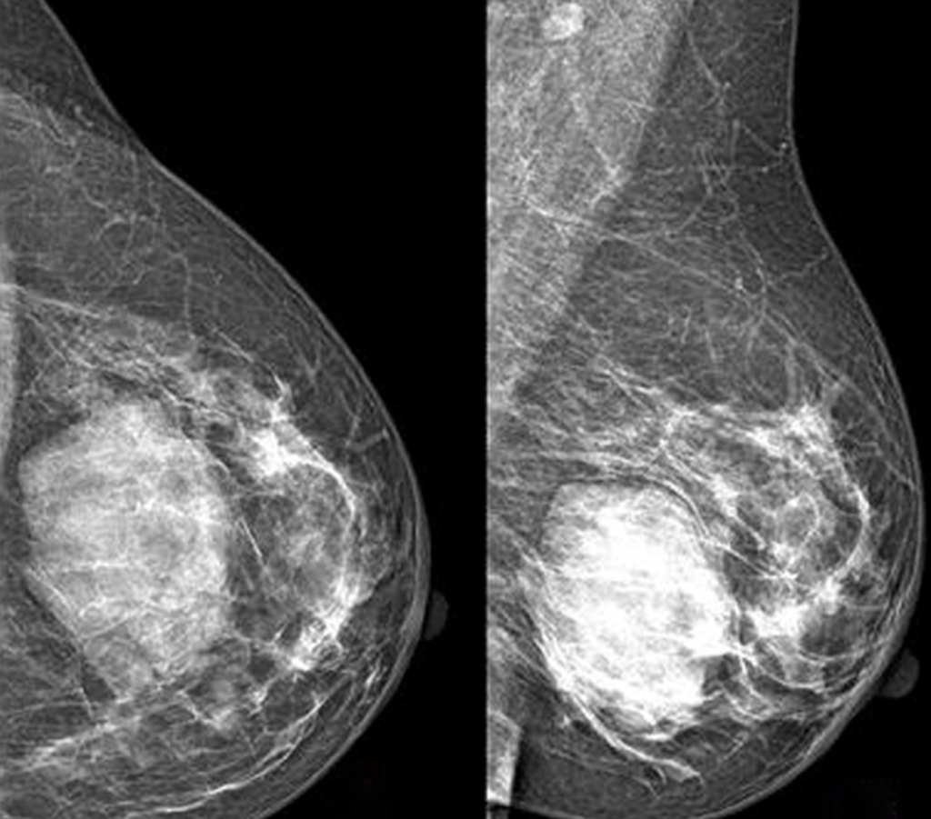
Image: Mammography performed with craniocaudal (A) and mediolateral oblique (B) incidences of the left breast shows a huge well-defined mass located between the inner and the outer inferior quadrants, with no suspicious features
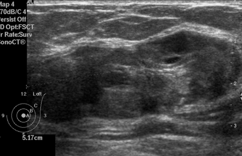
Image: Ultrasound of the left breast centered on the palpable lump showing an oval, heterogeneous lesions with smooth borders.
Mammography performed with craniocaudal (A) and mediolateral oblique (B) incidences of the left breast shows a huge well-defined mass located between the inner and the outer inferior quadrants, with no suspicious featuresUltrasound of the left breast centered on the palpable lump showing an oval, heterogeneous lesions with smooth borders.
References:
Celliers L, Wong DD, Bourke A. Pseudoangiomatous stromal hyperplasia: a study of the mammographic and sonographic features. Clin Radiol. 2010;65(2):145-149.
Guray M, Sahin AA. Benign breast diseases: classification, diagnosis, and management. Oncologist. 2006;11(5):435-439.
Hargaden GC, Yeh ED, Georgian-Smith D, Moore RH, Rafferty EA, et al. Analysis of the mammographic and sonographic features of pseudoangiomatous stromal hyperplasia. AJR Am J Roentgenol. 2008;191(2):359-363.
Salvador R, Lirola JL, Domínguez R, López M, Risueño N. Pseudo-angiomatous stromal hyperplasia presenting as a breast mass: imaging findings in three patients. Breast. 2004;13(5):431-435.
Gresik CM, Godellas C, Aranha GV, Rajan P, Shoup M. Pseudoangiomatous stromal hyperplasia of the breast: a contemporary approach to its clinical and radiologic features and ideal management. Surgery. 2010;148:752-757.
Virk RK, Khan A. Pseudoangiomatous stromal hyperplasia: an overview. Arch Pathol Lab Med. 2010;134:1070-1074.
Neal L, Sandhu NP, Hieken TJ, Glazebrook KN, Mac Bride MB, Dilaveri CA, et al. Diagnosis and management of benign, atypical, and indeterminate breast lesions detected on core needle biopsy. Mayo Clin Proc. 2014; 89:536-547.
Degnim AC , Frost MH, Radisky DC, Anderson SS, Vierkant RA, Boughey JC, et al. Pseudoangiomatous stromal hyperplasia and breast cancer risk. Ann Surg Oncol. 2010;17(12):3269-3277.
Is the standard of care for the majority of invasive breast cancers:
That are clinically node-negative regardless of patient age
Locally advanced cancers (T3-T4):
Are still considered contraindications to sentinel lymph node biopsy, thus inflammatory cancer (T4d):
Makes her only axillary staging option:
An axillary lymph node dissection
Sentinel node biopsy is prohibited by virtue of the inflammatorydiagnosis:
As the diagnosis suggests the dermal lymphatics are compromised with tumor
While some studies have shown a higher false negative rate in patients undergoing sentinel lymph node biopsy after neo-adjuvant chemotherapy:
This does not appear to affect axillary recurrence or survival:
Leading to current consensus guidelines that it is safe in this population
Several studies have shown that sentinel lymph node biopsy is safe in:
Multifocal and multicentric disease.
There is no accurate axillary imaging that currently can replace sentinel lymph node biopsy in breast cancer
References
Lyman GH, Temin S, Edge SB, Newman LA, Turner RR, Weaver DL, et al. Sentinel lymph node biopsy for patients with early-stage breast cancer: American Society of Clinical Oncology clinical practice guideline update. J Clin Oncol 2014;32(13):1365-1383.
Krag DN, Anderson SJ, Julian TB, Brown AM, Harlow SP, Costantino JP, et al. Sentinel-lymph-node resection compared with conventional axillary-lymph-node dissection in clinically node-negative patients with breast cancer: overall survival findings from the NSABP B-32 randomised phase 3 trial. Lancet Oncol. 2010;11(10):927-933.
Mamounas EP, Brown A, Anderson S, Smith R, Julian T, Miller B, Bear HD, et al. Sentinel node biopsy after neoadjuvant chemotherapy in breast cancer: results from National Surgical Adjuvant Breast and Bowel Project Protocol B-27. J Clin Oncol 2005;23(12):2694-2702.
Geng C, Chen X, Pan X, Li J. The feasibility and accuracy of sentinel lymph node biopsy in initially clinically node-negative breast cancer after neoadjuvant chemotherapy: a systematic review and meta-analysis. PLoS One. 2016;11(9):e0162605.
Knauer M, Konstantiniuk P, Haid A, Wenzl E, Riegler-Keil M, Pöstlberger S, et al. Multicentric breast cancer: a new indication for sentinel node biopsy–a multi-institutional validation study. J Clin Oncol.2006;24(21):3374-3380.
Can be a very useful tool in both the diagnosis and staging of breast cancer
Based on the Surveillance, Epidemiology, and End Results (SEER) Program database and Medicare data pre-operative breast MRI use:
Has increased from:
5.9% to 22.4% for ductal carcinoma in situ (DCIS)
7.0% to 24.3% for invasive cancer
In cases such as invasive lobular disease:
Pre-operative MRI has been shown to decrease the rate of re-excision:
However, data on the benefit of routine pre-operative breast MRI are mixed:
With some studies showing an increase in mastectomy rate:
While other studies have not shown an increase in mastectomy rate
Contralateral mastectomy has also been shown to be higher in those with pre-operative MRI
When all tumor types were considered:
There was consistently no difference in the rate of re-excision in those undergoing pre-operative MRI vs. no MRI
A recent meta-analysis showed that pre-operative MRI:
Was associated with increased odds of receiving ipsilateral mastectomy and contralateral prophylactic mastectomy with no difference in rates of re-excision, re-operation, or positive margins:
Therefore, use of MRI should be an individualized decision, and routine use in all patients is not appropriate
References
Henderson LM, Weiss J, Hubbard RA, O’Donoghue C, DeMartini WB, Buist DS, Kerlikowske K, Goodrich M, Virnig B, Tosteson AN, Lehman CD, Onega T. Factors Associated with Preoperative Magnetic Resonance Imaging Use among Medicare Beneficiaries with Nonmetastatic Breast Cancer. Breast J. 2016 Jan-Feb;22(1):24-34. doi: 10.1111/tbj.12522. Epub 2015 Oct 28.
Mann RM, Loo CE, Wobbes T, Bult P, Barentsz JO, Gilhuijs KG, Boetes C. The impact of preoperative breast MRI on the re-excision rate in invasive lobular carcinoma of the breast. Breast Cancer Res Treat. 2010 Jan;119(2):415-22. doi: 10.1007/s10549-009-0616-6.
Houssami N, Turner RM, Morrow M. Meta-analysis of pre-operative magnetic resonance imaging (MRI) and surgical treatment for breast cancer. Breast Cancer Res Treat. 2017;165(2):273-283.
Fancellu A, Soro D, Castiglia P, Marras V, Melis M, Cottu P, et al. Usefulness of magnetic resonance in patients with invasive cancer eligible for breast conservation: a comparative study. Clin Breast Cancer. 2014;14(2):114-121.
Fancellu A, Turner RM, Dixon JM, Pinna A, Cottu P, Houssami N. Meta-analysis of the effect of preoperative breast MRI on the surgical management of ductal carcinoma in situ. Br J Surg. 2015;102(8):883-893.
Parsyan A, Moldoveanu D, Balram B, Wong S, Zhang DD, Svadzian A, et al. Influence of preoperative magnetic resonance imaging on the surgical management of breast cancer patients. Am J Surg. 2016;211(6):1089-1094.
Fibroadenomas typically appear as smooth masses with high signal intensity on T2-weighted images and enhancement with the administration of gadolinium-based contrast agent
Fibroadenoma:
Is a common benign breast lesion:
Results from the excess proliferation of connective tissue
Fibroadenomas characteristically contain both:
Stromal and epithelial cells
Epidemiology:
They usually occur in women:
Between the ages of 10 and 40 years
It is the most common breast mass:
In the adolescent and young adult population :
Their peak incidence is between:
25 and 40 years
The incidence decreases after 40 years
Clinical presentation:
The typical presentation is in a woman of reproductive age:
With a mobile palpable breast lump:
Due to their hormonal sensitivity:
Fibroadenomas commonly enlarge during pregnancy and involute at menopause:
Hence, they rarely present after the age of 40 years
The lesions are well defined and well-circumscribed clinically and the overlying skin is normal
The lesions are not fixed to the surrounding parenchyma and slip around under the palpating fingers:
Hence the colloquial term a breast “mouse”
Pathology:
A fibroadenoma is a type of adenomatous breast lesion:
It contains epithelium:
Has minimal malignant potential
Multiple fibroadenomas occur in:
10% to 15% of patients:
Patients with multiple fibroadenomas:
Tend to have a strong family history of these tumors
They are assumed to be:
Aberrations of normal breast development (ANDI) or the product of hyperplastic processes:
Rather than true neoplasms
Fibroadenomas can be stimulated by estrogen and progesterone:
Some fibroadenomas also have receptors and respond to:
Growth hormone and epidermal growth factor
When found in an adolescent girl:
The term juvenile fibroadenoma is more appropriate
Location:
Although they can be located anywhere in the breast:
There may be a predilection for the upper outer quadrant
Associations:
Cyclosporin use o Cowden syndrome
Radiographic features:
Mammography:
Fibroadenomas have a spectrum of features:
Well-circumscribed discrete oval mass hypodense or isodense to the breast glandular tissue
Mass with macro-lobulation or partially obscured margin
Involuting fibroadenomas in older, typically postmenopausal patients may contain:
Calcification:
Often producing the classic, coarse popcorn calcification appearance
In some cases the whole lesion is calcified
Calcification may also present as crushed stone-like micro-calcification:
Which makes differentiation from malignancy difficult
Breast ultrasound:
Typically seen as a well-circumscribed, round to ovoid, or macro-lobulated mass with generally uniform hypoechogenicity
Sometimes a thin echogenic rim (pseudo capsule) may be seen sonographically
Breast MRI:
T1: typically hypo intense or isointense compared with adjacent breast tissue
T2: can be hypo- or hyper intense
T1 C+ (Gd): can be variable but a majority will show slow initial contrast enhancement followed by a persistent delayed phase (type I enhancement curve)
Non-enhancing internal septations may be seen
Diagnosis:
These lesions are easily biopsied under ultrasound guidance
When a lesion has the typical features of a fibroadenoma on ultrasound and there are no clinical red flags:
They can be safely followed clinically
When lesions enlarge or have atypical imaging findings:
Ultrasound-guided core biopsy is a minimally invasive outpatient procedure that will give a diagnosis with virtually no complications
There may be a maximum diameter above which a biopsy should be done if no previous imaging is available:
The reason for intervention based on size is that a phyllodes tumor may be indistinguishable from a fibroadenoma on ultrasound:
A maximum diameter of 2.5 cm may be a useful benchmark for biopsy if you have no previous imaging
Interval enlargement:
Is an indication for biopsy
Symptomatic, progressively enlarging masses or atypical presentations:
May warrant surgical excision
If a needle biopsy shows that a mass less than 2 centimeters in size is a fibroadenoma, with no other concerning features:
It does not have to be surgically removed
The patient’s core biopsy pathology demonstrating a fibroadenoma is consistent with the typical imaging findings of a smooth, round, hypoechoic mass:
As the biopsy is concordant, no further intervention is needed:
Follow-up for reassurance is acceptable
Treatment and prognosis:
They are benign lesions with minimal or no malignant potential
The risk of malignant transformation is extremely low:
Has been reported to range around 0.0125% to 0.3%
Indications for biopsy include:
Enlarging lesion
Atypical findings on ultrasound
A lesion above 2.5 cm and there are no previous studies for comparison
Patient peace of mind:
Some patients are simply not happy with a palpable mass in the breast without a histological diagnosis:
This is a valid and reasonable indication for biopsy
References
Tan BY, Tan PH A Diagnostic Approach to Fibroepithelial Breast Lesions. Surg Pathol Clin. 2018 Mar;11(1):17-42.
Fibromatosis may be very difficult to distinguish from scar / granulation tissue pathologically:
Especially in patients with prior resection and concern for recurrence
Beta-catenin nuclear staining:
Is characteristic of fibromatosis:
But is positive in only up to 70% of cases and may be difficult to demonstrate
Spindle cell proliferation formed of bland looking cell arranged in short fascicles with dissection of breast fat lobules at the periphery The cells showed diffuse positive nuclear staining for B-catenin
References
Lee A, Gobbi H. Desmoid type fibromatosis In: Lakhani SR, Ellis IO, Schnit SJ, Tan PH, van de Vijver MJ et al. eds. WHO Classification of Tumours of the Breast. Lyon: IARC Press, 2012:131-2.
Schnitt SJ, Collins L. Biopsy Interpretation of the Breast. 3rd edition. Philadelphia, PA: Wolters Kluwer, 2018: 412-416.
Kuba MG, Lester SC, Giess CS, Bertagnolli MM3, Wieczorek TJ4, Brock JE. Fibromatosis of the breast: diagnostic accuracy of core needle biopsy. Am J Clin Pathol. 2017;148(3):243-250.
Kim T, Jung EA, Song JY, Roh JH, Choi JS, Kwon JE. Prevalence of the CTNNB1 mutation genotype in surgically resected fibromatosis of the breast. Histopathology. 2012;60(2):347-356.
According to the World Health Organization definition:
Complex fibroadenoma is a fibroadenoma containing:
Cysts greater than 3 mm in size
Sclerosing adenosis
Epithelial calcifications
Papillary apocrine hyperplasia
While this is a benign diagnosis:
Some authors have reported a slightly increased risk of subsequent breast cancer development:
On par with other proliferative lesions of the breast:
About 2 to 3-fold
Thus, the terminology “complex fibroadenoma” is not always employed
The term “complex” does not imply any relation to radial scar or epithelial atypia in the setting of a fibroadenoma
References
Tan PH, Tse G, Lee A, et al. Fibroepithelial tumours. In: Lakhani SR, Ellis IO, Schnit SJ Tan PH, van de Vijver MJ. eds. WHO Classification of Tumours of the Breast. Lyon: IARC Press, 2012:142-143.
Dupont WD, Page DL, Parl FF, Vnencak-Jones CL, Plummer WD Jr, Rados MS, et al. Long-term risk of breast cancer in women with fibroadenoma. N Engl J Med. 1994;331(1):10-15.
Nassar A, Visscher DW, Degnim AC, Frank RD, Vierkant RA, Frost M, et al. Complex fibroadenoma and breast cancer risk: a Mayo Clinic Benign Breast Disease Cohort Study. Breast Cancer Res Treat.2015;153(2):397-405.
Krings G, Bean GR, Chen YY. Fibroepithelial lesions; The WHO spectrum. Semin Diagn Pathol. 2017;34(5):438-452.