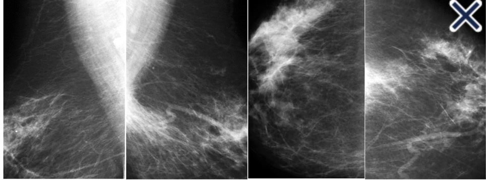
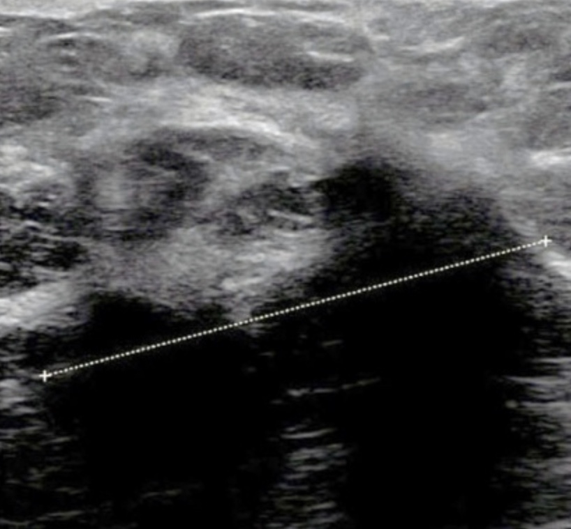
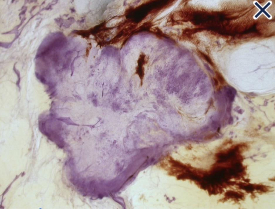
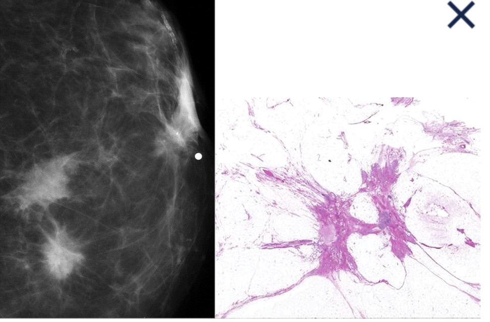
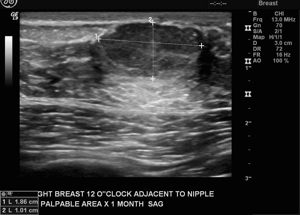
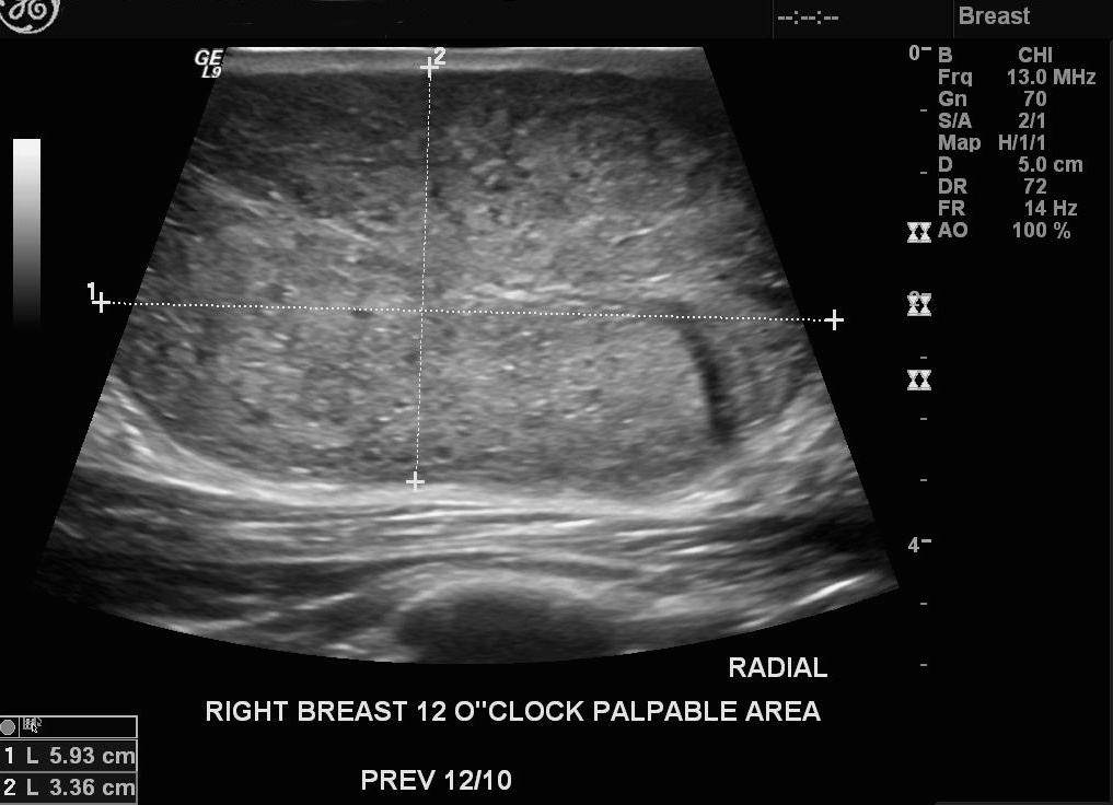
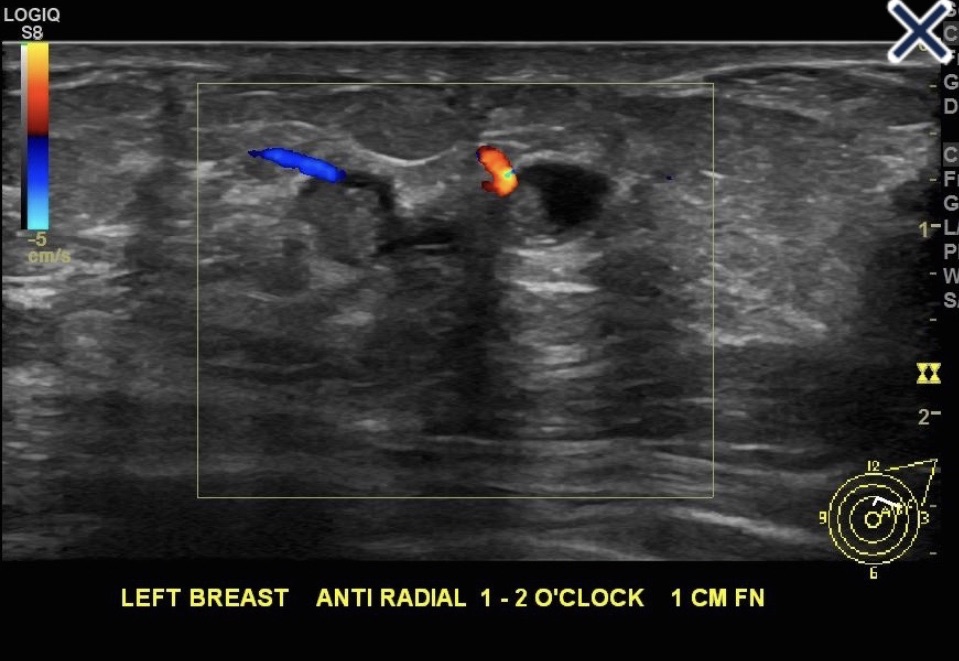
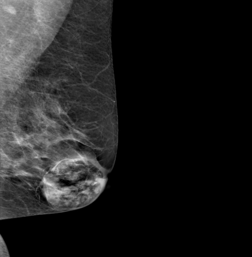
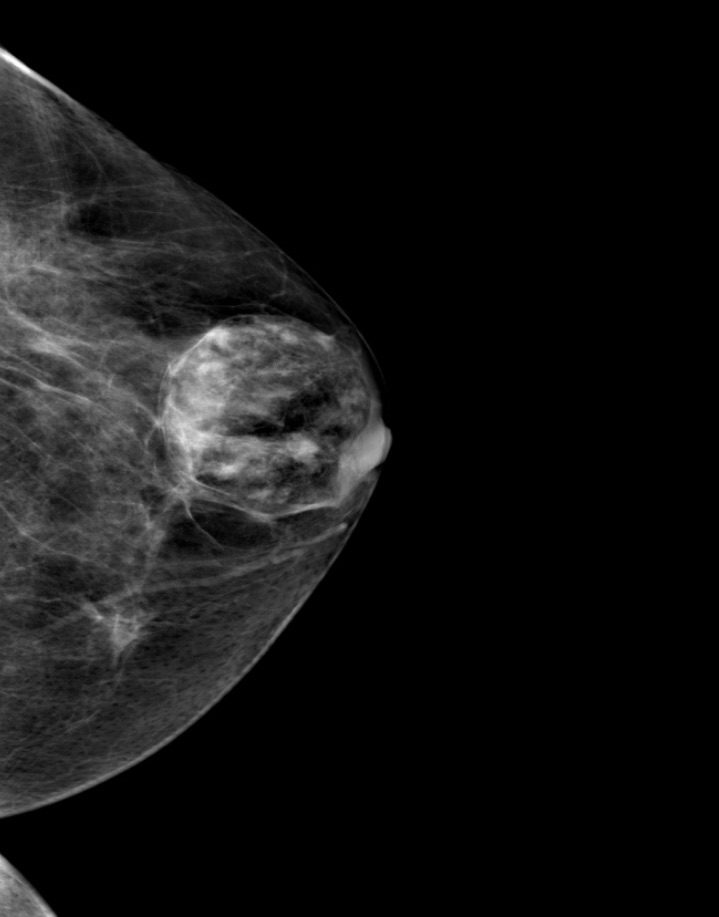
- Fibroadenomas:Are benign, solid neoplasms of the breast:Consisting of fibroepithelial elements
- Their size is hormonally influenced:As evidenced by fluctuation in size with the menstrual cycle and regression in postmenopausal women
- Fibroadenomas are often solitary masses:But in approximately 25% of patients present with multiple lesion’s
- They have a characteristic clinical presentation: Rubbery, mobile, and firm:Despite this, previous reports have indicated that diagnosis by clinical examination:Is accurate in only 50% to 75% of patient’s
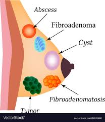
- One of the clinical dilemmas facing both surgeons and patients is the concern that the mass is something more ominous than a fibroadenoma:Both benign and malignant phylloides tumors may mimic fibroadenomas
- Additionally, published reports have described adenocarcinoma and ductal carcinoma in situ:Arising within fibroadenomas or misdiagnosed as fibroadenomas
- Because of the potential for more aggressive pathology masquerading as fibroadenomas:Management has been debated and recommendations changed several times in recent decades:Until the mid-1980s:Standard practice was excision of all fibroadenomas
-
- Subsequent studies in the 1980s and 1990s:Demonstrated the safety of observing the presumed fibroadenomas:In women under age 35:Who had a fine-needle aspirate biopsy that did not contain malignant or suspicious cells
- More recently, the question has been asked whether biopsy is even necessary:Smith and Burrows concluded:That patients under the age of 25 with benign ultrasound findings:Could be safely observed without a biopsy
-

- Criteria for excision of suspected fibroadenomas of the breast: Patients with an age greater than 35 years
- Immobile or poorly circumscribed mass
- Size greater than 2.5 cm
- Biopsy not definitive for fibroadenoma
- Fibroadenomas:Son neoplasias benignas y sólidas de la mama que consisten en elementos fibroepiteliales.
- Su tamaño está influenciado hormonalmente: Como lo demuestra la fluctuación en el tamaño con el ciclo menstrual y regresión en mujeres posmenopáusicas.
- Los fibroadenomas son a menudo tumores solitarios: Pero en aproximadamente el 25% de los pacientes presentan lesiones múltiples
- Tienen una presentación clínica característica: Gomoso, móvil y firme: A pesar de esto, informes anteriores han indicado que el diagnóstico mediante examen clínico: Es preciso en solo 50% a 75% de los pacientes
- Uno de los dilemas clínicos que enfrentan los cirujanos y los pacientes es la preocupación de que la tumoración sea algo más siniestra que un fibroadenoma: Los tumores filoides benignos y malignos pueden simular fibroadenomas:Además, los informes publicados han descrito el adenocarcinoma y el carcinoma ductal in situ: Surgen dentro de fibroadenomas o se diagnostican erróneamente como fibroadenomas.
- Debido al potencial de una patología más agresiva disfrazada de fibroadenomas: El manejo ha sido debatida y las recomendaciones cambiaron varias veces en las últimas décadas:Hasta mediados de la década de 1980: La práctica estándar fue la escisión de todos los fibroadenomas.
-
- Estudios posteriores en las décadas de 1980 y 1990: Demostró la seguridad de observar los presuntos fibroadenomas: En mujeres menores de 35 años que tuvieron una biopsia por aspiración con aguja fina que no contenía células malignas o sospechosas
-
- Más recientemente, se ha preguntado si la biopsia es necesaria: Smith y Burrows concluyeron: Que los pacientes menores de 25 años con hallazgos benignos de ultrasonido: Podría observarse con seguridad sin una biopsia.
-