
- Breast pain (mastalgia) is common in women and occasionally occurs in men
- Although it is usually mild and self-limited:
- Approximately 15% of affected women require treatment
- Evaluation of breast pain is important to determine whether the pain is due to:
- Normal physiological changes related to hormonal fluctuation or to a pathologic process such as breast cancer
- Breast pain:
- Is a rare symptom of breast cancer
- Women who present with breast pain but who have a normal exam and imaging studies:
- Can be reasonably assured that their risk of breast cancer is:
- Similar to that of a woman without breast pain
- Can be reasonably assured that their risk of breast cancer is:
- While cyclical breast pain has traditionally been attributed to fibrocystic changes, chronic cystic mastitis, and mammary dysplasia:
- Breast pain and nodularity are so common that the term fibrocystic “disease”:
- Has become obsolete:
- It should no longer be used
- Has become obsolete:
- Breast pain and nodularity are so common that the term fibrocystic “disease”:
- Epidemiology:
- Breast pain is common:
- Up to 70% of women in Western societies:
- Will experience it sometime during their lives
- One study of almost 1700 women (mean age 34 years) surveyed by online questionnaire:
- Found that over one-half (51.5%) had experienced breast pain
- Up to 70% of women in Western societies:
- Pain was more commonly reported among:
- Older women
- Those with larger breast sizes
- Those less fit and / or physically active
- Among women who reported symptoms:
- 41% and 35% reported negative impacts from breast pain on their sexual health and sleep, respectively
- 10% of those symptomatic had reported breast pain as an issue for over half of their lives
- Breast pain is common:
- The prevalence of breast pain appears to depend on the population studied:
- Breast pain is less common in Asian cultures:
- Affecting as few as 5% of women
- Breast pain is less common in Asian cultures:
- Classification and etiology:
- Breast pain can be classified into three categories:
- Cyclical
- Noncyclical
- Extramammary
- Clinically it is more important to differentiate between extra mammary and true breast pain than between cyclical and noncyclical pain:
- This is because management of cyclical and noncyclical breast pain is similar:
- While extramammary pain may require a different treatment
- This is because management of cyclical and noncyclical breast pain is similar:
- Breast pain can be classified into three categories:
- Cyclical breast pain:
- Affects two-thirds of patients with true mastalgia
- Cyclical pain is associated with hormonal fluctuations of the menstrual cycle:
- Usually presenting in the week prior to onset of menses
- It is frequently bilateral and most severe in the upper outer quadrant of the breasts
- Minor cyclical breast discomfort is normal:
- It begins during the late luteal phase and dissipates with the onset of menses
- This is usually bilateral and diffuse pain
- Cyclical breast discomfort is caused by normal hormonal changes associated with ovulation:
- That stimulate the proliferation of normal glandular breast tissue and result in pain
- The stimulation of ductal elements by estrogen, stimulation of the stroma by progesterone, and / or stimulation of ductal secretion by prolactin:
- All contribute to cyclical pain during the menstrual cycle
- Cyclical breast pain can also be associated with pharmacologic hormonal agents:
- Postmenopausal hormone therapy
- Oral contraceptive pills
- Noncyclical breast pain:
- Affects one-third of women with true mastalgia
- The pain does not follow the usual menstrual pattern:
- May be constant or intermittent
- More likely to be unilateral and variable in its location in the breast
- Noncyclical breast pain is more likely to be related to:
- A breast or chest wall lesion
- Possible etiologies include:
- Large pendulous breasts:
- May cause pain due to stretching of Cooper’s ligaments
- Neck, back, shoulder pain and headache may be present, as well as a rash under the pendulous breast in the inframammary fold
- Diet, lifestyle:
- A high-fat diet, smoking, and caffeine intake have been associated with breast pain:
- It is difficult to conduct randomized trials with appropriate blinding that will negate the placebo effect
- Hence, there is currently no high-quality evidence to suggest that a low-fat diet, smoking cessation, or caffeine avoidance reduces breast pain
- A high-fat diet, smoking, and caffeine intake have been associated with breast pain:
- Hormone replacement therapy :
- Up to one-third of menopausal women receiving postmenopausal hormone therapy experience some degree of noncyclical breast pain:
- Which may spontaneously resolve over time
- Up to one-third of menopausal women receiving postmenopausal hormone therapy experience some degree of noncyclical breast pain:
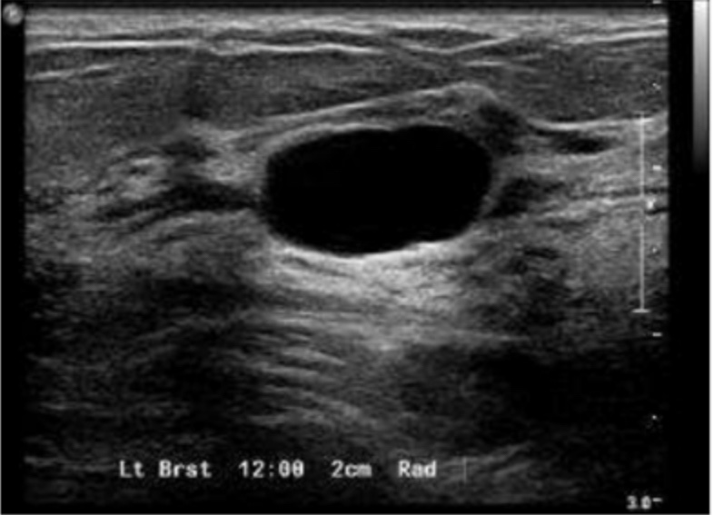
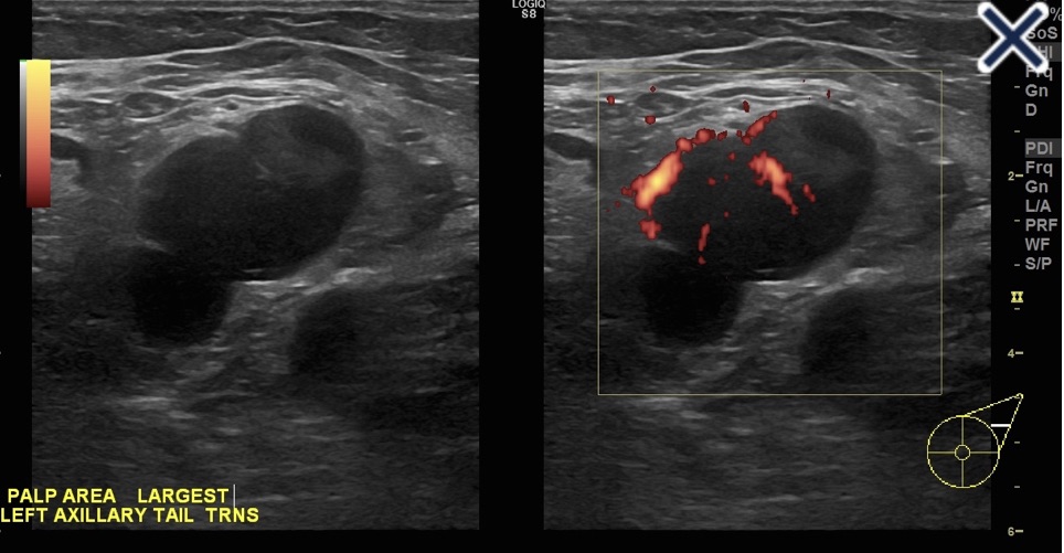
- Breast cysts:
- Solitary cysts:
- Particularly when the presentation is abrupt, are frequently painful
- Solitary cysts:
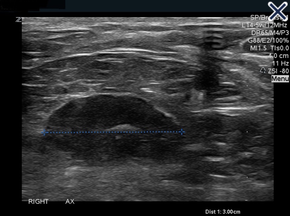
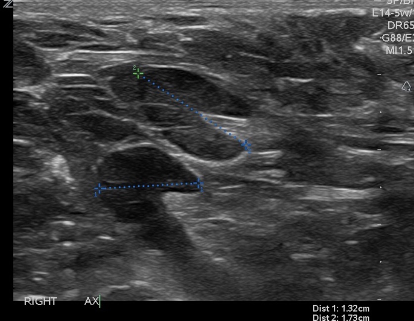
- Ductal ectasia:
- Is characterized by distention of subareolar ducts:
- Due to inflammation unrelated to infection
- Ductal ectasia may be associated with fever and acute local pain and tenderness:
- Caused by penetration of the duct wall by lipid material:
- Which may resolve to leave a subareolar nodule
- Caused by penetration of the duct wall by lipid material:
- In one study, the site and degree of duct dilatation correlated with the intensity of noncyclical breast pain
- Is characterized by distention of subareolar ducts:
- Mastitis:
- Mastitis or breast abscess typically presents as a painful, swollen, and red breast in a febrile woman
- Mastitis is more prevalent during lactation but can also occur in nonlactating women:
- Idiopathic granulomatous mastitis [IGM] or smokers
- Inflammatory breast cancer:
- Women with de novo inflammatory breast cancer (primary disease) may present with:
- Pain and a rapidly progressing tender, firm, enlarged breast
- The skin over the breast is warm and thickened, with a “peau d’orange” (orange skin) appearance, but there is often no fever or leukocytosis
- Women with de novo inflammatory breast cancer (primary disease) may present with:
- Hidradenitis suppurativa:
- Although primarily confined to the axilla:
- Can involve the breast and present as breast nodules and pain
- Although primarily confined to the axilla:
- Other etiologies of breast pain include:
- Pregnancy
- Thrombophlebitis (Mondor’s disease)
- Trauma
- Macrocysts
- Prior breast surgery
- A variety of medications:
- Hormones as well as some antidepressants, cardiovascular agents, and antibiotics
- Large pendulous breasts:
- Extramammary pain:
- Some women who present with breast pain actually have referred pain from sources other than the breasts
- The breast is innervated by:
- The anterolateral and anteromedial branches of the intercostal nerves (T3 to T5):
- Irritation of these nerves anywhere along their course can lead to pain that is felt in the breast or nipple
- The anterolateral and anteromedial branches of the intercostal nerves (T3 to T5):
- In some studies done in primary care and certain breast clinic settings, it has been found that women presenting with breast pain:
- More often have extramammary pain rather than true mastalgia
- Extramammary pain may be from:
- Musculoskeletal sources such as the chest wall, spinal or paraspinal disorders, trauma, or scarring from prior biopsy
- It may also be related to medical problems such as biliary, pulmonary, esophageal, or cardiac disease
- Chest wall pain:
- Is frequently due to pectoralis major muscle injury, related to repetitive activities such as water skiing, raking, rowing, or shoveling
- Chest wall pain that presents as bilateral parasternal discomfort can also arise from:
- Costochondritis:
- Typically the second through fifth costochondral junctions
- Tietze syndrome:
- Typically the second and third costochondral junctions
- Costochondritis:
- Other etiologies of chest wall pain include:
- Slipping and clicking ribs and arthritis
- Spinal and paraspinal disorders:
- Radicular chest wall pain may be due to:
- Cervical arthritis:
- This pain typically occurs in older women in whom vertebral, spinal, and paraspinal problems in the neck and upper thorax accumulate with age
- Paraspinal muscle spasm and other impingements on the free course of the sensory nerves from the neck and upper thorax:
- Can cause a radiculopathy leading to pain or hyperesthesia
- Burning pain, which is typical of nerve root pressure, is a common feature
- Imaging studies of the neck may reveal the etiology of the pain
- Cervical arthritis:
- Radicular chest wall pain may be due to:
- Trauma:
- Breast pain can be caused by local trauma, such as seat belt injury, child or pet kicking, or intimate partner violence, to the breasts or anterior chest wall
- Pain can also be caused by intercostal neuralgia due to a respiratory infection or underlying pleuritic lesions
- Additionally, gallbladder disease or ischemic heart disease may present as intermittent chest pain attributed to the breast
- Postthoracotomy syndrome:
- Is an unusual disorder in which a healing chest wound simulates the effect of a suckling infant
- It can be associated with:
- An elevated prolactin concentration
- Breast pain
- Milk production
- A similar effect can be seen with other forms of chest wall irritation, including burns and chafing from clothing overlying the nipple
- History:
- It may be helpful to ask women with cyclical pain to record the occurrence and severity of breast pain in a diary and note potential aggravating and ameliorating factors
- Questions the patient should be asked about her pain include:
- Where in the breast or axilla does the pain occur?
- Is the pain bilateral?
- What does the pain feel like?
- How severe is the pain?
- If premenopausal:
- Is it phasic, with peaks at midcycle and premenstrually?
- Is it associated with use of oral contraceptive pills or hormone replacement therapy?
- Did it begin after a recent birth or pregnancy loss or termination?
- Is it related to vigorous or repetitive use of the pectoral muscle group?
- Is there a concurrent neck, back, or shoulder problem?
- Are there systemic or other local symptoms, such as fever or erythema?
- Is there a history of recent trauma to the chest?
- Does the pain affect her ability to perform daily activities?
- In addition, a complete medical and surgical history and systematic review of systems should be obtained. Breast cancer risk should be assessed
- Chest wall pain is often lateral and may be burning or knifelike, and localized or diffuse
- Physical examination:
- Breast:
- The breast should be examined for signs of inflammation or infection, which would suggest an etiology of mastitis.
- Mastitis typically presents as a painful, swollen, and red breast in a febrile woman.
- Mastitis is more prevalent during lactation but can also occur in nonlactating women
- The key point in examining a woman with breast pain is to look for signs suggestive of breast malignancy:
- Such as a mass, skin changes, or bloody nipple discharge
- The four breast quadrants, subareolar areas, axillae, and supraclavicular and infraclavicular areas should be systematically examined with the woman both lying and sitting with her hands on her hips and then above her head
- The specific goals of the examination are to:
- Check for skin changes, noting the symmetry and contour of the breasts, position of the nipples; scars; skin retraction; dimpling; edema or erythema; ulceration or crusting of the nipple; and changes in skin color
- Check for enlarged or tender axillary, supraclavicular, or infraclavicular lymph nodes
- Delineate and document breast masses
- Check for nipple discharge
- Identify localized areas of tenderness and relate them to areas of pain noted by the woman and to other physical findings
- Women found to have a palpable breast mass, skin changes, or bloody nipple discharge should be referred to a breast specialist for further evaluation and imaging to treat or exclude breast cancer
- Chest wall:
- Physical exam should also aim at differentiating true breast pain from extramammary pain
- Features of breast pain that suggest an extramammary origin include:
- Unilateral, and brought on by activity
- Located very lateral or medial in the breast
- Reproducible by pressure on a specific area of the chest wall
- To specifically look for chest wall pain, women may be asked to lie on each side:
- These positions enable the breast to fall away from the chest wall, which permits palpation of the underlying chest wall muscles and ribs
- Women with pain in the lower aspect of their breast should have the breast elevated with one hand and the underlying chest wall palpated with the other
- Chest wall pain due to pectoralis major muscle injury can be reproduced by asking the patient to place her hand flat on the iliac wing and push inward
- Women found to have chest wall pain can be reassured that there is no serious underlying cause for the pain, and they can be treated according to the symptoms
- Breast:
- Imaging:
- For most women who present with breast pain, a thorough history and physical examination must be performed, and clinical judgment must be used in deciding upon any diagnostic imaging studies
- Suspicious physical findings present:
- Women of any age who have suspicious physical findings such as a mass, skin changes, or bloody nipple discharge should undergo:
- Mammography with or without ultrasound
- Women of any age who have suspicious physical findings such as a mass, skin changes, or bloody nipple discharge should undergo:
- Suspicious physical findings absent:
- Assuming they are up to date with breast cancer screening, women who have breast pain but no other suspicious findings on physical exam:
- May undergo breast imaging selectively based on their presentation and age
- Breast imaging, even with a negative result, has been credited with alleviating patient anxiety
- Seeking reassurance is often cited as the main reason for imaging in patients with breast pain
- Many women do not seek further medical attention after assurance that their pain is not due to breast cancer
- May undergo breast imaging selectively based on their presentation and age
- The imaging modalities most commonly used in these clinical scenarios are breast ultrasound and mammography
- There are no data to suggest the use of breast magnetic resonance imaging (MRI) for this patient population
- In a case-control study, there was no difference in breast cancer incidence in women undergoing mammography for a painful breast (0.5%) compared with the contralateral nonpainful breast (0.5%) and compared with women without breast pain (0.7%)
- Assuming they are up to date with breast cancer screening, women who have breast pain but no other suspicious findings on physical exam:
- Three studies of ultrasound for focal breast pain without a palpable mass detected cancer in 0%, 1.2%, and 4.6% of patients
- The American College of Radiology Appropriateness Criteria guidelines recommend the following approach to selecting an imaging modality:
- Women with cyclical or bilateral nonfocal breast pain:
- Usually do not require imaging:
- The yield of finding a specific cause with imaging is low
- Usually do not require imaging:
- Women with noncyclical, unilateral, or focal breast pain that is not extramammary (eg, chest wall pain), as determined by physical exam:
- Should undergo breast imaging to elucidate the underlying etiology and exclude breast cancer
- The choice of imaging modality is based on age:
- Women under 30 years of age:
- Should undergo ultrasound because it is more accurate than mammography for that age group
- Mammography is added if abnormality is found on the ultrasound and/or if a patient’s history or risk status justifies the radiation exposure
- Women between 30 and 39 years of age:
- Should also undergo ultrasound
- Unilateral or bilateral mammography should also be performed because in this age group some small cancers are found on mammography but not ultrasound
- Women age 40 and older:
- Should undergo both mammography and ultrasound
- Women under 30 years of age:
- For women who have breast pain but no abnormality on physical examination or imaging studies:
- The risk for breast cancer is low at approximately 0.5%
- Women with cyclical or bilateral nonfocal breast pain:
- Treatment:
- After obtaining normal findings on clinical and imaging studies, reassurance is often all that is required:
- A simple assurance that the patient does not have breast cancer provides adequate relief for 78% to 85% of women
- Such patients would also benefit from a follow-up visit in two to three months to exclude or treat recurrent/persistent pain
- For some women, however, breast pain can cause problems with their activities of daily living:
- As an example, in a study of 1171 healthy premenopausal women:
- 11% reported moderate-to-severe pain that interfered with sexual activity (48%), physical activity (37%), social activity (12%), and school activity (8%):
- Consequently, these women required treatment for their breast pain
- 11% reported moderate-to-severe pain that interfered with sexual activity (48%), physical activity (37%), social activity (12%), and school activity (8%):
- As an example, in a study of 1171 healthy premenopausal women:
- Approximately 15% of women seen in the breast clinic for breast pain:
- Require treatment beyond simple reassurance
- Breast pain is treated:
- Medically
- Breast surgery is not indicated to treat pain:
- In the absence of any breast pathology
- First-line therapy:
- First-line therapy for breast pain is conservative and typically includes:
- Reassurance that this is not a malignancy
- Physical support
- Over-the-counter analgesics
- Manipulation of hormone-based medications for those who take them
- First-line therapy for breast pain is conservative and typically includes:
- I prefer to treat with first-line therapy for six months before moving onto one of the second-line therapies:
- Which may be more effective but also have more side effects
- Some practitioners also endorse therapies such as caffeine abstinence or evening primrose oil (EPO):
- Although such therapies have not been proven effective by vigorous placebo controlled trials:
- They are generally harmless and may provide relief for some patients
- Although such therapies have not been proven effective by vigorous placebo controlled trials:
- Physical support:
- Support garments:
- A well-fitting brassiere to better support the breast is widely advocate
- The use of a support bra with steel underwire tends to reduce mastalgia in women with pendulous breasts
- In addition, use of a “sports bra” during exercise has been shown to reduce pain related to breast movement
- Wearing a soft, supportive bra at night stops the breast pulling down on the chest wall, supports tender breast tissues, and helps many women sleep
- Women with asymmetric breasts may benefit from specialized fitting to place extra padding on one side, which permits appropriate support of that side without overcompressing the contralateral side
- Compresses:
- Some women obtain relief from application of warm compresses or ice packs or gentle massage
- For those who breast feed:
- Ice packs are recommended during the obstructive (prebacterial) phase of puerperal mastitis:
- To decrease milk production regionally and thereby relieve ductal intraluminal pressure and subsequent pain
- Ice packs are recommended during the obstructive (prebacterial) phase of puerperal mastitis:
- Support garments:
- Acetaminophen or NSAID:
- Can be used to relieve breast pain
- Topical NSAIDs may also be useful:
- While the weaker types of topical NSAID (eg, ibuprofen gel) may not be effective in relieving breast pain
- Data from randomized trials demonstrated significant improvement in those treated with diclofenac gel with minimal side effects
- In the United States (US), two types of topical NSAIDs are available:
- Salicylate, the active ingredient in aspirin, is found in Aspercreme and Nuprin
- Diclofenac, which has the same active ingredient as the oral NSAID, is available as a patch, gel, or topical solution
- Second-line therapy:
- Treatment with one of the second-line therapies may be required in patients who still have debilitating breast pain despite first-line therapy for six months
- Some physicians prefer to use tamoxifen first because it has fewer side effect than danazol
- Treatment with tamoxifen or danazol for one to three months, until either pain subsides or side effects increase
- Tamoxifen:
- For patients with more severe mastalgia refractory to other treatments, tamoxifen can provide breast pain relief
- A meta-analysis of three randomized trials found tamoxifen to be more effective in relieving breast pain than placebo (relative risk 1.92, 95% CI 1.42-2.58)
- Tamoxifen is effective at both doses of 20 mg daily and 10 mg daily:
- The side effects are significantly reduced at the lower dose
- Thus, when used off-label to treat severe mastalgia, tamoxifen is usually given at 10 mg once daily for three months
- However, tamoxifen is associated with menopause-like symptoms such as:
- Hot flashes:
- Vaginal dryness
- Joint pain
- Leg cramps
- It can also increase the risk of:
- Blood clots
- Strokes
- Uterine cancer
- Cataracts
- Thus, tamoxifen is infrequently used to treat mastalgia
- Restricting tamoxifen to the luteal phase of the menstrual cycle has also been suggested to reduce side effects
- Danazol:
- Is an androgen, and for severe mastalgia, it is usually given at 200 mg once daily
- It should be noted that since 2018, the US Food and Drug Administration (FDA) no longer approves the use of danazol for the indication of fibrocystic breast disease
- Danazol is effective in relieving breast pain and tenderness:
- According to a meta-analysis of four randomized trials against placebo, it resulted in a 20 point mean reduction in pain score on a visual analogue scale (VAS) of 0 to 100
- However, the use of danazol is limited by its androgenic effects
- At the recommended dose of 200 mg daily, significant proportions of patients reported side effects such as:
- Weight gain (30%)
- Menstrual irregularity (50%)
- Deepening of the voice (10%)
- Hot flashes (10%)
- Restricting the use of danazol to the luteal phase of the menstrual cycle reduces the side effects without compromising its effectiveness
- Women on hormone-based medications:
- Postmenopausal hormone therapy that causes breast pain should be decreased or discontinued if at all possible:
- This should only be done if breast pain is intolerable and after discussing with the patient the risks and benefits or curtailing hormone replacement therapy
- It is not clear whether oral contraceptives cause or relieve cyclical mastalgia:
- Decreasing the dose of estrogen in an oral contraceptive regimen can be effective in controlling breast pain
- In other studies, oral contraceptives can reduce breast pain severity and duration in some women with cyclical symptoms:
- The impact of oral contraceptive pills on breast pain may largely depend on their compositions; alternatively, they may have different effects on different women
- Progestogens also improve breast pain symptoms in some women:
- While oral and topical (applied to the breast) progesterone did not show benefit in randomized trials:
- A vaginal cream of micronized progesterone (4 g of vaginal cream containing 2.5% natural progesterone used from the 19th to the 25th day of the cycle for six cycles) reduced breast pain in 65% of women compared with 22% of controls in a trial
- While oral and topical (applied to the breast) progesterone did not show benefit in randomized trials:
- Postmenopausal hormone therapy that causes breast pain should be decreased or discontinued if at all possible:
- Therapies not proven by randomized trial data:
- The role of diet and lifestyle in relieving cyclical breast pain is unclear, with a strong likelihood of a placebo response for many interventions
- However, some practitioners feel that some of these treatments (eg, caffeine abstinence and evening primrose oil [EPO]) are worth trying because they are generally harmless and may offer some women pain relief
- A low-fat (15% of calories), high complex carbohydrate diet has been effective in some observational studies and small randomized trials:
- However, the trials could not be blinded, which may invite a placebo effect
- Additionally, such low-fat diets are difficult to maintain beyond a few weeks
- Elimination of caffeine has not been effective in controlled trials, although it seems to be helpful in some women
- EPO or its active ingredient gamma linoleic acid (GLA) has been studied in multiple randomized trials of breast pain:
- Despite early enthusiasm, neither has been shown to be effective beyond the placebo effect
- Vitamin E has been shown in multiple randomized trials to be no better than placebo in the treatment of benign breast disease:
- Thus, vitamin E should not be prescribed to treat mastalgia
- Bromocriptine is a dopamine agonist that inhibits prolactin release:
- Although bromocriptine is effective in relieving pain compared with placebo, it is less effective than danazol, and up to 80% of women develop side effects such as headaches and dizziness
- Therefore, it is no longer used to treat breast pain
- Several other drugs that affect estrogen or prolactin secretion (including bromocriptine and other gonadotropin-releasing hormone [GnRH] agonists) have been studied but are not advocated for use in patients with severe mastalgia, because of unfavorable side effect profiles
- After obtaining normal findings on clinical and imaging studies, reassurance is often all that is required:
- Investigational therapies:
- Because of the unfavorable side effect profiles of the medications currently used to treat mastalgia (eg, danazol, tamoxifen), there is great interest in developing natural (herbal) products that could relieve breast pain:
- However, the benefits of most of these products remain unproven due to a lack of vigorous testing in randomized trials
- Phytoestrogens, such as genistein, isoflavones, and soy milk, have been investigated as treatments for breast pain:
- Soy milk has been tested against cow milk in a controlled trial, and although an improvement of symptoms was noted in 56% of test subjects versus 10% of controls, the trial was criticized for noncompliance due to the unpalatable taste of the soy milk
- Agnus castus, a fruit extract, has significantly lowered visual analogue pain scores against placebo in controlled trials and is well tolerated
- Matricaria chamomilla (chamomile) extract has also improved cyclical breast pain on a visual analogue scale compared with placebo in a controlled trial
- Chest wall pain:
- For women diagnosed with chest wall pain, local heat and analgesics such as acetaminophen or NSAIDs may relieve pain, but most women do not require therapy beyond reassurance that the source of pain is muscle strain or articular
- Patients should reduce or cease activities that brought on or aggravated their pain until the pain improves
- In severe cases in which the pain is localized but not relieved by over-the-counter pain medications, a trigger point injection with a mixture of a local anesthetic and corticosteroid may bring relief for the patient and can be repeated as necessary
- Because of the unfavorable side effect profiles of the medications currently used to treat mastalgia (eg, danazol, tamoxifen), there is great interest in developing natural (herbal) products that could relieve breast pain:
- Prognosis:
- In general, mastalgia has a natural history of remission and relapse, evidenced by the fact that improvement is seen in as many as 40% of women receiving placebo in randomized trials
- The prognosis of women who have breast pain is variable and influenced by the age of onset of pain and whether pain is cyclical or noncyclical:
- In one series, cyclical breast pain spontaneously resolved within three months of onset in 20% to 30% of women, but transient relapses were common
- In another series, noncyclical breast pain spontaneously resolved in 50% of patients
- Relief may be spontaneous or related to a hormonally mediated event:
- Such as pregnancy or menopause
- Associated conditions:
- Breast pain is usually a symptom, not a diagnosis
- Although most women who have breast pain will not have any associated conditions, some will, in which case their pain should be treated as a component of the associated condition
- Premenstrual syndrome:
- Is characterized by the presence of both physical and behavioral (including affective) symptoms that occur repetitively in the second half of the menstrual cycle and interfere with some aspects of the woman’s life
- Breast tenderness is one of the common symptoms of PMS
- A meta-analysis of 10 randomized trials of selective serotonin reuptake inhibitors (SSRIs) used in women with premenstrual symptoms showed SSRIs to be more effective than placebo at relieving breast pain
- Thus, women who have breast pain or tenderness as a component of PMS may benefit from SSRIs
- Breast cancer:
- The presence of a breast cancer in a patient who presents with only pain is extremely low, ranging from 0.5% to 33.%
- Breast pain may occur at the time of presentation of a breast cancer, although the pain is typically associated with adjacent benign, cystic breast tissue rather than the cancer
- One caveat in retrospective studies is that recall of breast pain might be increased after the diagnosis of breast cancer
- In addition, pain may also occur following the imaging and core biopsy of the cancer rather than being associated with the cancer itself.
- Prior breast surgery:
- Pain that develops after breast surgery is of a different etiology and treated differently from de novo breast pain







 Rodrigo Arrangoiz MS, MD, FACS, FSSO