¿Qué puedo hacer para reducir el riesgo de cáncer de mama?

img_3761img_3768-1

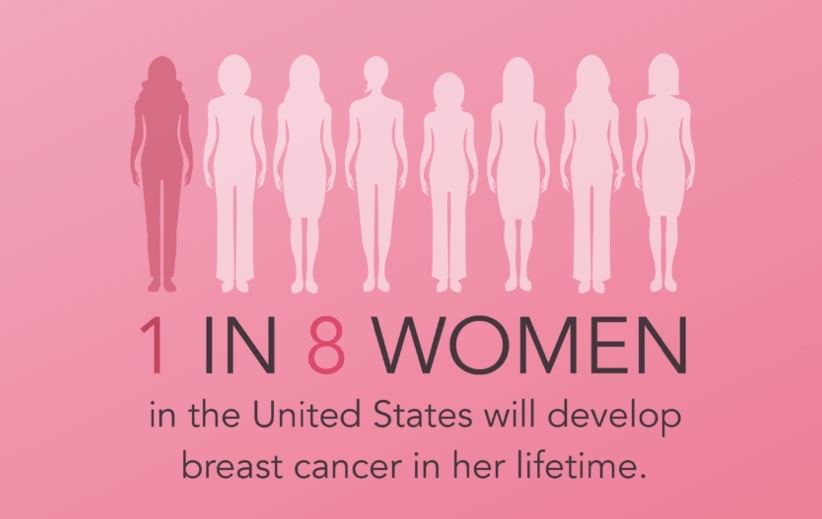
  • Existen muchos factores en el transcurso de la vida que pueden influir en el riesgo de que tenga cáncer de mama. Algunos factores no se pueden cambiar, como hacerse mayor o los antecedentes familiares, pero usted puede disminuir el riesgo de tener cáncer de mama al cuidar su salud de la siguiente manera:
    • Mantenga un peso saludable.
    • Haga ejercicio con regularidad.Foto de dos mujeres jóvenes corriendo
    • No tome alcohol o, si lo hace, limite las bebidas alcohólicas a no más de una por día.
    • Si está recibiendo, o le han dicho que reciba, terapia de reemplazo hormonalExternal o anticonceptivos orales  (píldoras anticonceptivas), consulte con su médico acerca de los riesgos y averigüe si es lo mejor para usted.
    • Si es posible, amamante a sus hijos.
    • Si tiene antecedentes familiares de cáncer de mama o cambios heredados en sus genes BRCA1 y BRCA2, hable con su médico acerca de otras maneras de reducir su riesgo.
  • Mantener una buena salud durante toda la vida disminuirá el riesgo de tener cáncer y mejorará las probabilidades de sobrevivir si se enferma de cáncer.

👉Rodrigo Arrangoiz MS, MD, FACS cirujano oncology y cirujano de mamá de Sociedad Quirúrgica S.C en el America British Cowdray Medical Center en la ciudad de Mexico:

  • Es experto en el manejo del cáncer de mama.

👉Es miembro de la American Society of Breast Surgeons:

Training:

• General surgery:

• Michigan State University:

• 2004 al 2010

• Surgical Oncology / Head and Neck Surgery / Endocrine Surgery:

• Fox Chase Cancer Center (Filadelfia):

• 2010 al 2012

• Masters in Science (Clinical research for health professionals):

• Drexel University (Filadelfia):

• 2010 al 2012

• Surgical Oncology / Head and Neck Surgery / Endocrine Surgery:

• IFHNOS / Memorial Sloan Kettering Cancer Center:

• 2014 al 2016

#Arrangoiz

#Surgeon

#Cirujano

#SurgicalOncologist

#CirujanoOncologo

#BreastSurgeon

#CirujanodeMama

#CancerSurgeon

#CirujanodeCancer

👉Plática del Dr. Arrangoiz sobre el cáncer de mama:

Rodrigo Arrangoiz MS, MD, FACS Cirujano Oncólogo de Mama

👉Rodrigo Arrangoiz MS, MD, FACS cirujano oncology y cirujano de mamá de Sociedad Quirúrgica S.C en el America British Cowdray Medical Center en la ciudad de Mexico:

  • Es experto en el manejo del cáncer de mama.

👉Es miembro de la American Society of Breast Surgeons:

Training:

• General surgery:

• Michigan State University:

• 2004 al 2010

• Surgical Oncology / Head and Neck Surgery / Endocrine Surgery:

• Fox Chase Cancer Center (Filadelfia):

• 2010 al 2012

• Masters in Science (Clinical research for health professionals):

• Drexel University (Filadelfia):

• 2010 al 2012

• Surgical Oncology / Head and Neck Surgery / Endocrine Surgery:

• IFHNOS / Memorial Sloan Kettering Cancer Center:

• 2014 al 2016

#Arrangoiz

#Surgeon

#Cirujano

#SurgicalOncologist

#CirujanoOncologo

#BreastSurgeon

#CirujanodeMama

#CancerSurgeon

#CirujanodeCancer

👉Plática del Dr. Arrangoiz sobre el cáncer de mama:

¿Cuáles son los factores de riesgo del cáncer de mama?

👉Algunos estudios han demostrado que el riesgo de tener cáncer de mama se debe a una combinación de factores.

👉Los principales factores que influyen en el riesgo de una persona incluyen ser mujer y hacerse mayor.

👉La mayoría de los cánceres de mama se detectan en mujeres de 50 años o más.

👉Algunas mujeres tienen cáncer de mama aunque no tengan ningún otro factor de riesgo conocido.

👉Tener un factor de riesgo no significa que la persona tendrá la enfermedad, y no todos los factores de riesgo tienen el mismo efecto.

👉La mayoría de las mujeres tienen algunos factores de riesgo, pero la mayoría de las mujeres no tienen cáncer de mama. Si usted tiene factores de riesgo de cáncer de mama, hable con Rodrigo Arrangoiz MS, MD, FACS cirujano de mama de Sociedad Quirurgica S.C. sobre las maneras en que puede disminuir el riesgo y acerca de las pruebas de detección de esta enfermedad.

Factores de riesgo que no pueden cambiar

  • Hacerse mayor. El riesgo de cáncer de mama aumenta con la edad; la mayoría de los cánceres de mama se diagnostica después de los 50 años de edad.
  • Mutaciones genéticas. Cambios (mutaciones) heredados en ciertos genes, tales como en el BRCA1 y el BRCA2. Las mujeres que han heredado estos cambios genéticos tienen mayor riesgo de presentar cáncer de mama y de ovario.
  • Historial reproductivo. Inicio temprano de la menstruación antes de los 12 años de edad y comienzo de la menopausia después de los 55 años de edad exponen a las mujeres a hormonas por más tiempo, lo cual aumenta el riesgo de cáncer de mama.
  • Tener mamas densas. La mamas densas tienen más tejido conjuntivo que tejido adiposo, lo cual, a veces, puede hacer difícil la detección de tumores en una mamografía. Las mujeres con mamas densas tienen más probabilidades de tener cáncer de mama.
  • Antecedentes personales de cáncer de mama o ciertas enfermedades de las mamas que no son cancerosas. Las mujeres que han tenido cáncer de mama tienen mayores probabilidades de tener esta enfermedad por segunda vez. Algunas enfermedades de las mamas que no son cancerosas, como la hiperplasia atípica o el carcinoma lobulillar in situ, están asociadas a un mayor riesgo de tener cáncer de mama.
  • Antecedentes familiares de cáncer de mama. El riesgo de una mujer de tener cáncer de mama es mayor si su madre, una hermana o una hija (parientes de primer grado) o varios integrantes de la familia por el lado paterno o materno han tenido cáncer de mama. Tener un pariente de primer grado de sexo masculino con cáncer de mama también aumenta el riesgo para la mujer.
  • Tratamientos previos con radioterapia. Las mujeres que han recibido radioterapia en el pecho o las mamas antes de los 30 años de edad (por ejemplo, para el tratamiento del linfoma de Hodgkin) tienen un riesgo mayor de presentar cáncer de mama más adelante en la vida.
  • También tienen mayor riesgo las mujeres que tomaron el medicamento dietilestilbestrol, que se les administró a algunas mujeres embarazadas en los Estados Unidos entre los años 1940 y 1971 para prevenir el aborto espontáneo. Las mujeres cuyas madres tomaron dietilestilbestrol cuando estaban embarazadas de ellas también tienen riesgo.

Factores de riesgo que pueden cambiar

  • No mantenerse físicamente activa. Las mujeres que no se mantienen físicamente activas tienen un mayor riesgo de tener cáncer de mama.
  • Tener sobrepeso o ser obesa después de la menopausia. Las mujeres mayores que tienen sobrepeso o que son obesas tienen mayor riesgo de tener cáncer de mama que las que tienen un peso normal.
  • Tomar hormonas. Algunas formas de terapia de remplazo hormonal (aquellas que incluyen tanto estrógeno como progesterona) que se toman durante la menopausia pueden aumentar el riesgo de cáncer de mama si se toman por más de cinco años. Ciertos anticonceptivos orales (píldoras anticonceptivas) aumentan el riesgo de cáncer de mama también.
  • Historial reproductivo. Quedar embarazada por primera vez después de los 30 años de edad, no amamantando y nunca tener un embarazo que llegue a término puede aumentar el riesgo de cáncer de mama.
  • Tomar alcohol. Algunos estudios muestran que el riesgo de la mujer de tener cáncer de mama aumenta cuanto mayor sea la cantidad de alcohol que tome.

👉Las investigaciones como el Dr. Arrangoiz sugieren que otros factores, tales como el tabaquismo, la exposición a sustancias químicas que pueden causar cáncer y cambios en otras hormonas debido al trabajar en el turno de la noche, también pueden aumentar el riesgo de cáncer de mama.

👉Rodrigo Arrangoiz MS, MD, FACS cirujano oncology y cirujano de mamá de Sociedad Quirúrgica S.C en el America British Cowdray Medical Center en la ciudad de Mexico:

  • Es experto en el manejo del cáncer de mama.

👉Es miembro de la American Society of Breast Surgeons:

Training:

• General surgery:

• Michigan State University:

• 2004 al 2010

• Surgical Oncology / Head and Neck Surgery / Endocrine Surgery:

• Fox Chase Cancer Center (Filadelfia):

• 2010 al 2012

• Masters in Science (Clinical research for health professionals):

• Drexel University (Filadelfia):

• 2010 al 2012

• Surgical Oncology / Head and Neck Surgery / Endocrine Surgery:

• IFHNOS / Memorial Sloan Kettering Cancer Center:

• 2014 al 2016

#Arrangoiz

#Surgeon

#Cirujano

#SurgicalOncologist

#CirujanoOncologo

#BreastSurgeon

#CirujanodeMama

#CancerSurgeon

#CirujanodeCancer

¿Cuáles son los síntomas del cáncer de mama?

👉Los signos de advertencia del cáncer de mama pueden ser distintos en cada persona.

👉Algunas personas no tienen ningún tipo de signos o síntomas.

👉Algunas señales de advertencia del cáncer de mama son:

  • Un bulto nuevo en la mama o la axila (debajo del brazo).
  • Aumento del grosor o hinchazón de una parte de la mama.
  • Irritación o hundimientos en la piel de la mama.
  • Enrojecimiento o descamación en la zona del pezón o la mama.
  • Hundimiento del pezón o dolor en esa zona.
  • Secreción del pezón, que no sea leche, incluso de sangre.
  • Cualquier cambio en el tamaño o la forma de la mama.
  • Dolor en cualquier parte de la mama.

👉Tenga en cuenta que estos signos de advertencia pueden darse con otras afecciones que no son cáncer.

Si tiene algún signo o síntoma que le preocupa, consulte a su médico de inmediato.

¿Qué es una mama normal?

👉No existe un solo tipo de mama.

👉Lo que puede ser normal en usted tal vez no lo sea en otra mujer.

👉La mayoría de las mujeres dicen que sienten bultos o superficies irregulares en las mamas.

👉La manera en que las mamas se ven y se sienten también puede ser afectada por la menstruación, la maternidad, subir o bajar de peso, y por ciertos medicamentos. Asimismo, las mamas tienden a cambiar con la edad.

👉Para obtener más información, pregúntenle a Rodrigo Arrangoiz MS, MD, FACS el cirujano de mama de Sociedad Quirúrgica S.C.

¿Qué significa si encuentro un bulto en mis mamas?

👉Muchas afecciones pueden causar bultos en las mamas, entre ellas el cáncer.

👉Sin embargo, la mayoría de los bultos en las mamas son causados por otras afecciones médicas.

👉Las dos causas más comunes de bultos en las mamas son la enfermedad fibroquística y los quistes.

👉La enfermedad fibroquística produce cambios en las mamas que no son cancerosos y que pueden causar bultos, sensibilidad al tacto y dolor.

👉Los quistes son pequeños sacos llenos de líquido que pueden formarse en las mamas.

👉Rodrigo Arrangoiz MS, MD, FACS cirujano oncology y cirujano de mamá de Sociedad Quirúrgica S.C en el America British Cowdray Medical Center en la ciudad de Mexico:

  • Es experto en el manejo del cáncer de mama.

👉Es miembro de la American Society of Breast Surgeons:

Training:

• General surgery:

• Michigan State University:

• 2004 al 2010

• Surgical Oncology / Head and Neck Surgery / Endocrine Surgery:

• Fox Chase Cancer Center (Filadelfia):

• 2010 al 2012

• Masters in Science (Clinical research for health professionals):

• Drexel University (Filadelfia):

• 2010 al 2012

• Surgical Oncology / Head and Neck Surgery / Endocrine Surgery:

• IFHNOS / Memorial Sloan Kettering Cancer Center:

• 2014 al 2016

#Arrangoiz

#Surgeon

#Cirujano

#SurgicalOncologist

#CirujanoOncologo

#BreastSurgeon

#CirujanodeMama

#CancerSurgeon

#CirujanodeCancer

Current Management of Ductal Carcinoma Insitu (DCIS)

👉The goal of multimodality care for DCIS, which may include surgery, radiotherapy, and hormone therapy, is to prevent the development of invasive breast cancer.
👉While mastectomy is associated with a decreased rate of local recurrence (approximately 1% at 10 years) compared to conservative surgery with or without radiation (approximately 10% and 30% respectively, at 10 years), it is rarely necessary in patients with smaller areas of DCIS.
👉An adequate negative margin for DCIS is 2 mm. 
👉Whole-breast radiotherapy delivered over five to six weeks following surgery reduces local recurrence rate by 50% to 60% and should be considered in the majority of patients who undergo conservative surgery for DCIS.
👉Efforts to identify subsets of patients with DCIS who undergo conservative surgery and can safely forgo radiotherapy are underway including utilization of the yet to be prospectively validated multigene expression tests such as the Oncotype DX DCIS score.
👉Detection of DCIS has dramatically increased following the widespread adoption of screening mammography.
👉Today approximately 90% of DCIS lesions are detected mammographically, and DCIS now accounts for approximately 20% of breast cancer incidence.
👉It is estimated that 14% to 50% of DCIS lesions will progress to invasive breast cancers if left untreated.
👉Identification of features that reliably predict the development of invasive cancer, however, remains elusive.
👉HER2 overexpression is more common in DCIS lesions than in invasive breast cancer.
👉It has been previously estimated that approximately 50% to 60% of high-grade lesions are HER2 / neu-positive.
👉However, preliminary results from the National Surgical Adjuvant Breast and Bowel Project (NSABP) B-43 trial found that 34.9% of patients undergoing breast conservation for DCIS were HER2-positive.
👉The NSABP B-43 trial is evaluating whether trastuzumab added to radiotherapy in patients with HER2-amplified DCIS decreases risk of recurrence compared with patients receiving standard radiotherapy alone.

REFERENCES

  1. Fisher B, Land S, Mamounas E, Dignam J, Fisher ER, Wolmark N. Prevention of invasive breast cancer in women with ductal carcinoma in situ: an update of the National Surgical Adjuvant Breast and Bowel Project experience. Semin Oncol. 2001;28:400-418. https://www.ncbi.nlm.nih.gov/pubmed/11498833
  2. Hughes LL, Wang M, Page DL, et al. Local excision alone without irradiation for ductal carcinoma in situ of the breast: a trial of the Eastern Cooperative Oncology Group. J Clin Oncol. 2009;27:5319-5324. http://www.ncbi.nlm.nih.gov/pubmed/19826126
  3. Katz SJ, Lantz PM, Janz NK, et al. Patterns and correlates of local therapy for women with ductal carcinoma-in-situ. J Clin Oncol. 2005;23:3001-3007. http://www.ncbi.nlm.nih.gov/pubmed/15860856
  4. Silverstein MJ, Lagios MD. Treatment selection for patients with ductal carcinoma in situ (DCIS) of the breast using the University of Southern California/Van Nuys (USC/VNPI) Prognostic Index. Breast J. 2015;21:127-132. https://www.ncbi.nlm.nih.gov/pubmed/25600630
  5. Solin LJ, Gray R, Baehner FL, et al. A multigene expression assay to predict local recurrence risk for ductal carcinoma in situ of the breast. J Natl Cancer Inst. 2013;105:701-710. http://www.ncbi.nlm.nih.gov/pubmed/23641039
  1. Ernster VL, Ballard-Barbash R, Barlow WE, et al. Detection of ductal carcinoma in situ in women undergoing screening mammography. J Natl Cancer Inst. 2002;94:1546-1554. http://www.ncbi.nlm.nih.gov/pubmed/12381707
  2. Kuerer HM, Albarracin CT, Yang WT, et al. Ductal carcinoma in situ: state of the science and roadmap to advance the field. J Clin Oncol. 2009;27:279-288. http://www.ncbi.nlm.nih.gov/pubmed/19064970
  3. NSABP B-43: Radiation therapy with or without trastuzumab in treating women with ductal carcinoma in situ who have undergone lumpectomy. https://clinicaltrials.gov/ct2/show/NCT00769379
  4. Siziopikou KP, Anderson SJ, Cobleigh MA, et al. Preliminary results of centralized HER2 testing in ductal carcinoma in situ (DCIS): NSABP B-43. Breast Cancer Res Treat. 2013;142:415-421. doi: 10.1007/s10549-013-2755-z. Epub 2013 Nov 8. http://www.ncbi.nlm.nih.gov/pubmed/24202240

👉Rodrigo Arrangoiz MS, MD, FACS cirujano oncology y miembro de Sociedad Quirúrgica S.C en el America British Cowdray Medical Center en la ciudad de Mexico:

  • Es experto en el manejo del cáncer de mama.

Training:

• General surgery:

• Michigan State University:

• 2004 al 2010

• Surgical Oncology / Head and Neck Surgery / Endocrine Surgery:

• Fox Chase Cancer Center (Filadelfia):

• 2010 al 2012

• Masters in Science (Clinical research for health professionals):

• Drexel University (Filadelfia):

• 2010 al 2012

• Surgical Oncology / Head and Neck Surgery / Endocrine Surgery:

• IFHNOS / Memorial Sloan Kettering Cancer Center:

• 2014 al 2016

#Arrangoiz

#Surgeon

#Cirujano

#SurgicalOncologist

#CirujanoOncologo

#BreastSurgeon

#CirujanodeMama

#CancerSurgeon

#CirujanodeCancer

Should All Women with Breast Cancer get a Staging MRI?

👉Magnetic resonance imaging is more sensitive than mammography in detecting breast cancers, especially in dense breasts, although it suffers from low specificity.

👉This results in additional biopsies of lesions seen on MRI that were not visualized by mammogram or ultrasound.

👉In one study of patients who underwent preoperative MRI, 12% of the subjects required additional biopsies, and 25% of these were malignant (the vastm majorty were benign lesions).

👉The prospective Comparative Effectiveness of MRI in Breast Cancer (COMICE) trial randomized 1623 women with breast cancer who were considered appropriate breast conservation candidates to either undergo preoperative MRI or to proceed to surgery with no further imaging.

👉The study showed that patients were more likely to undergo mastectomy as the first operation if they had a preoperative MRI (7%) versus those who did not (1%).

👉Re-excision rates following partial mastectomy were the same in the two groups (10% vs 11%).

👉These results have been replicated by the MR mammography of nonpalpable breast tumours (MONET) trial as well as several retrospective reviews.

👉Some single-institution trials have shown that MRI can identify contralateral cancers in patients with breast cancer.

👉However, many of these contralateral lesions are ductal carcinoma in situ or small, low-grade neoplasms.

👉The fact that the rate of contralateral cancers discovered by MRI is higher than the rate of development of a second contralateral cancer leads to the question of whether these cancers are clinically relevant or whether systemic chemotherapy and endocrine therapy given for the primary tumor may treat this occult neoplasm.

👉There are mixed data as to whether MRI decreases the rate of contralateral breast cancer after patients are treated for their primary cancer.

👉To date, no studies have shown that MRI improves survival.

👉Rodrigo Arrangoiz MS, MD, FACS cirujano oncology y miembro de Sociedad Quirúrgica S.C en el America British Cowdray Medical Center en la ciudad de Mexico:

  • Es experto en el manejo del cáncer de mama.

Training:

• General surgery:

• Michigan State University:

• 2004 al 2010

• Surgical Oncology / Head and Neck Surgery / Endocrine Surgery:

• Fox Chase Cancer Center (Filadelfia):

• 2010 al 2012

• Masters in Science (Clinical research for health professionals):

• Drexel University (Filadelfia):

• 2010 al 2012

• Surgical Oncology / Head and Neck Surgery / Endocrine Surgery:

• IFHNOS / Memorial Sloan Kettering Cancer Center:

• 2014 al 2016

#Arrangoiz

#Surgeon

#Cirujano

#SurgicalOncologist

#CirujanoOncologo

#BreastSurgeon

#CirujanodeMama

#CancerSurgeon

#CirujanodeCancer

Sentinel Lymph Node Biopsy (SLNB) Alone in Patients Treated with Total Mastectomy – Is it Indicated or More Studies Required?

👉The results of the ACOSOG Z0011 clearly demonstrate that patients with one to two positive sentinel lymph nodes who undergo a breast-conserving operation followed by whole breast radiation can safely forego completion ALND.

👉Patients treated with mastectomy were not included in this trial, and applicability of the Z0011 results to these patients has not been established.

👉A retrospective review examined the outcomes in a series of patients with a positive SLNB undergoing mastectomy who received no further axillary treatment.

👉Axillary recurrence rates were compared in 210 patients treated with total mastectomy and 325 patients treated with breast conservation with a positive SLNB who did not undergo ALND between 1997 and 2009.

👉The patients treated with total mastectomy were on average younger and had larger tumors than the patients who underwent breast-conserving operations.

👉Ninety-four percent of the breast conservation group and 5% of the total mastectomy group underwent postmastectomy radiation.

👉At 4 years, three patients in the mastectomy group (1%) and three patients in the breast conservation group (1%) had an axillary recurrence.

👉The authors concluded that early-stage breast cancer patients with minimal nodal disease who undergo mastectomy experience excellent outcomes without ALND.

👉Another recent report followed 58 highly selected node-positive mastectomy patients who did not undergo ALND or nodal radiation.

👉At a median follow-up of 5.5 years, two of the 58 patients (3.4%) developed regional recurrence, with only one axillary recurrence.

👉The conclusions of both studies are limited by the small numbers of patients with macrometastatic lymph node disease.

👉A study in the Netherlands (BOOG 2013-07) seeks to determine whether completion axillary treatment can be safely omitted in sentinel lymph node–positive breast cancer patients treated with mastectomy.

👉T1-T2, clinically node-negative patients are randomized following mastectomy to no further axillary-specific treatment, radiation, or completion ALND.

👉The results should help clarify the role of ALND in these patients.

👉ASCO guidelines currently do not recommend SLNB alone for mastectomy patients with a positive SLNB.

👉Rodrigo Arrangoiz MS, MD, FACS cirujano oncology y miembro de Sociedad Quirúrgica S.C en el America British Cowdray Medical Center en la ciudad de Mexico:

  • Es experto en el manejo del cáncer de mama.

Training:

• General surgery:

• Michigan State University:

• 2004 al 2010

• Surgical Oncology / Head and Neck Surgery / Endocrine Surgery:

• Fox Chase Cancer Center (Filadelfia):

• 2010 al 2012

• Masters in Science (Clinical research for health professionals):

• Drexel University (Filadelfia):

• 2010 al 2012

• Surgical Oncology / Head and Neck Surgery / Endocrine Surgery:

• IFHNOS / Memorial Sloan Kettering Cancer Center:

• 2014 al 2016

#Arrangoiz

#Surgeon

#Cirujano

#SurgicalOncologist

#CirujanoOncologo

#BreastSurgeon

#CirujanodeMama

#CancerSurgeon

#CirujanodeCancer

Parameters in Differentiated Thyroid Cancer

👉Pathologic features taken into account in American Joint Committee on Cancer (AJCC) and the American Thyroid Association (ATA) systems include aggressive histologic variants of papillary thyroid cancer (PTC), gross extra thyroid extension (ETE), angioinvasion, lymph node metastasis, extra nodal extension (ENE), and genetics.

👉As most DTC is indolent, identification and reporting of key features may facilitate more aggressive treatment for patients at high risk, spare patients at lower risk, and provide more accurate risk expectations for patients.

👉Rodrigo Arrangoiz MS, MD, FACS cirujano de tumores de cabeza y cuello y cirugía endocrina de Sociedad Quirúrgica S.C. es experto en el manejo de nódulos tiroides y del cáncer de tiroides.

Articulo publicado por el Dr. Arrangoiz sobre el manejo de nódulos tiroides:

https://file.scirp.org/pdf/IJOHNS_2018072717023407.pdf

Es pionero en México:

  • Cirugia tiroidea minimamente invasiva
  • La cirugia minimamente invasiva radio-guiada de paratiroides

Su entrenamiento fue el siguiente:

• Cirugia general y gastrointestinal:
• Michigan State University:
• 2004 al 2010image-48• Cirugia oncológica / tumores de cabeza y cuello / cirugia endocrina:
• Fox Chase Cancer Center (Filadelfia):
• 2010 al 2012image-39• Maestria en ciencias (Clinical research for healthprofessionals):
• Drexel University (Filadelfia):
• 2010 al 2012image-50• Cirugia de tumores de cabeza y cuello / cirugiaendocrina
• IFHNOS / Memorial Sloan Kettering Cancer Center:
• 2014 al 2016

image-6image-51 

http://www.sociedadquirurgica.com

http://www.hiperparatiroidismo.info

http://www.cirugiatiroides.com

#Arrangoiz

#CirugiadeTumoresdeCabezayCuello

#CirugiaEndocrina

#CirugiaOncologica

#HeadandNeckSurgery

#EndocrineSurgery

#SurgicalOncology

Should I Get a Prophylactic / Risk Reducing Contralateral Mastectomy?

img_3761

17093

  • In the last 10 years there has been a significant focus on the role of contralateral prophylactic mastectomy and the risk of contralateral breast cancer.
  • Single-institution studies and SEER data:
    • Have found contralateral prophylactic mastectomy rates:
      • To be as high as 25%:
        • And continue to increase:
          • Further, this trend seems to be limited to the United States
  • Nichols et al:
    • Reviewed the rates of contralateral breast cancer using the SEER database between 1976 and 2006:
      • Overall, they found the rate of contralateral breast cancer to be declining:
        • Approximately 3% per year since 1985
      • From 1990 forward:
        • They found this benefit to be restricted to those>
          • Presenting with an ER-positive breast cancer>
            • With annual incidence rates of contralateral breast cancer of 0.25% to 0.37% when the index cancer was diagnosed after age 30 years
        • The rates of contralateral breast cancer were higher in patients with ER-negative breast cancers:
          • But still remained reasonably low:
            • With annual incidence rates of contralateral breast cancer of:
              • 0.45% to 0.65% when the index cancer was diagnosed after age 40 years
  • Contralateral prophylactic mastectomy is attributed with approximately a:
    • 90% to 94% overall reduction in the risk of contralateral breast cancer:
      • Not 100%
    • Contralateral prophylactic mastectomy does not improve overall survival:
      • Recent modeling suggests:
        • The added absolute benefit of contralateral prophylactic mastectomy to overall survival:
          • Is less than 1.45%

👉Rodrigo Arrangoiz MS, MD, FACScirujano oncology y miembro de Sociedad Quirúrgica S.C en el America British Cowdray Medical Center en la ciudad de Mexico:

  • Es experto en el manejo del patología de mama:

prof_739_20190417135234

Training:

• General surgery:

• Michigan State University:

• 2004 al 2010

• Surgical Oncology / Head and Neck Surgery / Endocrine Surgery:

• Fox Chase Cancer Center (Filadelfia):

• 2010 al 2012

• Masters in Science (Clinical research for health professionals):

• Drexel University (Filadelfia):

• 2010 al 2012

• Surgical Oncology / Head and Neck Surgery / Endocrine Surgery:

• IFHNOS / Memorial Sloan Kettering Cancer Center:

• 2014 al 2016

#Arrangoiz

#Surgeon

#Cirujano

#SurgicalOncologist

#CirujanoOncologo

#BreastSurgeon

#CirujanodeMama

#CancerSurgeon

#CirujanodeCancer

Top Breast Cancer Papers of 2018

https://www.generalsurgerynews.com/In-the-News/Article/10-19/Surgeon-Reviews-Top-Breast-Cancer-Papers-of-2018/56184?sub=D05FEDE8A93BD6EED7F5E1777D724E7E32628044E96C5A21A5E3B0BFE3BD1AA2&enl=true&dgid=&utm_source=enl&utm_content=1&utm_campaign=20191107&utm_medium=button

👉Rodrigo Arrangoiz MS, MD, FACScirujano oncology y miembro de Sociedad Quirúrgica S.C en el America British Cowdray Medical Center en la ciudad de Mexico:

  • Es experto en el manejo del cáncer de mama.

Training:

• General surgery:

• Michigan State University:

• 2004 al 2010

• Surgical Oncology / Head and Neck Surgery / Endocrine Surgery:

• Fox Chase Cancer Center (Filadelfia):

• 2010 al 2012

• Masters in Science (Clinical research for health professionals):

• Drexel University (Filadelfia):

• 2010 al 2012

• Surgical Oncology / Head and Neck Surgery / Endocrine Surgery:

• IFHNOS / Memorial Sloan Kettering Cancer Center:

• 2014 al 2016

#Arrangoiz

#Surgeon

#Cirujano

#SurgicalOncologist

#CirujanoOncologo

#BreastSurgeon

#CirujanodeMama

#CancerSurgeon

#CirujanodeCancer