Breast Hamartoma

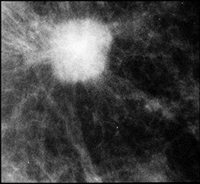
👉Breast hamartomas most likely result from an overgrowth of a portion of normal breast tissue, or from fibrous and glandular elements getting incorporated into a lipomatous growth.

👉Thus, they are sometimes called a “breast within a breast.”

👉They are also variously called adenolipofibroma, lipofibroadenoma, adenolipofibroma, and fibroadenolipoma, because they contain fibrous, fatty, and epithelial tissues.

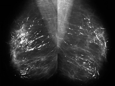
👉They may present as palpable masses, but are more commonly discovered on routine imaging.

👉They are round, oval, or lobulated and are usually well-circumscribed.

👉They have a mixture of water density and fatty elements and frequently have either a capsule or the appearance of a capsule resulting from surrounding compressed breast tissue.

👉A mammographically classic hamartoma does not require further imaging, short interval follow-up, or biopsy.

👉The presence of a breast hamartoma should prompt further questioning of the patient to be sure there is nothing to suggest she has multiple hamartoma syndrome (Cowden syndrome), a rare disorder caused by a deleterious mutation in the PTEN gene.

👉Patients with this disorder have an increased head circumference, multiple trichilemmoma skin lesions, intestinal hamartomas, and an increased risk of cancer of the breast, thyroid, endometrium and kidney.

👉If there is a personal or family history suggestive of the disorder, genetic testing should be done.

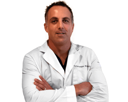
 👉Rodrigo Arrangoiz MS, MD, FACS cirujano oncology y cirujano de mamá de Sociedad Quirúrgica S.C en el America British Cowdray Medical Center en la ciudad de Mexico:

  • Es experto en el manejo del cáncer de mama.

 👉Es miembro de la American Society of Breast Surgeons:

Training:

• General surgery:

• Michigan State University:

• 2004 al 2010

• Surgical Oncology / Head and Neck Surgery / Endocrine Surgery:

• Fox Chase Cancer Center (Filadelfia):

• 2010 al 2012

• Masters in Science (Clinical research for health professionals):

• Drexel University (Filadelfia):

• 2010 al 2012

• Surgical Oncology / Head and Neck Surgery / Endocrine Surgery:

• IFHNOS / Memorial Sloan Kettering Cancer Center:

• 2014 al 2016

#Arrangoiz

#Surgeon

#Cirujano

#SurgicalOncologist

#CirujanoOncologo

#BreastSurgeon

#CirujanodeMama

#CancerSurgeon

#CirujanodeCancer

Simple Breast Cyst

👉The patient has a BI-RADS 2 simple cyst.

👉To be regarded as a simple cyst, a mass must meet three criteria.

1. The margins must be circumscribed, ie, a margin “that is well defined or sharp, with an abrupt transition between the lesion and surrounding tissue.”

2. It must be anechoic , ie, “without internal echoes.”

3. It must show posterior acoustical enhancement, (ie, “a column that is more echogenic deep to the mass.”

👉Simple cysts are almost never associated with cancer in the absence of other abnormalities seen on mammogram or ultrasound.

 👉Rodrigo Arrangoiz MS, MD, FACS cirujano oncology y cirujano de mamá de Sociedad Quirúrgica S.C en el America British Cowdray Medical Center en la ciudad de Mexico:

  • Es experto en el manejo del cáncer de mama.

 👉Es miembro de la American Society of Breast Surgeons:

Training:

• General surgery:

• Michigan State University:

• 2004 al 2010

• Surgical Oncology / Head and Neck Surgery / Endocrine Surgery:

• Fox Chase Cancer Center (Filadelfia):

• 2010 al 2012

• Masters in Science (Clinical research for health professionals):

• Drexel University (Filadelfia):

• 2010 al 2012

• Surgical Oncology / Head and Neck Surgery / Endocrine Surgery:

• IFHNOS / Memorial Sloan Kettering Cancer Center:

• 2014 al 2016

#Arrangoiz

#Surgeon

#Cirujano

#SurgicalOncologist

#CirujanoOncologo

#BreastSurgeon

#CirujanodeMama

#CancerSurgeon

#CirujanodeCancer

FISH-positive HER-2 testing results based on current American Society of Clinical Oncology / College of American Pathologists (ASCO/CAP) Guidelines

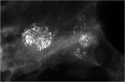
👉HER-2 expression in breast cancer is primarily assessed semiquantitatively by IHC.

👉The 2013 ASCO / CAP guidelines outline an IHC scoring method based on four classes:

– 0, 1+, 2+, and 3+.

– A score of 0 is negative, indicating no observed staining in invasive tumor cells.

-A score of 1+ is negative and indicates weak, incomplete membrane staining in any proportion of invasive tumor cells or weak, complete membrane staining in less than 10% of invasive tumor cells.

-A score of 2+ is equivocal, indicating circumferential membrane staining that is incomplete and/or weak/moderate and in more than 10% of invasive tumor cells or complete and circumferential membrane staining that is intense and in 10% or less of invasive tumor cells. All 2+ equivocal cases undergo subsequent testing by FISH.

-A score of 3+ is positive and indicates circumferential membrane staining that is complete and intense in a homogeneous and contiguous population, present in more than 10% of invasive tumor cells, and readily appreciated using a low-power objective. 

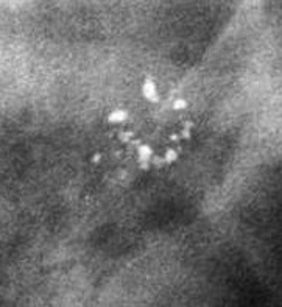
👉FISH is a sensitive and accurate method of scoring invasive breast tumor tissue for HER-2 expression.

👉Initial gene amplification studies by FISH assessment used chromosome 17 centromere (CEP17) or another gene on the same chromosome as an internal control, with a ratio of 2.0 or more considered evidence of HER-2 amplification.

👉These criteria were used as the cutoff for enrollment in trials evaluating HER-2 targeted therapies.

👉In 2007, the ASCO/CAP guidelines were changed to define HER-2 amplified as a ratio of 2.2 or more.

👉More recent guidelines have changed the ratio cutoff back to a ratio of 2.0 or more with the inclusion of criteria to account for HER-2 copy number per tumor cell.

👉Based on the recent guidelines, HER-2 is amplified in cases where the HER-2/CEP17 ratio is 2.0 or more with an average HER-2 copy number of less than 4.0 signals/cell or the HER-2/CEP17 ratio is less than 2.0 with an average HER-2 copy number of 6.0 or more signals/cell using a dual probe or a HER-2 copy number of 6.0 or more copies/cell using a single probe.

👉FISH testing is negative for HER-2 amplification with a HER-2/CEP17 ratio of less than 2.0 with an average HER-2 copy number of less than 4.0 signals/cell or an average HER-2 copy number of less than 4.0 signals/cell using a single probe.

 👉Rodrigo Arrangoiz MS, MD, FACS cirujano oncology y cirujano de mamá de Sociedad Quirúrgica S.C en el America British Cowdray Medical Center en la ciudad de Mexico:

  • Es experto en el manejo del cáncer de mama.

 👉Es miembro de la American Society of Breast Surgeons:

Training:

• General surgery:

• Michigan State University:

• 2004 al 2010

• Surgical Oncology / Head and Neck Surgery / Endocrine Surgery:

• Fox Chase Cancer Center (Filadelfia):

• 2010 al 2012

• Masters in Science (Clinical research for health professionals):

• Drexel University (Filadelfia):

• 2010 al 2012

• Surgical Oncology / Head and Neck Surgery / Endocrine Surgery:

• IFHNOS / Memorial Sloan Kettering Cancer Center:

• 2014 al 2016

#Arrangoiz

#Surgeon

#Cirujano

#SurgicalOncologist

#CirujanoOncologo

#BreastSurgeon

#CirujanodeMama

#CancerSurgeon

#CirujanodeCancer

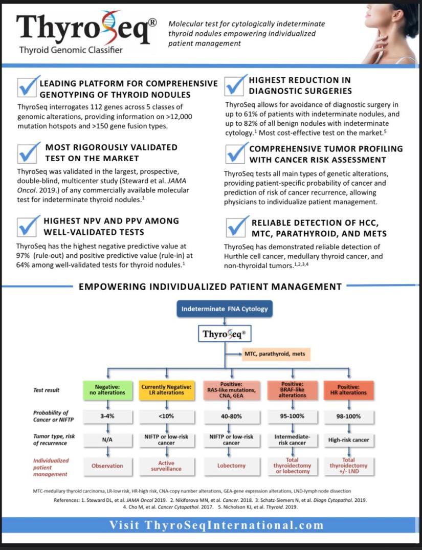
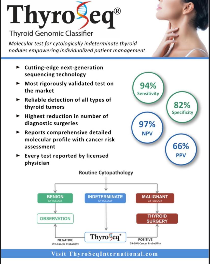
¡Ya Tenemos Thyroseq en México!

ab393bc5-542a-4d68-9e68-156a62075bb185e12396-e279-471a-b1ce-d2a7b530e18f

  • Para mayor informes de como solicitar el estudio por favor comunicarse por Inbox con el Dr. Rodrigo Arrangoiz o al teléfono 55 5432 5614


#Arrangoiz

#Thyroseq

#NódulosTiroideos

#CancerdeTiroides

#CirugiaTiroidea

#EstudioMolecular

#ThyroidCancer

#ThyroidNodules

#ThyroidSurgeon

#ThyroidExpert


Don’t Screen for Thyroid Problems in Asymptomatic Patients

👉The Canadian Task Force on Preventive Health Care has issued a strong recommendation against routine thyroid screening in nonpregnant adults without symptoms in the primary care setting, saying there is insufficient evidence of clinical benefit from the treatment of thyroid dysfunction in such individuals.

👉The guideline was published online November 17, 2019 in the Canadian Medical Association Journal (CMAJ) and was funded by the Public Health Agency of Canada.

👉”If you are a clinician who orders thyroid-stimulating hormone (TSH) tests as part of preventive health visits, we would like you to reconsider this practice.

👉The evidence isn’t there to suggest a health benefit for this type of screening as a routine part of care,” says Richard Birtwhistle, MD, chair of the Task Force Thyroid Dysfunction working group, in a press statement from CMAJ.

👉”Given the lack of clinical effectiveness and the burden on patients, including financial costs, screening patients without symptoms consumes resources that could be better used elsewhere,” added Birtwhistle, professor emeritus of family medicine and public health sciences at Queen’s University, in Kingston, Ontario, Canada.

👉This advice differs from the recommendations of other groups, as consensus on the issue is lacking.

👉”The recommendation represents a change in thinking about screening for thyroid dysfunction and contradicts other medical society recommendations that favor screening, particularly among older people,” write Juan Brito, MD, and Omar El Kawkgi, MD, of the Department of Endocrinology, Mayo Clinic, in Rochester, Minnesota.

👉Specifically, the position of the American Thyroid Association (ATA), published as joint guidelines with the American Association of Clinical Endocrinologists (AACE), is that screening for hypothyroidism in patients older than age 60 should be considered.

👉However, the US Preventive Services Task Force issued an updated guideline in 2015 recommending against screening for thyroid dysfunction based on insufficient evidence, they point out, and that position has also been endorsed by the American Academy of Family Physicians, editorialist El Kawkgi told Medscape Medical News.

#Arrangoiz

#Surgeon

#Cirujano

#SurgicalOncologist

#CirujanoOncologo

Surgery and Art

👉Surgery is a collision of Anatomy, Physiology, Pathology, Pharmacology, Psychology, Sociology, Engineering, Technology and just as importantly, Art.

👉Dr E. Levi

#Arrangoiz

#Surgeon

#Cirujano

#Teacher

#Profesor

#CancerSurgeon

#HeadandNeckSurgeon

#CirujanodeCabezayCuello

#SurgicalOncologist

#CirujanoOncologo

#BreastSurgeon

#CirujanodeMama

#CirujanodeCancer

Evaluation of Solid Ultrasound Breast Nodules

👉 The American College of Radiology BI-RADS classification subdivides:

– BI-RADS 0 incomplete evaluation, requieres additional imaging evaluation

– BI-RADS 1 normal, which has a 0% risk of malignancy

– BI-RADS 2 benign finding, which has a 0% risk of malignancy

– BI-RADS 3 probably benign, with a ≤2 risk of malignancy

– BI-RADS 4a mildly suspicious, which has a 2% to 10 % risk of malignancy,

– BI-RADS 4b moderately suspicious, which has 10% to 50 % risk of malignancy

– BI-RADS 4c highly suspicious, which has a 50% to 95% risk of malignancy

– BI-RADS 5 malignant, has 95% or greater chance of malignancy

👉 In evaluating a solid sonographic nodule:

– One should first look for any of the 10 signs of malignancy, and if even one of them is present, the lesion cannot be considered BI-RADS 3.

👉 The 10 signs of malignancy are:

– Shadowing

– Hypoechoic echotexture

– Spiculation

– Angular margins

– Thick echogenic halo

– Microlobulation

– Taller than wide

– Duct extension

– Branching pattern

– Calcifications

👉 It should be noted that Stavros compares the echogenicity of lesions to that of breast fat, not breast parenchyma:

– Therefore, a lesion with hypoechoic echotexture would be very hypoechoic if breast parenchyma is used as the reference

👉👉 Rodrigo Arrangoiz MS, MD, FACS cirujano oncology y cirujano de mamá de Sociedad Quirúrgica S.C en el America British Cowdray Medical Center en la ciudad de Mexico:

  • Es experto en el manejo del cáncer de mama.

👉 Es miembro de la American Society of Breast Surgeons:

Training:

• General surgery:

• Michigan State University:

• 2004 al 2010

• Surgical Oncology / Head and Neck Surgery / Endocrine Surgery:

• Fox Chase Cancer Center (Filadelfia):

• 2010 al 2012

• Masters in Science (Clinical research for health professionals):

• Drexel University (Filadelfia):

• 2010 al 2012

• Surgical Oncology / Head and Neck Surgery / Endocrine Surgery:

• IFHNOS / Memorial Sloan Kettering Cancer Center:

• 2014 al 2016

#Arrangoiz

#Surgeon

#Cirujano

#SurgicalOncologist

#CirujanoOncologo

#BreastSurgeon

#CirujanodeMama

#CancerSurgeon

#CirujanodeCancer

Management of the Axilla in Clinically Node-Positive Patients Following Neoadjuvant Chemotherapy (NAC)

cancer-of-the-vulva-38-638

  • Management of the axilla in clinically node-positive (cN1) patients following neoadjuvant chemotherapy (NAC) has evolved significantly over the past several years:
    • Historically:
      • The use of sentinel lymph node biopsy (SLNB) in initially node-positive patients after NAC was avoided because available data demonstrated :
        • False-negative rates approaching 25%:
          • Therefore, all patients were treated with axillary lymph node dissection post-NAC regardless of response to treatment.
    • Three recently published prospective studies evaluated the accuracy of sentinel node biopsy following NAC in patients presenting with node-positive disease:
      • Among 525 patients enrolled in the ACOSOG Z1071 trial with at least two sentinel nodes removed:
        • The false-negative rate was 12.6%>
          • However:
            • The false-negative rate was reduced to:
              • 10.8% when the SNB was performed with dual mapping (radioactive tracer and blue dye)
              • 9.1% when 3 or more sentinel nodes were removed.
      • Similarly, the overall false-negative rate among 226 clinically node-positive patients enrolled in the SENTinel NeoAdjuvant (SENTINA) prospective study:
        • Was 14.2%:
          • Which decreased to 8.6% with the use of dual mapping
          • 7.3% when 3 or more sentinel nodes were retrieved.
      • Although a false-negative rate of less than 10% is acceptable in the adjuvant setting:
        • Additional methods to decrease false-negative rates in this population are of clinical interest:
          • As the long-term consequences of leaving behind potentially “chemotherapy-resistant” cells are unknown:
            • One method to potentially improve the accuracy of SNB in this setting is:
              • To clip the cN1 positive node:
                • Then post-NAC localize the node and ensure removal of the clipped positive node at the time of SNB.
                • In a subset analysis of patients enrolled in the ACOSOG Z1071 trial with cN1 disease and at least two sentinel nodes removed, the false-negative rate of sentinel node biopsy was 6.8% when the clipped node was one of the sentinel nodes, 19% when the clipped node was found in the axillary dissection specimen, and identified, compared to 14.3% when the clip was placed but location was unknown.
                • The false-negative rate was 13.4% when no clip was placed.
        • Caudle et al. recently reviewed their experience with SNB and retrieval of the clipped node, also known as targeted axillary dissection, in clinically node-positive patients receiving NAC:
          • Among 85 patients who underwent targeted axillary dissection followed by axillary lymph node dissection, the false-negative rate was 2.0% compared to 10.6% for SNB alone in this cohort (p=0.13).
          • Importantly however, the mean number of sentinel lymph nodes removed in this cohort was 2.7 and dual tracer mapping was only used in 55% of the patients.
            • Further study of this technique is ongoing.
        • Other methods to minimize false-negative rates include documentation of treatment effect in the node:
          • Which may also serve as a surrogate that the original positive node(s) has/have been retrieved.
        • Although controversy exists as to the best method to evaluate the axilla post-NAC in patients presenting with node-positive disease:
          • SNB with localization of the clipped node and/or SNB with dual mapping and retrieval of 3 or more sentinel nodes are both acceptable approaches to the axilla following NAC.

REFERENCES

  1. Boileau JF, Poirier B, Basik M, et al. Sentinel node biopsy after neoadjuvant chemotherapy in biopsy-proven node-positive breast cancer: the SN FNAC study. J Clin Oncol. 2015:20;33:258-264. http://www.ncbi.nlm.nih.gov/pubmed/25452445
  2. Boughey JC, Suman VJ, Mittendorf EA, et al; Alliance for Clinical Trials in Oncology. Factors affecting sentinel lymph node identification rate after neoadjuvant chemotherapy for breast cancer patients enrolled in ACOSOG Z1071 (Alliance). Ann Surg. 2015;261:547-552. https://www.ncbi.nlm.nih.gov/pubmed/25664534
  3. Boughey JC, Suman VJ, Mittendorf EA, et al; Alliance for Clinical Trials in Oncology. Sentinel lymph node surgery after neoadjuvant chemotherapy in patients with node-positive breast cancer: the ACOSOG Z1071 (Alliance) clinical trial. JAMA. 2013;310:1455-1461. http://www.ncbi.nlm.nih.gov/pubmed/24101169
  4. Caudle AS, Yang WT, Krishnaumurthy S, et al. Improved axillary evaluation following neoadjuvant therapy for patients with node-positive breast cancer using selective evaluation of clipped nodes: implementation of targeted axillary dissection. J Clin Oncol. 2016;34:1072-1078. https://www.ncbi.nlm.nih.gov/pubmed/26811528
  5. Kuehn T, Bauerfeind I, Fehm T, et al. Sentinel-lymph-node biopsy in patients with breast cancer before and after neoadjuvant chemotherapy (SENTINA): a prospective, multicentre cohort study. Lancet Oncol. 2013;14:609-618. http://www.ncbi.nlm.nih.gov/pubmed/23683750
  6. Shen J, Gilcrease MZ, Babiera GV, et al. Feasibility and accuracy of sentinel node biopsy after preoperative chemotherapy in breast cancer patients with documented axillary metastases. Cancer. 2007;109:1255-1263. https://www.ncbi.nlm.nih.gov/pubmed/17330229

👉Rodrigo Arrangoiz MS, MD, FACS cirujano oncology y cirujano de mamá de Sociedad Quirúrgica S.C en el America British Cowdray Medical Center en la ciudad de Mexico:
  • Es experto en el manejo del cáncer de mama.

👉Es miembro de la American Society of Breast Surgeons:

Training:

• General surgery:

• Michigan State University:

• 2004 al 2010

• Surgical Oncology / Head and Neck Surgery / Endocrine Surgery:

• Fox Chase Cancer Center (Filadelfia):

• 2010 al 2012

• Masters in Science (Clinical research for health professionals):

• Drexel University (Filadelfia):

• 2010 al 2012

• Surgical Oncology / Head and Neck Surgery / Endocrine Surgery:

• IFHNOS / Memorial Sloan Kettering Cancer Center:

• 2014 al 2016

#Arrangoiz

#Surgeon

#Cirujano

#SurgicalOncologist

#CirujanoOncologo

#BreastSurgeon

#CirujanodeMama

#CancerSurgeon

#CirujanodeCancer

Decision Making – The Science Behind Been a Surgeon

👉Rodrigo Arrangoiz MS, MD, FACS cirujano oncology y cirujano de mamá de Sociedad Quirúrgica S.C en el America British Cowdray Medical Center en la ciudad de Mexico:
  • Es experto en el manejo del cáncer de mama.

👉Es miembro de la American Society of Breast Surgeons:

Training:

• General surgery:

• Michigan State University:

• 2004 al 2010

• Surgical Oncology / Head and Neck Surgery / Endocrine Surgery:

• Fox Chase Cancer Center (Filadelfia):

• 2010 al 2012

• Masters in Science (Clinical research for health professionals):

• Drexel University (Filadelfia):

• 2010 al 2012

• Surgical Oncology / Head and Neck Surgery / Endocrine Surgery:

• IFHNOS / Memorial Sloan Kettering Cancer Center:

• 2014 al 2016

#Arrangoiz

#Surgeon

#Cirujano

#SurgicalOncologist

#CirujanoOncologo

#BreastSurgeon

#CirujanodeMama

#CancerSurgeon

#CirujanodeCancer