Mammographic Images of Breast Cancer

Figure 1 Powdery Calcifications
Figure 2 Crushed Stone Type Calcifications
Figure 3 Spiculated Mass
Figure 4 Round Tumor

👉The cancers that caused all four of the mammographic abnormalities have the word “ductal” associated with them (Figure 1 and Figure 2: grade 1 and 2 “ductal carcinoma in situ,” Figure 3 and Figure 4 invasive ductal cancers), but it is now thought that 75% of all breast cancers actually originate in the acini of the lobules, and they have a better long-term prognosis than cancers that originate in the lactiferous ducts.

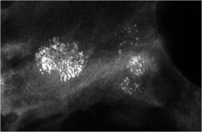
👉The powdery calcifications (Figure 1) are caused by a process most pathologists would term low nuclear grade ductal carcinoma in situ.

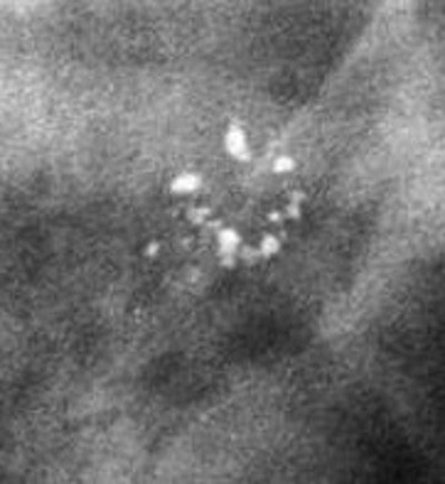
👉The “crushed stone” type calcifications (Figure 2) are caused by what would be termed intermediate nuclear grade ductal carcinoma in situ.

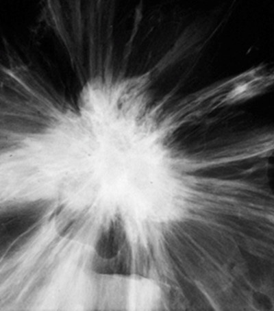
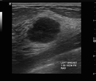
👉The spiculated mass (Figure 3) is the mammographic image of a tumor that would be termed low or intermediate grade infiltrating ductal carcinoma.

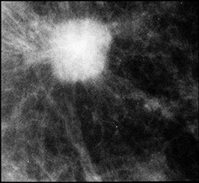
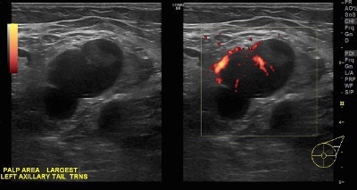
👉The round mass (Figure 4) is caused by a process called high-grade infiltrating ductal carcinoma, but it could also be a mucinous carcinoma or an invasive cribriform cancer.

👉Even the high-grade invasive carcinoma has an excellent prognosis if it is screen-detected when it is 14 mm or smaller in size.

👉Since 75% of all breast cancers (described above) originate within the acini of the breast lobules (terminal ductal lobular units), it is misleading to term them “ductal” in situ or invasive “ductal” carcinoma.

👉About 20% of all breast cancers originate in the major (lactiferous) ducts. These are diffuse from the outset since the ducts and their branches are usually filled with grade 3 cancer cells.

👉The graph shows the 25-year follow-up of patients in the Swedish Two-County mammographic screening trial segregated according to mammographic presentation.

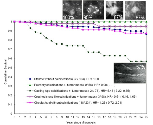
👉The cancers in the group with the poor prognosis originated in the ducts, were diffuse at the time of diagnosis, and had extensive ductal (casting type) calcifications.

👉The similarity of breast and prostate histology is known to all first-year medical students.

👉For years pathologists have classified prostate cancer by its anatomic site of origin.

👉Cancers that begin in the acini are called acinar adenocarcinoma of the prostate and have an excellent prognosis.

👉Those that begin in the ducts are called ductal adenocarcinoma of the prostate and have a poor prognosis.

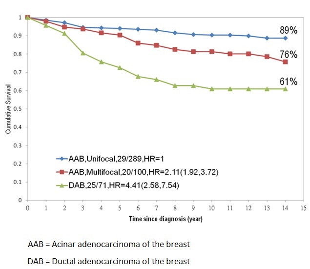
👉In recent years, Tabár and colleagues have called attention to a similar pattern of origin and prognosis for carcinomas of the breast, coining the terms acinar adenocarcinoma of the breast and ductal adenocarcinoma of the breast.

👉The TNM classification system does not always accurately reflect the prognosis of some breast cancers.

👉A 9-mm infiltrating carcinoma would have the same TNM classification regardless of whether it was a unifocal lesion, an extensive multifocal lesion with the largest focus being 9 mm, or a 9-mm infiltrating carcinoma associated with extensive casting calcifications, but the prognosis of each would be markedly different (Graph).

👉Rodrigo Arrangoiz MS, MD, FACS cirujano oncology y cirujano de mamá de Sociedad Quirúrgica S.C en el America British Cowdray Medical Center en la ciudad de Mexico:

  • Es experto en el manejo del cáncer de mama.

 👉Es miembro de la American Society of Breast Surgeons:

Training:

• General surgery:

• Michigan State University:

• 2004 al 2010

• Surgical Oncology / Head and Neck Surgery / Endocrine Surgery:

• Fox Chase Cancer Center (Filadelfia):

• 2010 al 2012

• Masters in Science (Clinical research for health professionals):

• Drexel University (Filadelfia):

• 2010 al 2012

• Surgical Oncology / Head and Neck Surgery / Endocrine Surgery:

• IFHNOS / Memorial Sloan Kettering Cancer Center:

• 2014 al 2016

#Arrangoiz

#Surgeon

#Cirujano

#SurgicalOncologist

#CirujanoOncologo

#BreastSurgeon

#CirujanodeMama

#CancerSurgeon

#CirujanodeCancer

Axillary Lymph Nodes on Ultrasound

👉A normal axillary lymph node is elliptical in shape (or bean shaped) and has a narrow, symmetrical, hypoechoic cortex surrounding an isoechoic to hyperechoic fatty hilum (Image).

👉The cortex of a normal node is composed largely of lymphatic tissue and fluid-filled cortical sinuses, thus the hypoechoic echogenicity.

👉The hilum contains alternating medullary cords and sinusoids that have innumerable acoustic interfaces, thus the higher degree of echogenicity.

👉When a lymph node is completely replaced with metastatic cancer, it will be rounded, hypoechoic, and the hilum will be completely obliterated. (Image).

👉Before the node reaches the stage of complete replacement, it can have an asymmetric, thickened cortex, with an eccentric hilum (Image).

👉Another presentation of a metastatic node is an asymmetric cortex where the tumor can be seen invading the hilum with convex indentations that look like “rat bites” (Image).

👉A metastatic node can also have severe compression of a central hilum resulting in a slit-like central hyperechoic band (Image).

👉Reactive nodes can be difficult to distinguish from metastatic nodes (Image).

👉In general, benign causes of nodal enlargement tend to thicken the cortex diffusely.

👉In addition, it is not uncommon to have a metastatic node adjacent to a normal node, but all nodes in a region tend to be reactive when the cause is benign.

👉Finally, a reactive node has blood supply on Doppler examination through a single hilum, whereas metastatic nodes tend to have multiple transcapsular vessels (Image).

👉Rodrigo Arrangoiz MS, MD, FACS cirujano oncology y cirujano de mamá de Sociedad Quirúrgica S.C en el America British Cowdray Medical Center en la ciudad de Mexico:

  • Es experto en el manejo del cáncer de mama.

 👉Es miembro de la American Society of Breast Surgeons:

Training:

• General surgery:

• Michigan State University:

• 2004 al 2010

• Surgical Oncology / Head and Neck Surgery / Endocrine Surgery:

• Fox Chase Cancer Center (Filadelfia):

• 2010 al 2012

• Masters in Science (Clinical research for health professionals):

• Drexel University (Filadelfia):

• 2010 al 2012

• Surgical Oncology / Head and Neck Surgery / Endocrine Surgery:

• IFHNOS / Memorial Sloan Kettering Cancer Center:

• 2014 al 2016

#Arrangoiz

#Surgeon

#Cirujano

#SurgicalOncologist

#CirujanoOncologo

#BreastSurgeon

#CirujanodeMama

#CancerSurgeon

#CirujanodeCancer


Check out these Thyroid-related articles

A Novel Technique for Performing Transoral Endoscopic Thyroidectomy Vestibular Approach (TOETVA): A Single-port Platform. Park JH, Bilegsaikhan SE, Suh YJ. Surg Laparosc Endosc Percutan Tech. 2019 Nov 26. PMID: 31790081 https://www.ncbi.nlm.nih.gov/pubmed/31790081

Subset analysis of the Japanese risk classification guidelines for papillary thyroid carcinoma. Ito Y, Miyauchi A, Yamamoto M, Masuoka H, Higashiyama T, Kihara M, Miya A. Endocr J. 2019 Nov 27. PMID: 31776303 https://www.ncbi.nlm.nih.gov/pubmed/31776303

American Thyroid Association American Association of Clinical Endocrinologists Endocrine Society ThyCa, Inc. Graves’ Disease and Thyroid Foundation

#ThyroidExpert

#Arrangoiz

#ThyroidSurgeon

#HeadandNeckSurgeon

#ThyroidCancer

#Teacher

#CancerSurgeon

Check out these Parathyroid-related articles

Increased risk for tooth extraction in primary hyperparathyroidism and hypercalcemia: a population study. Koman A, Näsman P, Discacciati A, Ekbom A, Nilsson IL, Sandborgh-Englund G. Clin Oral Investig. 2019 Dec 2. PMID: 31792613 https://www.ncbi.nlm.nih.gov/pubmed/31792613

Metabolic profile of human parathyroid adenoma. di Masi A, Leboffe L, Sodo A, Tabacco G, Cesareo R, Sbroscia M, Giovannoni I, Taffon C, Crucitti P, Longo F, Manfrini S, Ricci MA, Ascenzi P, Crescenzi A, Palermo A. Endocrine. 2019 Nov 30. PMID: 31786773 https://www.ncbi.nlm.nih.gov/pubmed/31786773

Long-term remission of disseminated parathyroid cancer following immunotherapy. Sarquis M, Marx SJ, Beckers A, Bradwell AR, Simonds WF, Bicalho MAC, Daly AF, Betea D, Friedman E, De Marco L. Endocrine. 2019 Nov 28. PMID: 31782130 https://www.ncbi.nlm.nih.gov/pubmed/31782130

Effects of parathyroidectomy on tumoral calcinosis in uremic patients with secondary hyperparathyroidism. Wang J, Zeng M, Yang G, Huang Y, Wu B, Guo J, Wang N, Xing C. BMC Surg. 2019 Sep 11;19(1):133. PMID: 31510980 https://www.ncbi.nlm.nih.gov/pubmed/31510980

#Arrangoiz

#ParathyroidExpert

#ParathyroidSurgeon

#Parathyroid

#Hyperparathyroidism

Ultrasonographic Image of a Medullary Breast Carcinoma

👉The sonographic image in this patient shows a fairly round, well-circumscribed, microlobulated lesion with posterior enhancement.

👉The most reliable sonographic feature of medullary carcinoma is enhanced through transmission.

👉This results from the highly cellular lesion with a paucity of desmoplastic fibrous tissue, in addition to areas of cystic necrosis and hemorrhage.

👉Medullary carcinomas are classically well-circumscribed, but most of them have some area of angularity.

👉High-grade, colloid (mucinous), and papillary carcinomas typically have a sonographic appearance similar to medullary carcinoma.

👉Rodrigo Arrangoiz MS, MD, FACS cirujano oncology y cirujano de mamá de Sociedad Quirúrgica S.C en el America British Cowdray Medical Center en la ciudad de Mexico:

  • Es experto en el manejo del cáncer de mama.

 👉Es miembro de la American Society of Breast Surgeons:

Training:

• General surgery:

• Michigan State University:

• 2004 al 2010

• Surgical Oncology / Head and Neck Surgery / Endocrine Surgery:

• Fox Chase Cancer Center (Filadelfia):

• 2010 al 2012

• Masters in Science (Clinical research for health professionals):

• Drexel University (Filadelfia):

• 2010 al 2012

• Surgical Oncology / Head and Neck Surgery / Endocrine Surgery:

• IFHNOS / Memorial Sloan Kettering Cancer Center:

• 2014 al 2016

#Arrangoiz

#Surgeon

#Cirujano

#SurgicalOncologist

#CirujanoOncologo

#BreastSurgeon

#CirujanodeMama

#CancerSurgeon

#CirujanodeCancer

Mastografía

👉Una mamografía es una imagen de la mama tomada con rayos X. Los médicos usan las mamografías para buscar signos de cáncer de mama en sus etapas iniciales. Las mamografías habituales son las mejores pruebas con que cuentan los médicos para detectar el cáncer de mama en sus etapas iniciales, a veces hasta tres años antes de que se pueda sentir.

¿Le preocupa el costo? Los CDC ofrece mamografías gratuitas o a muy bajo costo. 

¿Cómo se hace una mamografía?

👉Usted se colocará de pie frente a una máquina especial de rayos X. Una tecnóloga colocará su mama sobre una placa de plástico transparente. Después cubrirá esa mama con otra placa, presionando firmemente. Las placas aplanarán la mama y la mantendrán inmóvil mientras se toma la radiografía. Usted sentirá algo de presión. Los pasos se repiten para tomar una imagen del costado de la mama. Posteriormente, le sacarán una radiografía a la otra mama de la misma forma. Deberá esperar a que la tecnóloga revise las cuatro radiografías para asegurarse de que no haya necesidad de volver a tomarlas. Tenga presente que la tecnóloga no puede decirle los resultados de su mamografía. Las mamografías de cada mujer pueden verse un poco distintas porque no hay mujeres con mamas idénticas.

¿Qué se siente al hacerse una mamografía?

👉Para la mayoría de las mujeres, hacerse una mamografía es un proceso molesto. Algunas mujeres lo encuentran doloroso. Sin embargo, una mamografía solo toma unos minutos y las molestias desaparecen pronto. Lo que usted sienta depende de la destreza de la tecnóloga, el tamaño de las mamas y la cantidad de presión que se les tenga que aplicar. Sus mamas pueden estar más sensibles si tiene la menstruación o está a punto de tenerla. Un médico con capacitación especial, llamado radiólogo, interpretará la mamografía. Ese profesional revisará la radiografía en busca de signos de cáncer de mama en sus etapas iniciales u otros problemas.

👉Consejos para cuando se haga una mamografía
  • Intente no hacerse una mamografía la semana previa a su menstruación o mientras la tenga. Las mamas pueden estar sensibles al tacto o hinchadas en esos momentos.
  • El día de la mamografía, no se pongan desodorante, perfume ni talco. Estos productos pueden aparecer en la radiografía como manchas blancas.
  • Algunas mujeres prefieren usar una blusa con falda o pantalón en vez de un vestido. Tendrá que desvestirse de la cintura para arriba para hacerse la mamografía.

¿Cuándo tendré los resultados de mi mamografía?

👉Por lo general, recibirá los resultados dentro de unas cuantas semanas, aunque eso depende del lugar donde se haya hecho la mamografía. El radiólogo interpretará la mamografía y le enviará los resultados a usted y a su médico. Si hay algún motivo para preocuparse, el centro donde se haya hecho la mamografía se pondrá en contacto con usted antes del plazo previsto. Comuníquese con su proveedor de atención médica o con el centro donde se haya hecho la mamografía si no recibe un informe con los resultados dentro de 30 días después de la prueba.

¿Qué pasa si la mamografía es normal?

👉Continúe haciéndose las mamografías de acuerdo con las recomendaciones para su caso. Las mamografías son más útiles si pueden compararse con las que se hayan hecho antes. Esto le permite al radiólogo compararlas para detectar cambios en sus mamas.

Ejemplo de una mamografía con resultados normales

👉Un ejemplo de una mamografía normal. En virtud de que no hay mujeres con mama idénticas, las mamografías de cada mujer pueden verse un poco distintas.

¿Qué pasa si la mamografía es anormal?

👉Una mamografía anormal no siempre significa que haya cáncer. Sin embargo, le tendrán que hacer mamografías, pruebas o exámenes adicionales para que su médico pueda tener alguna certeza. También es posible que la remitan a un médico especialista en mamas o a un cirujano. Esto no significa necesariamente que usted tenga cáncer o que necesite una cirugía. Estos médicos son expertos en el diagnóstico de problemas mamarios. Los médicos harán pruebas de seguimiento para diagnosticar cáncer de mama o para determinar que no hay cáncer.

👉Rodrigo Arrangoiz MS, MD, FACS cirujano oncology y cirujano de mamá de Sociedad Quirúrgica S.C en el America British Cowdray Medical Center en la ciudad de Mexico:

  • Es experto en el manejo del cáncer de mama.

 👉Es miembro de la American Society of Breast Surgeons:

Training:

• General surgery:

• Michigan State University:

• 2004 al 2010

• Surgical Oncology / Head and Neck Surgery / Endocrine Surgery:

• Fox Chase Cancer Center (Filadelfia):

• 2010 al 2012

• Masters in Science (Clinical research for health professionals):

• Drexel University (Filadelfia):

• 2010 al 2012

• Surgical Oncology / Head and Neck Surgery / Endocrine Surgery:

• IFHNOS / Memorial Sloan Kettering Cancer Center:

• 2014 al 2016

#Arrangoiz

#Surgeon

#Cirujano

#SurgicalOncologist

#CirujanoOncologo

#BreastSurgeon

#CirujanodeMama

#CancerSurgeon

#CirujanodeCancer

BRAFV600E mutation: a potential predictor of more than a Sistrunk’s procedure in patients with thyroglossal duct cyst carcinoma and a normal thyroid gland

👉To assess the utility of mutational markers in determining the most appropriate initial surgery for patients with thyroglossal duct cyst carcinoma (TGDCCa) and a normal thyroid gland.

👉Our sample comprised 15 patients with a diagnosis of TGDCCa and a thyroid gland histologically negative for any malignant involvement, who underwent surgery between the years 1994 and 2017.

👉Clinical records were reviewed and tissue specimens were genetically tested for the presence of the most commonly encountered mutational markers in differentiated thyroid cancer: BRAF, N-RAS, and H-RAS.

👉The primary outcome of interest was the correlation between mutational marker positivity and the T-stage of the primary tumor and its potential implication on therapeutic decision making.

👉All 15 cases were papillary carcinomas with a mean tumor size of 17 mm (2–40 mm). According to the 7th edition of the American Joint Committee on Cancer TNM staging system, these represented: T (n= 3), T (n= 1), and T (n= 11). Cancerous invasion of the pericystic soft tissue and/or hyoid bone was considered T3. BRAF was the only mutational marker identified (7 in 15 cases). All BRAF-positive lesions were T, necessitating radioactive iodine ablation (RIA) therapy, therefore, total thyroidectomy. The correlation between BRAF positivity and extracystic cancerous extension was statistically significant [1.0 (7/7) vs. 0.5 (4/8); p value = 0.0035]. BRAF positivity seems to be predictive of locally advanced disease mandating RIA therapy.

👉Therefore, it could serve as a preoperative tool that predicts the need for total thyroidectomy, in addition to Sistrunk’s procedure.

#Arrangoiz

#ThyroidSurgeon

#ThyroidExpert

#Surgeon

#HeadandNeckSurgeon

Parathyroid Related Articles

Check out these Parathyroid-related articles published online or in print last week!

The Significance of Histologically “Large Normal” Parathyroid Glands in Primary Hyperparathyroidism. Krawitz R, Glover A, Koneru S, Jiang J, Di Marco A, Gill AJ, Aniss A, Sywak M, Delbridge L, Sidhu S. World J Surg. 2019 Nov 26. PMID: 31773224 https://www.ncbi.nlm.nih.gov/pubmed/31773224

Robust, quick, and convenient intraoperative method to differentiate parathyroid tissue. Kikumori T, Inaishi T, Miyajima N, Shibata M, Takeuchi D. Surgery. 2019 Nov 18. PMID: 31753324 https://www.ncbi.nlm.nih.gov/pubmed/31753324

PRIMARY HYPERPARATHYROIDISM DURING PREGNANCY: A CASE SERIES OF 8 PATIENTS. Song A, Wang W, Chen S, Wang Y, Liu S, Nie M, Jiang Y, Li M, Xia W, Meng X, Xing X, Wang O. Endocr Pract. 2019 Nov;25(11):1127-1136. PMID: 31414909 https://www.ncbi.nlm.nih.gov/pubmed/31414909

Timing of Parathyroidectomy Does Not Influence Renal Function After Kidney Transplantation. van der Plas WY, El Moumni M, von Forstner PJ, Koh EY, Dulfer RR, van Ginhoven TM, Rotmans JI, Appelman-Dijkstra NM, Schepers A, Hoorn EJ, Plukker JTM, Vogt L, Engelsman AF, Nieveen van Dijkum EJM, Kruijff S, Pol RA, de Borst MH; Dutch Hyperparathyroidism Study Group. World J Surg. 2019 Aug;43(8):1972-1980. PMID: 30798418 https://www.ncbi.nlm.nih.gov/pubmed/30798418

American Association of Clinical Endocrinologists Endocrine Society

Rodrigo Arrangoiz MS, MD, FACS a head and neck surgeon / endocrine surgeon / surgical oncologist and is a member of Sociedad Quirúrgica S.C at the America British Cowdray Medical Center in Mexico City:


prof_739_20190417135234

Training:

• General surgery:

• Michigan State University:

• 2004 al 2010

• Surgical Oncology / Head and Neck Surgery / Endocrine Surgery:

• Fox Chase Cancer Center (Filadelfia):

• 2010 al 2012

• Masters in Science (Clinical research for health professionals):

• Drexel University (Filadelfia):

• 2010 al 2012

• Surgical Oncology / Head and Neck Surgery / Endocrine Surgery:

• IFHNOS / Memorial Sloan Kettering Cancer Center:

• 2014 al 2016

#Arrangoiz

#Teacher

#Surgeon

Rodrigo Arrangoiz MS, MD, FACS Surgeon

👉Rodrigo Arrangoiz MS, MD, FACS cirujano oncology y cirujano de mamá de Sociedad Quirúrgica S.C en el America British Cowdray Medical Center en la ciudad de Mexico:

  • Es experto en el manejo del cáncer de mama.

👉👉Es miembro de la American Society of Breast Surgeons:

Training:

• General surgery:

• Michigan State University:

• 2004 al 2010

• Surgical Oncology / Head and Neck Surgery / Endocrine Surgery:

• Fox Chase Cancer Center (Filadelfia):

• 2010 al 2012

• Masters in Science (Clinical research for health professionals):

• Drexel University (Filadelfia):

• 2010 al 2012

• Surgical Oncology / Head and Neck Surgery / Endocrine Surgery:

• IFHNOS / Memorial Sloan Kettering Cancer Center:

• 2014 al 2016

#Arrangoiz

#Surgeon

#Cirujano

#SurgicalOncologist

#CirujanoOncologo

#BreastSurgeon

#CirujanodeMama

#CancerSurgeon

#CirujanodeCancer

Rodrigo Arrangoiz MS, MD, FACS

👉Rodrigo Arrangoiz MS, MD, FACS es especialista en Cirugia de Mama / Cirugía de Tumores de Cabeza y Cuello / Cirugía Endocrina y Cirugía Oncológica compleja.

👉Fue egresado como Médico General de la Universidad Anahuac Suma Cum Laude.

👉Es miembro de Sociedad Quirúrgica S.C. que es el único grupo quirúrgico en México en donde todos los socios se entrenaron en las mejores instituciones académicas de los Estados Unidos de América. 

👉El Doctor Arrangoiz es experto en el manejo del Cáncer de Tiroides, Patología Quirúrgica de Tiroides, Hiperparatiroidismo Primario, Tumores de Cabeza y Cuello y Cáncer de Mama. 

👉Fue entrenado en las mejores instituciones académicas de los Estados Unidos y mantiene certificaciones por los Consejos de Cirugía General y Cirugía Oncológica en México y en los Estados Unidos de América. 
 

 👉Es miembro de la American Society of Breast Surgeons:

👉Es miembro de la American Thyroid Association:

👉Su entrenamiento es el siguiente:

• General surgery:

• Michigan State University:

• 2004 al 2010

• Surgical Oncology / Head and Neck Surgery / Endocrine Surgery:

• Fox Chase Cancer Center (Filadelfia):

• 2010 al 2012

• Masters in Science (Clinical research for health professionals):

• Drexel University (Filadelfia):

• 2010 al 2012

• Surgical Oncology / Head and Neck Surgery / Endocrine Surgery:

• IFHNOS / Memorial Sloan Kettering Cancer Center:

• 2014 al 2016

#Arrangoiz

#Surgeon

#Cirujano

#SurgicalOncologist

#CirujanoOncologo

#BreastSurgeon

#CirujanodeMama

#CancerSurgeon

#CirujanodeCancer