Autofluirescence Imaging of Parathyroid Glands

👉Interested in the utility of autofluorescence imaging for parathyroid glands?

👉Check out this study: https://www.sciencedirect.com/science/article/pii/S0039606019305604?via%3Dihub

#Arrangoiz

#Surgeon

#ParathyroidSurgeon

#EndocrineSurgery

#HeadandNeckSurgeon

Conferencia Magistral – Dr. Yuri Nikiforov – Utilidad de las Pruebas Moleculares en los Nódulos Tiroideos Indeterminados

  • Se va llevar acabo en el Auditorio A del Hospital ABC de Santa Fe
  • También va ser transmitido al Hospital ABC Observatorio.
    • La conferencia va ser de 10 am a 12 pm
    • Sin costo
    • Cupo Limitado
    • Dirigida a médicos, residentes, enfermeras que manejen patología de la glándula tiroides.

img_4687

#Arrangoiz

#HeadandNeckSurgeon

#ThyroidSurgeon

#ThyoidExpert

#ThyroidNodules

#EndocrineSurgery


Evaluation of a Breast Nodule by Ultrasound

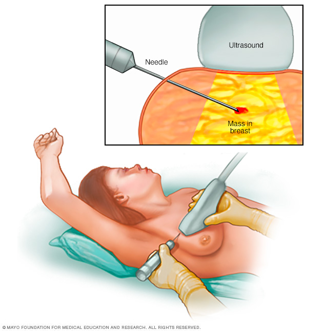
    1. You first evaluate the lesion for any of the ten malignant signs identified on ultrasound:
    • Shadowing
    • Hypoechoic echotexture
    • Spiculation
    • Angular margins
    • Thick echogenic capsule
    • Taller than wider
    • Microlobulation
    • Duct extension
    • Branching pattern
    • Calcifications

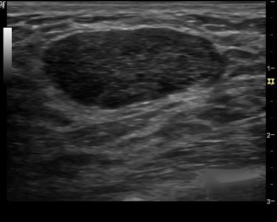
2. Finding none, you move on to the second step in the evaluation process and specifically look for one of the three strictly defined benign signs, and if any of them is found, the lesion can be considered BI-RADS 3.

      • The three benign findings defined by Stavros are:
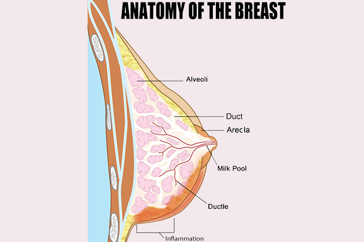
        1. A purely hyperechoic lesion with no hypoechoic area larger than a normal duct or lobule.
        2. Elliptical, wider than tall, well-circumscribed and thin echogenic capsule.
        3. Gently lobulated, wider than tall, well-circumscribed and thin echogenic capsule.

– Combining the elliptical or gently lobulated shapes with the presence of a complete, thin echogenic capsule is necessary because many circumscribed carcinomas and most ductal carcinoma in situ are encompassed in a thin, echogenic capsule.

👉Rodrigo Arrangoiz MS, MD, FACS cirujano oncology y cirujano de mamá de Sociedad Quirúrgica S.C en el America British Cowdray Medical Center en la ciudad de Mexico:

  • Es experto en el manejo del cáncer de mama.

👉Es miembro de la American Society of Breast Surgeons:

Training:

• General surgery:

• Michigan State University:

• 2004 al 2010

• Surgical Oncology / Head and Neck Surgery / Endocrine Surgery:

• Fox Chase Cancer Center (Filadelfia):

• 2010 al 2012

• Masters in Science (Clinical research for health professionals):

• Drexel University (Filadelfia):

• 2010 al 2012

• Surgical Oncology / Head and Neck Surgery / Endocrine Surgery:

• IFHNOS / Memorial Sloan Kettering Cancer Center:

• 2014 al 2016

#Arrangoiz

#Surgeon

#Cirujano

#SurgicalOncologist

#CirujanoOncologo

#BreastSurgeon

#CirujanodeMama

#CancerSurgeon

#CirujanodeCancer

Which factors predict late recurrence of PHPT after curative surgery?

👉Which factors predict late recurrence of PHPT after curative surgery?

-Six month calcium greater than 9.7 mg/dL

-Eucalcemic parathyroid hormone elevation at 6 months

👉Check out this paper from @PittEndoSurg https://www.surgjournal.com/article/S0039-6060(19)30464-7/fulltext #Arrangoiz

#ParathyroidSurgery

#ParathyroidSurgeon

#HeadandNeckSurgeon

#EndocrineSurgery

Acute Pancreatitis


images

  • Introduction:
    • Acute pancreatitis:
      • Is an acute inflammatory process of the pancreas:
        • Mortality ranges from:
          • Three percent in patients with interstitial edematous pancreatitis to 17% in patients who develop pancreatic necrosis
  • Classification of Acute Pancreatitis:
    • According to the Atlanta classification:
      • Acute pancreatitis can be divided into two broad categories:
        • Interstitial edematous acute pancreatitis:
          • Which is characterized by:
            • Acute inflammation of the pancreatic parenchyma and peripancreatic tissues
            • But without recognizable tissue necrosis
        • Necrotizing acute pancreatitis:
          • Which is characterized by inflammation associated with pancreatic parenchymal necrosis and/or peripancreatic necrosis
    • According to the severity, acute pancreatitis is divided into the following:
      • Mild acute pancreatitis:
        • Which is characterized by the absence of organ failure and local or systemic complications
      • Moderately severe acute pancreatitis:
        • Which is characterized by no organ failure or transient organ failure (less than 48 hours) and/or local complications
      • Severe acute pancreatitis:
        • Which is characterized by persistent organ failure (greater than 48 hours) that may involve one or multiple organs
  • Assessment of Disease Severity:
    • At initial evaluation:
      • The severity of acute pancreatitis should be assessed by:
        • Clinical examination to assess for:
          • Early fluid losses
          • Organ failure:
            • Particularly cardiovascular, respiratory, or renal compromise

Modified_Marshall_scoring_system_for_organ_dysfunction

            • Measurement of the systemic inflammatory response syndrome (SIRS) score

SIRS

    • I perform the following laboratory tests to help establish the severity of acute pancreatitis and guide management:
      • These include a:
        • Complete metabolic panel
        • Serum calcium
        • Complete blood count
        • Serum triglycerides
        • Lactate
        • Although measurement of serum amylase and lipase is useful for diagnosis of pancreatitis:
          • Serial measurements in patients with acute pancreatitis are not useful to predict disease severity, prognosis, or for altering management
    • Routine abdominal computed tomography (CT) scan:
      • Is not recommended at initial presentation in patients with acute pancreatitis unless there is diagnostic uncertainty:
        • Because there is no evidence that CT improves clinical outcomes:
          • And the complete extent of pancreatic and peripancreatic necrosis may only become clear 72 hours after the onset of acute pancreatitis
    • Several other scoring systems also exist to predict the severity of acute pancreatitis based upon:
      • Clinical, laboratory, radiologic risk factors, and serum markers but can be used only 24 to 48 hours after disease onset and have not been shown to be consistently superior to assessment of SIRS or the APACHE II score
  • Indications for monitored or intensive care:
    • Admission to an intensive care unit setting is indicated in the following patients:
      • Patients with severe acute pancreatitis
      • Patients with acute pancreatitis and one or more of the following parameters:
        • Pulse less than 40 or  greater than 150 beats/minute
        • Systolic arterial pressure  less than 80 mmHg or mean arterial pressure less than 60 mmHg or diastolic arterial pressure greater than 120 mmHg
        • Respiratory rate greater than 35 breaths/minute
        • Serum sodium less than 110 mmol/L or greater than 170 mmol/L
        • Serum potassium less than 2.0 mmol/L or greater than 7.0 mmol/L
        • PaO2 less than 50 mmHg
        • pH less than 7.1 or greater than 7.7
        • Serum glucose greater than 800 mg/dL
        • Serum calcium greater than 15 mg/dL
        • Anuria
        • Coma
  • In patients with severe acute pancreatitis:
    • Intensive care unit monitoring and support of pulmonary, renal, circulatory, and hepatobiliary function may minimize systemic sequelae:
      • Transfer to a monitored or intensive care unit may be considered in the following patients, although it is not uncommon for these patients to be treated on the floor in centers with significant expertise in acute pancreatitis:
        • Persistent (greater than 48 hours) SIRS
        • Elevated hematocrit (greater than 44%)
        • Blood urea nitrogen (BUN) (greater than 20 mg/dL)
        • Creatinine (greater than 1.8 mg/dL)
        • Age  greater than 60 years
        • Underlying cardiac or pulmonary disease, obesity

Rodrigo Arrangoiz MS, MD, FACS cirujano oncology y miembro de Sociedad Quirúrgica S.C en el America British Cowdray Medical Center en la ciudad de Mexico:

  • Es experto en el manejo de patología quirúrgica compleja:

prof_739_20190417135234

Training:

• General surgery:

• Michigan State University:

• 2004 al 2010

• Surgical Oncology / Head and Neck Surgery / Endocrine Surgery:

• Fox Chase Cancer Center (Filadelfia):

• 2010 al 2012

• Masters in Science (Clinical research for health professionals):

• Drexel University (Filadelfia):

• 2010 al 2012

• Surgical Oncology / Head and Neck Surgery / Endocrine Surgery:

• IFHNOS / Memorial Sloan Kettering Cancer Center:

• 2014 al 2016

#Arrangoiz

#Surgeon

#Cirujano

#SurgicalOncologist

#CirujanoOncologo

#BreastSurgeon

#CirujanodeMama

#CancerSurgeon

#CirujanodeCancer

#Teacher

#Surgeon

#GeneralSurgeon

Can Sentinel Lymph Node Biopsy (SLNB) be Performed in Pregnant Women with Breast Cancer?

showimage.ashx

  • SLNB is the standard of care:
    • In patients with localized, clinically node-negative breast cancer
  • SLNB, compared to ALND:
    • Incurs lower risk of complications, including:
      • Arm pain
      • Paresthesias
      • Lymphedema
  • The role of SLNB during pregnancy:
    • Is controversial:
      • Largely due to the lack of data on the impact of vital blue dyes and radiocolloid tracer on fetal well-being.
  • Currently, if one chooses SLNB:
    • There are more data on the safety of radiocolloid as opposed to blue dyes:
      • One recent retrospective review of 81 clinically node-negative pregnant patients treated at a single institution:
        • Revealed a fairly even split between SLNB (53%) and upfront ALND (43%):
          • The remaining 4% had no lymph node surgery
      • Identification of a SLN was successful in all patients who underwent SLNB.
      • Technetium (99-Tc) alone:
        • Was used in the majority of those undergoing SLNB (16/25 patients).
      • Methylene blue dye alone:
        • Was used in 7/25 patients.
      • No maternal complications ensued, and gestational outcomes were excellent:
        • With 24/25 neonates born healthy:;
          • One neonate was born with a cleft palate in the context of other maternal risk factors.
    • Four other institutional studies have also demonstrated similar success and excellent maternal–fetal outcomes with SLNB during pregnancy:
      • Most of which used 99-Tc alone or in conjunction with blue dye.
    • Both isosulfan blue and methylene blue are pregnancy class C drugs:
      • Concern exists over the risk of allergic reaction and even anaphylaxis with isosulfan blue.
    • Models of fetal radiation exposure have calculated that:
      • The dose received by the fetus with 99-Tc for SLNB ranges from only 1.14 microGy to 4.3 microGy:
        • Whereas risk of fetal malformation is associated with levels exceeding 100 microGy.
  • Based on this data:
    • It appears that SLNB is likely safe in pregnancy, with the caveat that no large, prospective trials have been or are likely to be performed given the rarity of breast cancer in this population.
  • Despite this, the recently updated American Society of Clinical Oncology Guidelines on SLNB:
    • Still advise against SLNB in pregnant women due to insufficient data.
  • Forthright conversations with pregnant patients must address the potential risks and benefits associated with each of the options presented above.


  • REFERENCES

    • Gropper AB, Calvillo KZ, et al. Sentinel lymph node biopsy in pregnant women with breast cancer. Ann Surg Oncol. 2014;21:2506-2511.
    • Lyman GH, Temin S, Edge SB, Sentinel lymph node biopsy for patients with early-stage breast cancer: American Society of Clinical Oncology clinical practice guideline update. J Clin Oncol. 2014;32):1365-1383.
    • Spanheimer PM, Graham MM, Sugg SL, et al. Measurement of uterine radiation exposure from lymphoscintigraphy indicates safety of sentinel lymph node biopsy during pregnancy. 


👉Rodrigo Arrangoiz MS, MD, FACScirujano oncology y miembro de Sociedad Quirúrgica S.C en el America British Cowdray Medical Center en la ciudad de Mexico:

  • Es experto en el manejo del patología de mama:

prof_739_20190417135234

Training:

• General surgery:

• Michigan State University:

• 2004 al 2010

• Surgical Oncology / Head and Neck Surgery / Endocrine Surgery:

• Fox Chase Cancer Center (Filadelfia):

• 2010 al 2012

• Masters in Science (Clinical research for health professionals):

• Drexel University (Filadelfia):

• 2010 al 2012

• Surgical Oncology / Head and Neck Surgery / Endocrine Surgery:

• IFHNOS / Memorial Sloan Kettering Cancer Center:

• 2014 al 2016

#Arrangoiz

#Surgeon

#Cirujano

#SurgicalOncologist

#CirujanoOncologo

#BreastSurgeon

#CirujanodeMama

#CancerSurgeon

#CirujanodeCancer

Can Sentinel Lymph Node Biopsy (SLNB) be Performed in a Patient that has had Breast Conservation with SLNB?

bc-3650984-001-8colcancer-of-the-vulva-38-638breast-cancer

  • In 2014, updated The American Society of Clinical Oncology (ASCO) Guidelines were published on the use of SLNB for early-stage breast cancer:
      • A strong recommendation was made for reoperative SLNB:
        • In women who have undergone prior breast or axillary surgery.
      • There are now multiple reports of successful second SLNB in patients with a local breast cancer recurrence following a previous SLNB or even axillary lymph node dissection (ALND):
        • A recent systematic review and meta-analysis of studies on repeat SLNB in locally recurrent breast cancer found that sentinel node identification:
          • Was successful in 452 of the 692 patients (65.3%):
            • This percentage was significantly higher in patients who had undergone previous SLNB compared to previous ALND:
              • 81.0% vs 52.2%.
          • In most of these studies:
            • A dual tracer technique, with use of blue dye and radiocolloid, was performed.
          • Aberrant drainage pathways:
            • Were demonstrated in 43% of patients:
              • More frequently after previous ALND than after previous SLNB:
                • 69.2% vs 17.4%.
              • Lymphoscintigraphy, including examination of the contralateral axilla, should be performed if repeat SLNB is planned:
                • Given that these patients often have alternate drainage patterns.
  • REFERENCES
    • Lyman GH, Temin S, Edge SB, et al. Sentinel lymph node biopsy for patients with early-stage breast cancer: American Society of Clinical Oncology Clinical Practice Guideline update. J Clin Oncol. 2014; 32:1365-1383.
    • Maaskant-Braat AJ, Voogd AC, Roumen RM, et al. Repeat sentinel node biopsy in patients with locally recurrent breast cancer: a systematic review and meta-analysis of the literature. Breast Cancer Res Treat. 2013;138:13-20.

👉Rodrigo Arrangoiz MS, MD, FACScirujano oncology y miembro de Sociedad Quirúrgica S.C en el America British Cowdray Medical Center en la ciudad de Mexico:

  • Es experto en el manejo del patología de mama:

prof_739_20190417135234

Training:

• General surgery:

• Michigan State University:

• 2004 al 2010

• Surgical Oncology / Head and Neck Surgery / Endocrine Surgery:

• Fox Chase Cancer Center (Filadelfia):

• 2010 al 2012

• Masters in Science (Clinical research for health professionals):

• Drexel University (Filadelfia):

• 2010 al 2012

• Surgical Oncology / Head and Neck Surgery / Endocrine Surgery:

• IFHNOS / Memorial Sloan Kettering Cancer Center:

• 2014 al 2016

#Arrangoiz

#Surgeon

#Cirujano

#SurgicalOncologist

#CirujanoOncologo

#BreastSurgeon

#CirujanodeMama

#CancerSurgeon

#CirujanodeCancer

Bloody Nipple Discharge During Pregnancy

Presentation1

  • Bloody nipple discharge:
    • May occur in up to 20% of pregnancies.
  • Although disconcerting and warranting evaluation, it is generally self-limited and benign:
    • A history and physical examination is required:
      • Ultrasound examination should be ordered.
  • Epithelial proliferation and hypervascularity:
    • Are common during pregnancy and become most pronounced during the second and third trimesters.
  • If bloody nipple discharge:
    • Persists 1 to 2 months after delivery:
      • Further intervention would be warranted.

👉Rodrigo Arrangoiz MS, MD, FACScirujano oncology y miembro de Sociedad Quirúrgica S.C en el America British Cowdray Medical Center en la ciudad de Mexico:

  • Es experto en el manejo del patología de mama:

prof_739_20190417135234

Training:

• General surgery:

• Michigan State University:

• 2004 al 2010

• Surgical Oncology / Head and Neck Surgery / Endocrine Surgery:

• Fox Chase Cancer Center (Filadelfia):

• 2010 al 2012

• Masters in Science (Clinical research for health professionals):

• Drexel University (Filadelfia):

• 2010 al 2012

• Surgical Oncology / Head and Neck Surgery / Endocrine Surgery:

• IFHNOS / Memorial Sloan Kettering Cancer Center:

• 2014 al 2016

#Arrangoiz

#Surgeon

#Cirujano

#SurgicalOncologist

#CirujanoOncologo

#BreastSurgeon

#CirujanodeMama

#CancerSurgeon

#CirujanodeCancer

Calcium Supplementation Following Thyroid Surgery

👉Routine Ca supplementation post-total thyroidectomy appears to be the least costly option in a data-driven theoretical model.

👉More at: https://www.surgjournal.com/article/S0039-6060(19)30467-2/fulltext

#Arrangoiz

#ThyroidSurgeon

#ThyroidExpert

#HeadandNeckSurgeon

#Teacher

Bloody Nipple Discharge

mastitis

A 53-year-old postmenopausal woman presents with recurrent, spontaneous, single duct, bloody discharge from the left nipple over the last three weeks. You evaluate her and find no palpable abnormality, but confirm blood from a single duct. Mammogram and ultrasound are both normal.

  • This patient has evidence of pathologic (not physiologic) nipple discharge:
    • As it presents from a:
      • Single duct
      • Unilateral
      • Is spontaneous
      • Bloody, or serous
  • Physiologic nipple discharge:
    • Can be green or white
    • Bilateral
    • From multiple ducts
    • POresent only with manual expression of the breast
  • When pathologic discharge is present:
    • The most common etiology remains:
      • A benign intraductal papilloma.
  • Mammography in this setting:
    • Is limited by its low sensitivity for intraductal lesions
  • Ultrasound in conjunction with mammography:
    • Can improve sensitivity for intraductal masses:
      • But imaging alone cannot reliably distinguish benign from malignant lesions
  • When imaging identifies an abnormal finding in the setting of nipple discharge:
    • Needle biopsy:
      • Is helpful to determine etiology and guide future excision
  • When imaging work-up is otherwise negative:
    • Investigators have attempted to identify patients with nipple discharge in whom operation can be avoided:
      • One study showed 192 patients with unilateral, spontaneous, bloody, or serous nipple discharge and a normal mammogram:
        • Had a  less than 3% chance of malignancy:
          • When they had a normal subareolar ultrasound as well:
            • No patient had a cancer, but the numbers were small
      • On the other hand, a separate study found conflicting results:
          • As researchers identified cancer:
            • In 10% of patients who had normal physical examination, mammography, and ultrasound
          • The same group found that when additional evaluation was done:
            • With cytology, ductography, or MRI:
              • 7% (19/287) of patients were still diagnosed with cancer on duct excision
  • Ductogram is a challenging technique that requires breast imaging expertise:
    • It is often helpful in localizing an intraductal lesion and guiding percutaneous vacuum-assisted or surgical removal:
      • But it does not reliably exclude a malignant lesion
  • Negative imagin:
    • Does not obviate the need for surgical excision in a woman with:
      • Spontaneous, unilateral, single duct, bloody nipple discharge
  • The standard management for unilateral bloody nipple discharge that is without an imaging abnormality therefore remains:
    • Duct excision that is selective of the offending duct or as a major duct excision
  • When a retroareolar imaging abnormality is identified in the setting of nipple discharge:
    • It should be localized and removed.


#Arrangoiz

#Surgeon

#BreastSurgeon

#CancerSurgeon