Thyroidectomy and Voice Quality

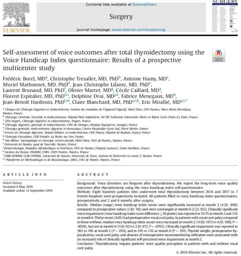
👉Self-rated voice quality is affected post-thyroidectomy even in patients without RLN injury.

👉Read more at:https://www.surgjournal.com/article/S0039-6060(19)30559-8/fulltext

#Arrangoiz

#ThyroidSurgeon

#HeadandNeckSurgeon

Hyperparathyroidism

👉Need some help staying organized when seeing your doctor for primary hyperparathyroidism?

👉Use the handy worksheet from the American Association of Endocrine Surgeons website patient resources found here:

https://collectedmed.com/index.php/article/article/demo_article_display/7552/83/2/1

#Arrangoiz

#ParathyroidSurgeon

#ParathyroidExpert

Rodrigo ARRANGOIZ MS, MD, FACS member of the American Head and Neck Society

👉I have been accepted as a member of the American Head and Neck Society

#Arrangoiz

#HeadandNeckSurgeon

#CancerSurgeon

Surgery or Active Surveillance Thyroid Microcarcinoma

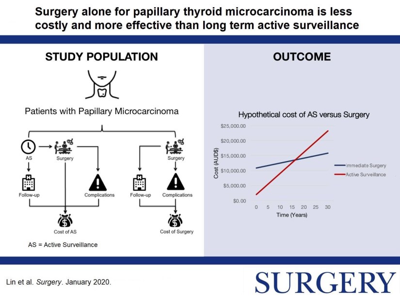
👉Surgery or active surveillance for #thyroid papillary microcarcinoma?

👉More in this study by Dr. Sywak et. al: https://www.surgjournal.com/article/S0039-6060(19)30468-4/fulltext

#Arrangoiz

#HeadandNeckSurgeon

#ThyroidSurgeon

#ThyroidExpert

#ThyroidCancer

#CancerSurgeon

#SurgicalOncologist

#Teacher

#Surgeon

#ThyroidCancer

Axillary Node Metastases with occult Primary Breast Cancer

Incidence and Differential Diagnosis

  • Occult primary breast cancer was first recognized by William Halsted:
    • Who described three patients presenting with axillary tumors that were eventually found to represent breast cancer.
  • In modern series:
    • Occult breast cancer accounts for 0.1% to 0.8%  of all newly diagnosed breast cancers:
      • The incidence has not decreased with improvements in breast imaging.
  • Differential diagnosis:
    • In general:
      • Palpable axillary nodes are more often related to benign rather than malignant disorders:
        • However, when cancer is identified:
          • The most common tumor causing axillary lymphadenopathy is:
            • Breast cancer.
      • In several series:
        • The incidence of breast cancer in mixed populations of men and women with metastatic axillary adenopathy:
          • Is 50% or higher:
            • The vast majority are women:
              • Although occult primary breast cancer has been reported in men:
                • It is very rare.
    • Other neoplasms that may present with axillary nodal involvement are:
      • Lymphomas
      • Melanomas
      • Sarcomas
      • Thyroid cancers
      • Skin cancers
      • Lung cancers
      • Less often:
        • Uterine, ovarian, sweat gland, or gastric cancers.
    • In approximately 30% of cases:
      • The primary site is never identified.
  • Initial Diagnostic Workup:
    • Biopsy:
      • The first step in the diagnostic workup of a patient with unexplained axillary adenopathy is:
        • A biopsy:
          • Besides standard light microscopic examination of hematoxylin and eosin-stained sections:
            • Other techniques such as;
              • Immunohistochemistry and sometimes electron microscopy.
                • Can help to narrow the differential diagnosis.
      • Immunohistochemistry:
        • The pathologic examination of a biopsy specimen for an isolated axillary lymph node metastatic adenocarcinoma or poorly differentiated carcinoma in a woman should include immunohistochemical staining for the following markers:
          • Carcinoembryonic antigen (CEA)
          • Cytokeratins 7 and 20
          • Estrogen receptor (ER) and progesterone receptor (PR)
          • Gross cystic disease fluid protein-15:
            • GCDFP:
              • Is identified by staining with the monoclonal antibody BRST2.
          • Mammaglobin
          • Thyroid transcription factor (TTF-1)
          • CA125
          • Men:
            • Should have routine staining for prostate cancer markers as well.
        • While none of these markers is sufficiently sensitive or specific to be used alone, certain patterns of expression favor the diagnosis of an occult breast cancer:
          • Positive staining for:
            • CEA, CK7, ER/PR, mammaglobin, CA125, and BRST2.
          • Negative staining for:
            • CK20 and TTF-1.
        • CEA is a sensitive marker for:
          • Adenocarcinomas of the breast, lung, and gastrointestinal tract:
            • But does not help to distinguish among these sites of origin.
        • On the other hand, differential expression of cytokeratins (CKs) can assist in this differentiation:
          • CK20 is a low molecular weight cytokeratin:
            • That is normally expressed in the gastrointestinal epithelium, urothelium, and in Merkel cells.
          • CK7 is expressed by tumors of the:
            • Lung, ovary, endometrium, and breast:
              • Not in the lower gastrointestinal tract.
                • The pattern of CK20 and CK7 may be particularly helpful in suggesting a primary site:
                  • The presence of CK7 and absence of CK20:
                    • Favors a diagnosis of breast cancer.
        • TTF-1:
          • Is rarely positive in breast cancers:
            • While it is positive in 70% to 80%  of nonsquamous cancers arising in the lung.
        • CA-125:
          • Is commonly positive in ovarian carcinomas
            • But is positive in about 10% of breast cancers.
        • ER/PR:
          • Its presence in an axillary node, particularly in conjunction with other compatible IHC findings:
            • Lends support to a diagnosis of an occult breast primary.
          • Although positive staining for ER and/or PR supports a possible diagnosis of breast cancer:
            • These markers are nonspecific and they may also be expressed in:
              • Ovarian, uterine, lung, stomach, thyroid, and hepatobiliary cancers:
                • However:
                  • ER/PR staining of an axillary node is compelling evidence of a primary breast cancer.
        • Other breast cancer-specific IHC stains are:;
          • BRST2 (for GCDFP) and mammaglobin:
            • BRST2 is positive in 65% to 80% of cases:
              • Is relatively specific for breast cancer:
                • Rarely, it is positive in:;
                  • Skin adnexal tumors, endometrial cancers, and salivary gland tumors.
            • While mammaglobin is more sensitive, it is less specific for breast cancer:
              • Gynecologic, lung, urothelial, thyroid, colon and hepatobiliary tumors may stain positive:
                • Both stains are thus typically used together.
        • HER2 immunostaining:
          • Is not generally useful for the differential diagnosis of a carcinoma arising in the axillary nodes as it lacks specificity:
            • Furthermore, only 18% to 20%  of breast cancers overexpress this protein:
              • However, assay for HER2 overexpression by IHC or fluorescent in situ hybridization (FISH) is a routine component of the evaluation of all breast cancers:
                • As it permits the identification of those women who are most likely to respond to treatments targeting HER2 (eg, the therapeutic monoclonal antibody trastuzumab). 

Rodrigo Arrangoiz MS, MD, FACS cirujano oncology y miembro de Sociedad Quirúrgica S.C en el America British Cowdray Medical Center en la ciudad de Mexico:

  • Es experto en el manejo del cáncer de mama.

Training:

• General surgery:

• Michigan State University:

• 2004 al 2010

• Surgical Oncology / Head and Neck Surgery / Endocrine Surgery:

• Fox Chase Cancer Center (Filadelfia):

• 2010 al 2012

• Masters in Science (Clinical research for health professionals):

• Drexel University (Filadelfia):

• 2010 al 2012

• Surgical Oncology / Head and Neck Surgery / Endocrine Surgery:

• IFHNOS / Memorial Sloan Kettering Cancer Center:

• 2014 al 2016

#Arrangoiz

#Surgeon

#Cirujano

#SurgicalOncologist

#CirujanoOncologo

#BreastSurgeon

#CirujanodeMama

#CancerSurgeon

#CirujanodeCancer

TOTAL THYROIDECTOMY- ENSURING COMPLETENESS OF RESECTION

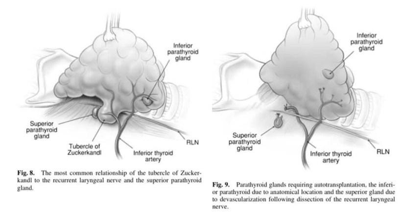
👉The technique of subtotal thyroidectomy formerly employed relied on the anatomical removal of visibly abnormal thyroid tissue, leaving behind bilateral posterior remnants.

👉The move to total thyroidectomy has meant not only that more thyroid tissue is now removed, but that rather there has been a progressive awareness of the vagaries of embryological development of the thyroid, with dissection now focused on those changes in order to ensure completeness of resection, and thus efficacy of the procedure.

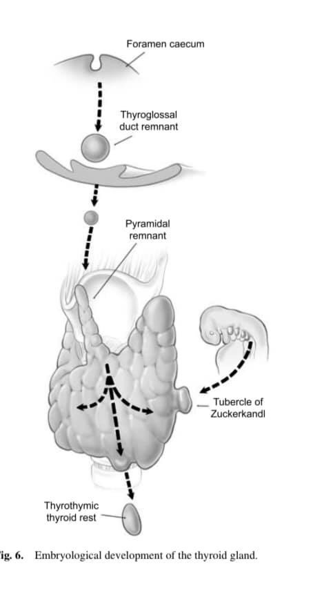
👉Thyroid development involves the midline descent of thyroid tissue from the foramen caecum to the level of the larynx along the thyroglossal tract, at which stage the left and right lobes develop.

👉What is not as well appreciated is that thyroid descent may well continue into the thyrothymic region (and even into the anterior mediastinum) forming a prolongation of thyroid tissue or even completely separate thyrothymic thyroid rests.

👉In addition the lateral thyroid component arising from the 4th branchial cleft and ultimobranchial body fuses with the median component to form the tubercle of Zuckerkandl, a distinct anatomical structure.

👉This fusion is not only the source of the thyroid’s C-cells, but is also essential in the process of follicular development.

👉Thyroglossal tract and pyramidal remnants – Routine dissection of the pyramidal area up to the laryngeal cartilage should be part of every total thyroidectomy. Even minimal recurrence in this area can be very apparent to the patient. More importantly, in patients with Graves’ disease, a small pyramidal lobe remnant can contain sufficient thyroid tissue to cause clinical recurrence even after an apparent ‘total’ thyroidectomy.

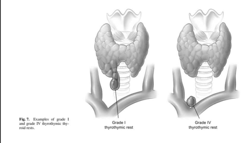
👉Thyrothymic thyroid rests – Thyrothymic thyroid rests are present in over 50% of patients, although most are small. They are often mistaken for small lymph nodes, or even parathyroid glands, and mostly cause no real problems. They are classified according to the nature of their connection to the thyroid gland proper. Grade I is a protrusion of thyroid tissue from the lower edge of the thyroid lobe, grade II is a thyroid rest connected by a bridge of thyroid tissue while grade III is connected by only a fibrovascular core. Grade IV has no connection at all with the thyroid proper. Their clinical significance lies in the fact that, if not removed at the initial operation, they may well reappear as retrosternal recurrence after apparent ‘total’ thyroidectomy. Routine dissection of the thyrothymic area down to the thoracic inlet looking for such rests should therefore be part of every total thyroidectomy.

👉The tubercle of Zuckerkandl – This structure, first described by Zuckerkandl in 1902, is a distinct anatomical entity, and can be found in nearly two-thirds of patients undergoing thyroid surgery. It is classified according to size (grades 0 to grades 3) using a system developed by Pelizzo et al. The tubercle of Zuckerkandl is often the source of local pressure or obstructive symptoms, especially when the thyroid itself is relatively small. The importance of the tubercle of Zuckerkandl, once again, is that if not looked for and removed during thyroid surgery, it may be a source of persistent unrelieved symptoms or recurrence. An understanding of the anatomy of the tubercle of Zuckerkandl is also central to safe surgical dissection. It usually enlarges lateral to the RLN, with the nerve appearing to pass into a cleft medial to it – a situation that some surgeons used to describe as the nerve passing into the thyroid substance. Early elevation of the tubercle of Zuckerkandl usually allows the recurrent nerve to be easily and safely ‘encountered’ even though not initially visible. However, an uncommon but high risk situation is where the RLN runs lateral to an enlarged tubercle of Zuckerkandl, placing it at increased risk of damage during dissection. Another important point is that the normal superior parathyroid gland, also being derived from the fourth branchial cleft, is commonly found in close association, cephalad to the tubercle of Zuckerkandl.

#Arrangoiz

#ThyroidCancer

#ThyroidSurgeon

#ThyroidExpert

#EndocrineSurgery

#HeadandNeckSurgeon

Ways to Reduce Breast Cancer Risk

#Arrangoiz

#BreastSurgeon

#CancerSurgeon

#SurgicalOncologist

#Surgeon

#Teacher

#BreastCancer

Novel Gene Panel to Predict Lymph Node Metastasis and Recurrence in Thyroid Cancer

👉A novel 25 gene panel can be used to predict LN metastasis in early #papillary thyroid cancer (OR = 8.06, P < .001) and disease-free survival (HR = 2.64, P = .043).

👉Read more at:

https://www.surgjournal.com/article/S0039-6060(19)30584-7/fulltext

#Arrangoiz

#ThyroidSurgeon

#ThyroidCancer

#ThyroidExpert

#HeadandNeckSurgeon

#CancerSurgeon

#Teacher

Etiology of Papillary Thyroid Cancer?

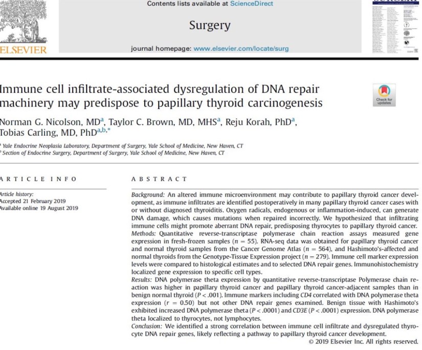
👉A strong correlation b/w immune cell infiltrate and dysregulated thyrocyte DNA repair gene expression suggests a potential mechanism for # papillary thyroid ca development.

More at:https://www.surgjournal.com/article/S0039-6060(19)30459-3/fulltext

#Arrangoiz

#ThyroidSurgeon

#ThyroidExpert

#CancerSurgeon

#Surgeon

When Should a Woman Being Screening Mammography?

  • The U.S. Preventive Services Task Force (USPSTF) recommends:

img_0087

  • The American College of Radiology and the Society of Breast Imaging recommend:
    • Annual screening mammography beginning at age 40 years.
    • They state that screening mammography may conclude:
      • When life expectancy is less than 5 to 7 years on the basis of age or comorbid conditions, or when abnormal results would not be acted upon.

img_0085

  • The American Cancer Society recommends:
    • Regular screening mammography for women beginning at age 45 years:
      • With qualified recommendations for women aged 45 to 54 years to be screened annually.
    • Women 55 years and older:
      • To transition to biennial screening or have the option to continue annual screening.
    • For women aged 40 to 44 years to have the option to begin annual screening.
    • ACS recommends women continue screening mammography:
      • As long as their overall health is good and life expectancy is 10 years or longer.

img_3768-1

  • The American Society of Breast Surgeons recommends:
    • That average risk women ages 40 to 44 years discuss the risks and benefits of screening with their physicians.
    • Annual mammographic screening is recommended:
      • For women ages 45 to 54 years.
    • Annual or biennial screening is recommended:
      • For women 55 years and older:
        • Based on a shared decision-making discussion of risks and benefits.
    • Biennial screening is recommended for women age 75 years and older if estimated life expectancy is at least 10 years.
  • References:
    1. The American Society of Breast Surgeons. Consensus statement on screening mammography. https://www.breastsurgeons.org/new_layout/about/statements/PDF_Statements/Screening_Mammography.pdf. Accessed May 8, 2016.
    2. Lee CH, Dershaw DD, Kopans D, et al. Breast cancer screening with imaging: recommendations from the Society of Breast Imaging and the ACR on the use of mammography, breast MRI, breast ultrasound, and other technologies for the detection of clinically occult breast cancer. J Am Coll Radiol. 2010;7:18-27.
    3. Oeffinger KC, Fontham ET, Etzioni R, et al; American Cancer Society. Breast cancer screening for women at average risk: 2015 guideline update from the American Cancer Society. JAMA. 2015;314:1599-1614.
    4. Siu AL; U.S. Preventive Services Task Force. Screening for breast cancer: U.S. Preventive Services Task Force recommendation statement. Ann Intern Med. 2016;164:279-296.

#Arrangoiz

#BreastSurgeon

#SurgicalOncologist

#CancerSurgeon