NSABP B-14

  • NSABP B-14:
    • Randomized patients after surgery to:
      • Five years of tamoxifen or
      • 5 years of placebo:
        • To determine if there was a:
          • Significant survival advantage with the addition of endocrine therapy to:
            • ER-positive tumors
    • After 10 years of follow-up:
      • A statistically significant DFS benefit was derived:
        • With the use of tamoxifen for 5 years:
          • 69% vs 57%
            • P<0.0001
      • With a 37% reduction:
        • In the rate of contralateral breast cancer (P=0.007)
      • The most recent update of this trial:
        • Continues to demonstrate this survival benefit at 15 years:
          • Irrespective of age
          • Menopausal status, and
          • Tumor ER concentration
      • A follow-up question to protocol B-14:
        • Asked the recommended duration of tamoxifen therapy beyond 5 years:
          • The same patient population was then re-randomized to:
            • Five additional years of tamoxifen or
            • Five years of placebo
          • There was a significant disadvantage in:
            • DFS:
              • 86% vs 92%, P= 0.003 and
            • Distant DFS:
              • 90% vs 96%, P=0.01:
                • For patients who continued tamoxifen for more than 5 years versus those who took it for only 5 years
                  • The lack of benefit with additional tamoxifen use was independent of patient age

#Arrangoiz #BreastSurgeon #CancerSurgeon #SurgicalOncologist #Teacher
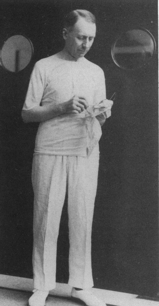
Thomas Peel Dunhill Pioneer of Thyroid Surgery

👉One of the pioneers in thyroid surgery in the twentieth century, Thomas Peel Dunhill, began his work on the problems of goiter and thyrotoxicosis in Melbourne in 1910 had performed 312 operations, 200 of which were for exophthalmic goiter.

Dunhill adopted a technique of total lobectomy on one side and subtotal on the other side after reading Frank Harley’s paper which was published in 1907.

👉He used his technique under local anesthesia at first and later under light general anesthesia. At that time the mortality for this operation was 30% in hospitals in London and with his method, he achieved a mortality of less than 3%, despite accepting the most severely ill patients suffering from uncontrolled atrial fibrillation.

👉The technique that he adopted — total lobectomy by a pericapsular dissection technique
— is even now considered by many surgeons the optimal method of resection.

👉He also described later operation on retrosternal goiter by splitting the sternum and in 1920 he produced his outstanding paper in an early issue of British Journal of Surgery.

👉In the same year, Sistrunk described his radical operation for the thyroglossal tract that included the resection of the middle third of hyoid at the base of the tongue.

#Arrangoiz #ThyroidSurgeon #ThyroidExpert

Chemotherapy for Breast Cancer During Pregnancy

  • Breast cancers during pregnancy:
    • Are often locally advanced due to delays in diagnosis, and many of these young patients will require chemotherapy
  • Systemic treatment of breast cancer during pregnancy:
    • Involves special consideration of both the mother and the baby
  • Data from a single-institution prospective study:
    • Reported on 40 pregnant women initially and updated their series years later to include 57 women:
      • Treated with FAC chemotherapy (5-FU, doxorubicin, and cyclophosphamide):
        • During the second and third trimesters
    • Investigators concluded:
      • This regimen to be safe as fetal malformations occurred only 1.3% of the time:
        • Which was similar to the rate of malformations seen in fetuses not exposed to chemotherapy
  • Methotrexate is contraindicated in all trimesters:
    • As its method of action is as an antimetabolite and anti-folate agent
  • Although limited data exist on taxane-based therapies:
    • The NCCN recommends that if needed:
      • It should be given with weekly dosing
  • Anti-HER2 therapy with either trastuzumab or pertuzamab:
    • Is contraindicated during pregnancy:
      • With case reports associating oligohydramnios or anhydramnios with therapy
  • First-trimester chemotherapy:
    • Is contraindicated based on:
      • An increased risk of:
        • Fetal malformation
        • Stillbirth
        • Miscarriage
  • Second- and third-trimester chemotherapy:
    • Appears to be safe:
      • But may be associated with:
        • Intrauterine growth retardation
        • Prematurity
        • Low birth weight
  • Chemotherapy:
    • Should not be given after 35 weeks’ gestation or within 3 weeks of planned delivery:
      • The median gestational age is 38 weeks with more than 50% of babies born by vaginal delivery
  • Breastfeeding while on chemotherapy:
    • Is contraindicated due to excretion of drugs into breast milk

REFERENCES

  1. National Comprehensive Cancer Network. NCCN Clinical Practice Guidelines for Oncology: Breast Cancer. https://www.nccn.org/professionals/physician_gls/f_guidelines.asp Published January 2016. Accessed January 29, 2017.
  2. Amant F, Deckers S, Van Calsteren K, et al. Breast cancer in pregnancy: Recommendations of an international consensus meeting. Eur J Cancer. 2010;46:3158-3168.
  3. Briggs GG, Freeman RK, Yaffe SJ. Drugs in Pregnancy and Lactation. A Reference Guide to Fetal and Maternal Risk. 6th ed. Philadelphia, Pa: Lippincott Williams and Wilkins; 2006.
  4. Gwyn K, Theriault R. Breast cancer during pregnancy. Oncology (Williston Park). 2001; 15(1):39-46.
  5. Hahn KM, Johnson PH, Gordon N, et al. Treatment of pregnant breast cancer patients and outcomes of children exposed to chemotherapy in utero. Cancer. 2006:107(6):1219-1226.

#Arrangoiz #Surgeon #CancerSurgeon #SurgicalOncologist #BreastSurgeon #BreastCancer #Teacher

Compliance and Resistance

  • Resistance:
    • Is the impedance of flow:
      • In the tubing and airways and therefore:
        • Can only occur when there is:
          • Airflow
    • According to Ohm’s Law:
      • Resistance (R) = Δ pressure /Δ volume
        • R = (Peak inspiratory pressure – Plateau pressure) / Tidal volume
          • R = (PIP- Pplat) / (TV)
  • Assuming a constant tidal volume:
    • The resistance equation can be simplified to:
      • R ≈ (PIP- Pplat)
  • Normal airway resistance:
    • Should be ≤ 5 cmH20
  • Resistance is a factor in ventilating all patients but can become particularly important:
    • When ventilating patients with COPD or asthma:
      • The resistance in a system:
        • Increases with decreasing diameter
          • While common examples include:
            • A very small endotracheal tube (ETT) or bronchospasm leading to narrowing of the airways:
              • Recall that a “decrease in the diameter” can also occur at just one point:
                • Such as with kinking or biting of the ETT, or a mucous plug in a large airway
  • Compliance refers to:
    • The distensibility of the system and is the inverse of elastance:
      • In other words:
        • It a measure of the lung’s ability to stretch and expand:
          • The more elastic a system, or higher the “recoil,”:
            • The lower the compliance:
              • A common analogy to understand the concepts of elastance is to analyze the recoil of springs:
                • Imagine a very tightly wound and stiff spring
                • This spring is difficult to stretch and wants to stay in the coiled position
                • This spring would have high elastance and low compliance
              • Envision a second, loosely coiled spring:
                • Very little force is required to stretch out this spring, and therefore, it has low elastance but high compliance
  • Although compliance commonly is used to describe the lung parenchyma:
    • Remember that compliance actually involves all components of the system:
      • In other words:
        • A patient with pulmonary edema may have low compliance:
          • Due to an issue with the lung parenchyma
        • But another patient may have similarly low compliance due to severe chest wall stiffness after a third-degree burn
        • Clinically, knowing the exact cause of decreased compliance in a given patient can be challenging:
          • Physicians should not, therefore, always assume that it is always related to “stiff lungs.”
  • In the figure below, the top “lungs” are healthy:
    • The lungs on the left have a resistance problem or impairment in airflow
    • The lungs on the right have a compliance problem or impairment in stretch and recoil
    • In this picture:
      • Both figures could have elevated peak inspiratory pressures (PIP):
        • Due to the excess pressure generated in the system:
          • However:
            • Only the right-hand figure would have an elevate plateau pressure (Pplat):
              • Since this process occurs when there is an absence of airflow
  • Compliance (C) = ∆ volume / ∆ pressure
    • C = Tidal volume / Plateau pressure – Peak inspiratory pressure
      • C = (TV) / (Pplat – PEEP)
  • Therefore, when troubleshooting high-pressures on the ventilators:
    • Two values are needed:
      • The peak inspiratory pressure (PIP):
        • Is the maximum pressure in the system and includes both:
          • Resistance and compliance:
            • An inspiratory pause stops all airflow:
              • Thereby removing resistance, and only leaving compliance, as illustrated in this diagram below:
                • The plateau pressure, or Pplat, is, therefore:
                  • A measure of compliance
  • These values can be displayed on the ventilator screen:
    • On most ventilators:
      • The PIP is always seen
      • While the Pplat is seen by pushing the “inspiratory hold” or “inspiratory pause” button on the ventilator
    • An elevated PIP and normal Pplat is:
      • Indicative of increased airway resistance
    • An elevated PIP and elevated Pplat is:
      • Indicative of abnormal compliance
  • Determining whether the patient has a:
    • Resistance problem or a compliance problem:
      • Can assist in the differential diagnosis of respiratory failure:
        • High Resistance:
          • High PIP, Low / Normal Pplat:
            • Kinked / obstructed ETT
            • Mucus plugging
            • Brochospasm
            • Endotracheal tube to narrow:
              • Small
            • Coughing
            • Bronchospasm:
              • Obstructive lung disease
    • Low Compliance:
      • High PIP, High Pplat:
        • Atelectasis
        • Pulmonary edema
        • ARDS
        • Hemothorax /pneumothorax
        • Pneumonia
        • Pulmonary fibrosis:
          • Restrictive lung disease
        • Air-trapping with accumulated auto-PEEP
        • Obesity
        • Abdominal compartment syndrome
        • Circumferential burns of the chest
        • Scoliosis
        • Supine position
  • Air trapping:
    • Also referred to as breath-stacking:
      • Can lead to the development of auto-PEEP, or intrinsic PEEP (iPEEP):
        • These pressures should be differentiated from the set PEEP, or extrinsic PEEP (ePEEP):
          • ePEEP refers to the additional end-expiratory positive pressure set during mechanical ventilation:
            • To prevent alveolar collapse and derecruitment
      • In contrast:
        • Auto-PEEP, or iPEEP:
          • Is a pathophysiological process:
            • That can occur when the ventilator initiates the next breath prior to complete exhalation:
              • While this is most common in patients with prolonged expiratory phases, such as asthma or COPD:
                • It can also occur in patients:
                  • Who have a fast respiratory rate or
                  • Those who are being ventilated with large tidal volumes
              • The amount of auto-PEEP can be measured by:
                • Pressing the “expiratory hold” or “expiratory pause” button on the ventilator:
                  • When this button is pressed, the ventilator will display the total PEEP:
                    • The auto-PEEP is the difference between the total PEEP and the set PEEP:
                      • Auto-PEEP (iPEEP) = Total PEEP – ePEEP
  • The Figure represents the effects of air trapping:
  • Air trapping, or autoPEEP:
    • Can lead to significant adverse cardiopulmonary effects
    • The increased intrathoracic pressure from autoPEEP can:
      • Decrease venous return and lead to hemodynamic instability, even cardiac arrest in severe cases
    • The increased pressures may also result in:
      • A pneumothorax or pneumomediastium
    • Additionally, air trapping can lead to:
      • Ineffective ventilation due to:
        • Collapse of the capillaries responsible for gas exchange:
          • With worsening hypercarbia and hypoxemia
      • While this may seem like a paradox:
        • As one may assume that increasing the minute ventilation, or moving more air:
          • Will improve ventilation, there is a limit to the beneficial effects:
            • Once the lungs are overdistended, gas exchange is ineffective
            • In these circumstances, allowing the patient sufficient time to exhale can decrease CO2 retention

#Arrangoiz #Surgeon #Teacher

Prognostic and Predictive Factors in Breast Cancer

  • Prognostic factors:
    • Provide information regarding:
      • Clinical outcome
    • The prognostic factors in breast cancer that have been validated in clinical testing and related to risk of relapse and survival are:
      • Lymph node status
      • Tumor size
      • Grade
      • Lymphovascular invasion
      • ER/PR status:
        • ER-negative and PR-negative tumors:
          • Are associated with a worse prognosis compared to ER-positive tumors
  • Predictive factors:
    • Provide information on likelihood of benefit from a given therapy
    • An example of a predictive factor:
      • ER/PR receptor expression:
        • Identifies those patients likeliest to benefit from hormonal therapies
      • Of note:
        • Tumor grade:
          • May be predictive of response to therapy, but not necessarily of survival benefit
  • REFERENCES
    • Cianfrocca M, Goldstein LJ. Prognostic and predictive factors in early stage breast cancer. Oncologist. 2004;9(6):606-616.
    • Kleer C, Sabel M. Prognostic and predictive factors in breast cancer. In: Kuerer HM, ed. Breast Surgical Oncology. New York, NY: McGraw Hill; 2010:243-249.

#Arrangoiz #CancerSurgeon #SurgicalOncologist #CancerSurgeon #BreastSurgeon #BreastCancer

NSABP B-35

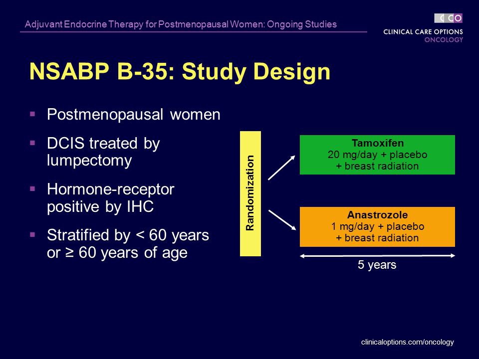
  • The NSABP B-35:
    • Is a phase III clinical trial:
      • That randomized postmenopausal women with ER-positive DCIS to:
        • 5 years of anastrozole or tamoxifen:
          • Following breast-conserving surgery and radiation
      • The trial sought to determine:
        • How effective anastrozole is compared to tamoxifen in preventing a breast cancer occurrence:
          • As well as the quality of life of patients taking anastrozole
    • The trial is currently closed after meeting its accrual goal of:
      • 3100 patients
    • Median follow-up of 9 years
    • Investigators found:
      • Significantly fewer breast cancer events in the anastrozole group (n = 90) than in the tamoxifen group (n = 122):
        • Hazard ratio, 0.73; confidence interval, 0.56–0.96, P=0.0234
    • The estimated 10-year breast-cancer-free interval rates were:
      • 93.5% for anastrozole versus
      • 89.2% for tamoxifen
        • This recorded difference in breast cancer-free interval:
          • Was attributable almost entirely to younger postmenopausal women:
            • Less than 60 years of age
    • Interestingly:
      • The difference between treatments did not become apparent until after 5 years of follow-up:
        • Likely due to the low number of events in both groups
    • There was no difference in overall survival (OS):
      • Between the two treatment groups

#Arrangoiz #BreastSurgeon #CancerSurgeon #SurgicalOncologist #Teacher #Surgeon

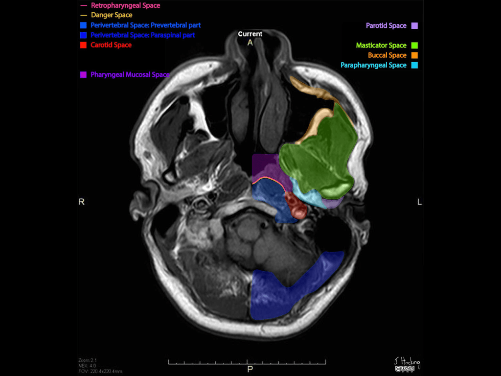
The Parapharyngeal Space

  • The parapharyngeal space:
    • Also known as the:
      • Prestyloid parapharyngeal space:
        • Is one of the seven deep compartments of the head and neck
  • It consists largely of:
    • Fatty areolar tissue
    • Contains branches of the:
      • Trigeminal nerve
    • Deep blood vessels
  • Two naming conventions exist in the literature:
    • In the first definition:
      • Familiar to most head and neck surgeons, the parapharyngeal space is divided into:
        • The prestyloid compartment and
        • Poststyloid (retrostyloid) compartment
    • In the second definition:
      • Introduced by some radiologists:
        • The prestyloid parapharygeal space is simply termed:
          • The parapharyngeal space, and
        • The poststyloid pharapharygeal space is termed:
          • The carotid space
  • Gross anatomy:
    • The parapharyngeal space:
      • Is shaped like a pyramid:
        • An inverted pyramid with:
          • Its base:
            • At the skull base
          • Its apex:
            • Inferiorly:
              • Pointing to the greater cornu of the hyoid bone
    • Contents:
      • Fat:
        • Main component
      • Deeps blood vessels:
        • Internal maxillary artery
        • Ascending pharyngeal artery
        • Pterygoid venous plexus:
          • Only small portion:
            • Because it is mainly within:
              • The masticator space
      • Nerve:
        • Small branch of the mandibular division of the trigeminal nerve (cranial nerve V):
          • Supplying the tensor veli palatini muscle
      • Salivary glands – depends on the definition:
        • Some say that it contains no salivary glands, others
        • Minor or ectopic salivary gland / rests
        • Retromandibular portion of the deep lobe of parotid gland
      • Lymph nodes
  • Boundaries:
    • The parapharyngeal space has complex fascial margins:
      • Occupying the space between the muscles of mastication and the muscles of deglutition:
        • Superior margin:
          • Base of skull
        • Inferior margin:
          • Greater cornu of the hyoid bone:
            • Although some state the space functionally ends higher:
              • With the styloglossus muscle:
                • At the level of the angle of the mandible
        • Medial margin:
          • Middle (pretracheal) layer of the deep cervical fascia:
            • Covering the:
              • Superior pharyngeal constrictor
              • Levator palatini muscle and
              • Tensor veli palatini muscle
        • Lateral margin:
          • Investing fascia (superficial layer) of the deep cervical fascia:
            • Covering the deep lobe of the parotid
        • Anterior margin:
          • Investing fascia (superficial layer) of the deep cervical fascia:
            • Covering the medial pterygoid muscle
        • Posterior margin:
          • Prevertebral layer of the deep cervical fascia
  • Relations:
    • Medial:
      • To the masticator space
    • Lateral:
      • To the pharyngeal mucosal space
    • Anterior:
      • To the prevertebral space
    • Posterior:
      • To the medial pterygoid

#Arrangoiz #HeadandNeckSurgeon #CancerSurgeon #SurgicalOncologist #Teacher

Cervical Fascia

  • Superficial cervical fascia:
    • Primarily includes the:
      • Platysma and
      • Subcutaneous fat and vessels:
        • However:
          • As with other fascia in the body:
            • The use of the terminology of the superficial cervical fascia has declined in favor of:
              • Subcutaneous tissue:
                • Thus, an unspecified reference to cervical fascia:
                  • Mainly refers to the deep cervical fascia
  • The deep cervical fascia:
    • Consists of three separate but related fascial layers:
      • That encircle structures in the neck and
      • Allow anatomic compartmentalisation into:
        • The deep spaces of the head and neck
      • Each layer contributes:
        • To the carotid sheath
    • Layers of the deep cervical fascia:
      • Superficial layer of the deep cervical fascia
      • Middle layer of the deep cervical fascia
      • Deep layer of the deep cervical fascia
  • The superficial layer of the deep cervical fascia:
    • Also known as:
      • The investing layer / fascia
    • Is the one of three layers of the deep cervical fascia:
      • That surrounds all of the neck:
      • That is deep to the platysma
      • The layer includes:
        • The masticator fascia
        • Submandibular fascia, and
        • Sternocleidomastoid-trapezius fascia
        • The fascia that superficially covers the parotid gland:
          • May also be derived from this layer:
            • But this inclusion is controversial
    • Gross anatomy – attachments:
      • Posteriorly:
        • Ligamentum nuchae
        • Cervical vertebral spinous processes
        • External occipital protuberance
      • Superiorly
        • Mandible:
          • The layer thickens to form the extrinsic ligaments of the mandible:
            • Sphenomandibular ligament and
            • Stylomandibular ligament
        • Mastoid process and styloid process
        • Central skull base
        • Zygomatic arch
        • Superior temporal line or temporal ridge
    • Inferiorly
      • Manubrium
      • Clavicle
      • Acromion and spine of the scapula
    • Contents:
      • The superficial layer of the deep cervical fascia:
        • Encircles everything in the neck:
          • Apart from the skin and superficial cervical fascia (subcutaneous tissue)
      • In addition, it splits to enclose the following structures:
        • Two salivary glands
          • Submandibular gland
          • Parotid glands
        • Two spaces:
          • Masticator space
          • Suprasternal space
        • Two muscles (other than those in the masticator space):
          • Sternocleidomastoid muscle
          • Trapezius muscle
    • In addition:
      • All layers of the deep cervical fascia:
        • Contribute to the carotid sheath
  • The middle layer of the deep cervical fascia:
    • Is the one of the three layers of the deep cervical fascia:
      • It most closely surrounds:
        • The visceral organs
    • This layer consists:
      • Anteriorly of the:
        • Strap muscle fascia:
          • Comprised of the:
            • Sterno-omohyoid layer and
            • Sternothyroid layer
            • Thyrohyoid layer
      • Posteriorly:
        • Visceral fascia:
          • Also commonly known as the:
            • Pharyngobasilar and
            • Buccopharyngeal fascia
              • Particularly in the suprahyoid neck, or, less commonly:
                • Pharyngomucosal fascia
          • The alternative term:
            • Pretracheal fascia:
              • May refer to either:
                • The visceral fascia or
                • The sternothyroid-thyrohyoid layer of strap muscle fascia:
                  • Which lies anterior to the trachea
    • Gross Anatomy – attachments:
      • Superiorly:
        • Skull base
      • Anteriorly:
        • Hyoid bone
        • Thyroid cartilage
        • Manubrium
      • Inferiorly:
        • Fibrous pericardium
        • Adventitia of the aortic arch
    • Contents:
      • Infrahyoid (strap) muscles:
        • Sternohyoid
        • Omohyoid
        • Sternothyroid
        • Thyrohyoid
      • Visceral space and pharyngeal mucosal space:
        • Buccinator muscle
        • Pharynx and pharyngeal constrictor muscles:
          • Superior pharyngeal constrictor muscle
          • Middle pharyngeal constrictor muscle, and
          • Inferior pharyngeal constrictor muscle
        • Cervical esophagus
        • Thyroid gland* and parathyroid glands
        • Trachea*
        • Larynx*
        • Visceral lymph nodes
        • Recurrent laryngeal nerve
          • *Some sources consider these structures to have their own fascia not derived from the middle layer of the deep cervical fascia:
            • But they are nevertheless considered part of the visceral space
    • In addition:
      • All layers of the deep cervical fascia:
        • Contribute to the carotid sheath:
          • However:
            • In the suprahyoid neck:
              • Above the carotid bifurcation:
                • The contribution of the middle layer is inconsistent
  • The deep layer of the deep cervical fascia:
    • Is one of the three layers of the deep cervical fascia:
      • It encases the:
        • Paravertebral muscles and
        • Forms the perivertebral space
      • It consists of the:
        • Perivertebral fascia:
          • The anterior part of which is called:
            • The prevertebral fascia and
            • The alar fascia
    • Gross Anatomy – attachments:
    • Medially:
      • Ligamentum nuchae
      • Cervical vertebral spinous processes and transverse processes
    • Laterally:
      • Carotid sheath
      • First rib:
        • From a portion of the layer called:
          • Sibson fascia
      • Axillary sheath
    • Superiorly
      • The skull base
    • Inferiorly
      • Coccyx:
        • For the prevertebral fascia)
      • Endothoracic fascia:
        • For the alar fascia
    • On each side:
      • A flap attaches to the transverse processes of the cervical vertebrae and:
        • Divides the peri-vertebral spaces into:
          • A pre-vertebral compartment:
            • Anteriorly and
          • A para-spinal compartment:
            • Posteriorly
    • Anteromedial to the scalene muscles:
      • The deep layer splits into two leaves:
        • The ventral leaf being:
          • The alar fascia, and
        • The dorsal leaf being:
          • The prevertebral fascia:
            • With the prevertebral space:
              • Space between the prevertebral fascia and the spine
          • The space between the alar fascia and the prevertebral fascia:
            • Is the danger space
          • The space between the alar fascia and the posterior aspect of the middle layer of the deep cervical fascia:
            • Is the retropharyngeal space
    • Contents:
      • Danger space:
        • Space between prevertebral and alar fascia
      • Prevertebral space:
        • Space between the prevertebral fascia and the spine:
          • Anterior component:
            • Of the perivertebral space
      • Longus colli and capitis muscles
      • Rectus capitis anterior and lateralis muscles
      • Scalenus anterior, medius, and posterior muscles
      • Sheath for subclavian artery and vein, brachial plexus
      • Vertebral column
      • Spinal cord and associated thecal sac, nerve roots, and vessels
      • Vertebrae and associated discs and ligaments
      • Paraspinal / paravertebral space:
        • Posterior component:
          • Of the perivertebral space
      • Levator scapulae
      • Deep cervical back muscles
      • Pierced by the four cutaneous branches of the cervical plexus:
        • Greater auricular nerve
        • Lesser occipital nerve
        • Transverse cervical nerve
        • Supraclavicular nerve
    • In addition:
      • All layers of the deep cervical fascia:
        • Contribute to the carotid sheath

#Arrangoiz #HeadandNeckSurgeon #CancerSurgeon #SurgicalOncologist #Teacher

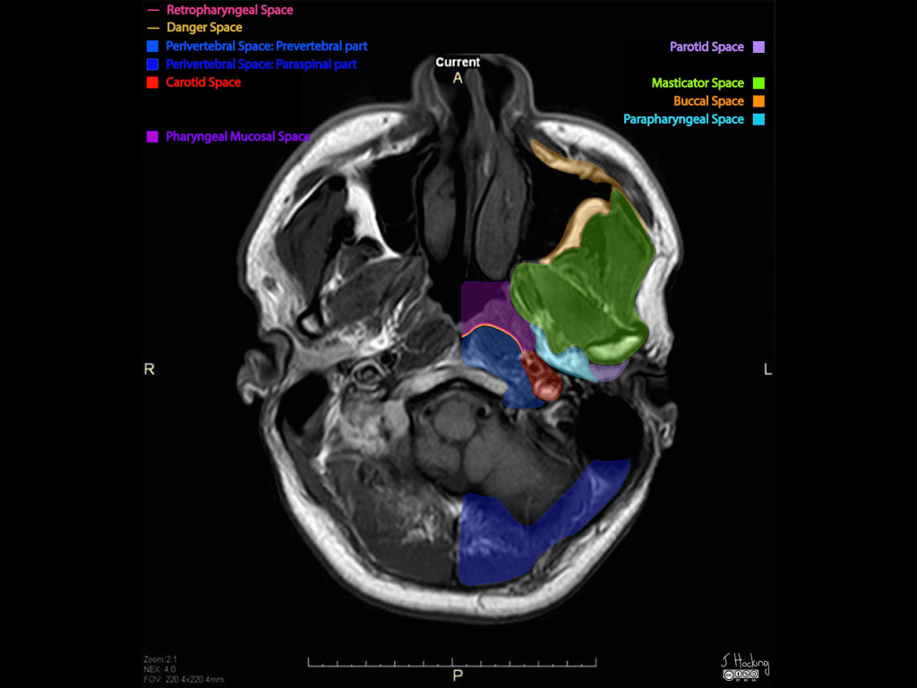
Masticator Space

  • The masticator space:
    • Is one of the seven deep compartments of the head and neck
  • Gross anatomy:
    • The masticator space are:
      • Paired suprahyoid cervical spaces:
        • On each side of the face
    • Each space is enveloped by:
      • The superficial layer of the deep cervical fascia:
        • The superficial layer of deep cervical fascia:
          • Splits into two at the lower border of the mandible:
            • The inner layer:
              • Running deep to the medial pterygoid muscle and attaches to the skull base medial to foramen ovale and
            • The outer layer:
              • Covering the masseter and temporalis muscles and attaches to the parietal calvaria superiorly
  • Contents:
    • Muscles of mastication
    • Ramus and body of mandible
    • Inferior alveolar nerve
    • Inferior alveolar vein and artery
    • Mandibular division of the trigeminal nerve (V3):
      • Enters the masticator space:
        • Via the foramen ovale
    • Pterygoid venous plexus
  • Boundaries and relations:
    • Anteriorly:
      • The buccal space
    • Posterolaterally:
      • Parotid space
    • Medially:
      • Parapharyngeal space
  • Communications:
    • Masticator space malignancies can spread perineurally:
      • Via the mandibular division of the trigeminal (V3) nerve:
        • Into the middle cranial fossa

#Arrangoiz #HeadandNeckSurgeon #CancerSurgeon #surgicalOncologist #Teacher