Breast Screening

Breast cancer screening means checking a woman’s breasts for cancer before there are signs or symptoms of the disease. All women need to be informed by their health care provider about the best screening options for them. When you are told about the benefits and risks of screening and decide with your health care provider whether screening is right for you—and if so, when to have it—this is called informed and shared decision-making.

Although breast cancer screening cannot prevent breast cancer, it can help find breast cancer early, when it is easier to treat. Talk to your doctor about which breast cancer screening tests are right for you, and when you should have them.

Breast Cancer Screening Recommendations

The United States Preventive Services Task Forceexternal icon (USPSTF) is an organization made up of doctors and disease experts who look at research on the best way to prevent diseases and make recommendations on how doctors can help patients avoid diseases or find them early.

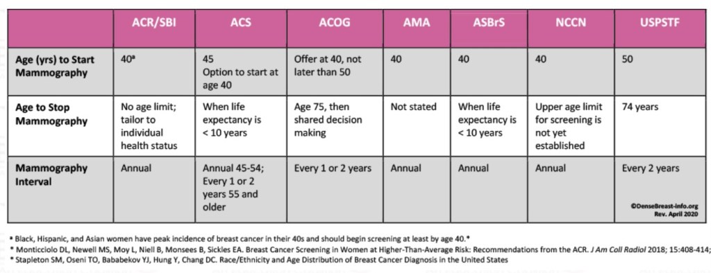
The USPSTF recommends that women who are 50 to 74 years old and are at average risk for breast cancer get a mammogram every two years. Women who are 40 to 49 years old should talk to their doctor or other health care professional about when to start and how often to get a mammogram. Women should weigh the benefits and risks of screening tests when deciding whether to begin getting mammograms before age 50.

The Breast Cancer Screening Guidelines for Women compares recommendations from several leading organizations.

Breast Cancer Screening Tests

Where Can I Go to Get Screened?

You can get screened for breast cancer at a clinic, hospital, or doctor’s office. If you want to be screened for breast cancer, call your doctor’s office. They can help you schedule an appointment.

Most health insurance plans are required to cover screening mammograms every one to two years for women beginning at age 40 with no out-of-pocket cost (like a co-pay, deductible, or co-insurance).

Mammogram

mammogram is an X-ray of the breast. For many women, mammograms are the best way to find breast cancer early, when it is easier to treat and before it is big enough to feel or cause symptoms. Having regular mammograms can lower the risk of dying from breast cancer. At this time, a mammogram is the best way to find breast cancer for most women.

Breast Magnetic Resonance Imaging (MRI)

A breast MRI uses magnets and radio waves to take pictures of the breast. MRI is used along with mammograms to screen women who are at high risk for getting breast cancer. Because breast MRIs may appear abnormal even when there is no cancer, they are not used for women at average risk.

Other Exams

Clinical Breast Exam

clinical breast exam is an examination by a doctor or nurse, who uses his or her hands to feel for lumps or other changes.

Breast Self-Awareness

Being familiar with how your breasts look and feel can help you notice symptoms such as lumps, pain, or changes in size that may be of concern. These could include changes found during a breast self-exam.You should report any changes that you notice to your doctor or health care provider.

Having a clinical breast exam or doing a breast self-exam has not been found to lower the risk of dying from breast cancer.

Benefits and Risks of Screening

Every screening test has benefits and risks, which is why it’s important to talk to your doctor before getting any screening test, like a mammogram.

Benefit of Screening

The benefit of screening is finding cancer early, when it’s easier to treat.

Risks of Screening

Harms can include false positive test results, when a doctor sees something that looks like cancer but is not. This can lead to more tests, which can be expensive, invasive, time-consuming, and may cause anxiety.

Tests also can lead to overdiagnosis,when doctors find a cancer that would not have gone on to cause symptoms or problems, or even may go away on its own. Treatment of these cancers is called overtreatment. Overtreatment can include treatments recommended for breast cancer, such as surgery or radiation therapy. These can cause unnecessary and unwanted side effects. Other potential harms from breast cancer screening include pain during the procedure and radiation exposure from the mammogram test itself. While the amount of radiation in a mammogram is small, there may be risks with having repeated X-rays.

#Arrangoiz #BreastSurgeon

Breast Conserving Surgery (BCS) after Neoadjuvant Chemotherapy

  • Neoadjuvant chemotherapy:
    • Usually has been administered in cases of:
      • Inoperable or locally advanced breast cancer:
        • To downsize the primary tumor and nodal disease:
          • To facilitate local-regional therapy:
            • With surgery and / or radiation
    • Given the success of this approach in locally advanced disease:
      • Combined with the known benefits of adjuvant systemic therapy:
        • Neoadjuvant chemotherapy has also been assessed for the management of patients with operable breast cancer
  • A well-recognized role of neoadjuvant chemotherapy:
    • Is the ability to improve surgical options for patients by:
      • Downsizing tumors and increasing the chances for breast conservationĀ 
  • In the landmark National Surgical Adjuvant Breast and Bowel Project (NSABP) B-18 trial:
    • 1523 patients with primary operable breast cancer
    • Were randomized to either preoperative or postoperative systemic therapy with:
      • Four cycles of standard doxorubicin (60 mg/m2) plus cyclophosphamide (600 mg/m2) (AC) given every three weeks
    • The administration of preoperative therapy in this trial:
      • Increased the proportion of patients able to receive breast conservation surgery by 12%:
        • The breast conservation therapy rate increased from:
          • 60% to 68%
    • This result has been confirmed in other studies:
      • Suggesting that neoadjuvant chemotherapy can:
        • Enable downsizing of tumors and reduce mastectomy rates in favor of breast conservation therapy
    • Critical to the success and widespread adoption of this approach was:
      • The demonstration of comparable distant disease control with neoadjuvant chemotherapy vs adjuvant chemotherapy
    • In the NSABP B-18 trial:
      • At a mean of 9.5 years of follow-up:
        • No significant differences were seen in:
          • Disease-free and overall survival rates between the two randomized groups:
            • 69% vs 70%, P = .80; 55% vs 53%, P = .50, respectively
    • Similar results have been observed in other randomized studies, and a recent pooled meta-analysis demonstrated that:
      • Both approaches provide equivalent survival outcomes for patients:
        • Consequently, neoadjuvant chemotherapy is a safe alternative to adjuvant therapy, especially in patients in whom breast conservation therapy is desired
  • Data from multiple studies has shown that when the same regimens are utilized:
    • No survival advantage has been identified between neoadjuvant and adjuvant chemotherapy
  • The two landmark trials that established that there is no survival advantage between neoadjuvant and adjuvant chemotherapy were the:
    • NSABP B-18:
      • Roughly 1500 patients treated with adriamycin and cyclophosphamide, 16 years follow-up
    • NSABP B-27:
      • Approximately 2300 patients treated with adriamycin, cyclophosphamide, and a taxanes, 8.5 years follow-up
  • Evolving evidence is demonstrating that:
    • The degree of pathologic response:
      • Correlates with both disease-free survival (DFS) and overall survival (OS) outcomes
      • These has been shown to be breast cancer subtype dependent:
        • With the more aggressive subtypes, like triple negative breast cancers (TNBC) and HER2 positive breast cancers:
          • Having a much higher pathologic response rates compared to hormone receptor positive cancers
        • TNBC have a pathologic complete response (pCR) rate of :
          • 34%
        • HER2 positive hormone receptor negative cancers have a pCR rate of:
          • 50%
        • HER2 positive hormone receptor positive cancers have a pCR rate of:
          • 30%
        • Hormone receptor positive cancers have a pCR rate of:
          • 7% to 16%
      • This data will help refine adjuvant therapy options in patients with TNBC and HER2 positive breast cancers
  • An advantage of neoadjuvant chemotherapy is that:
    • It allows for down-staging of the disease making breast conservation surgery (BCS) a possible option in patients with large tumors and it reduces the need for axillary node dissection (Table 1):
      • In these trials tumor shrinkage was seen in 79% of patients with 36% have a clinical complete response rate (cCR) and 43% having a clinical partial response rate (cPR
      • The NSABP B-18 identified that the patients who had the largest tumors (5 cm or greater):
        • Had the best benefit of neoadjuvant therapy in terms of BCTĀ (Table 2)
  • Multiple studies have shown:
    • That BCT after neoadjuvant chemotherapy is safe and did not result in higher rates of local or regional recurrence:
      • The long-term results of NSABP B-18 and B-27 showed no difference in local and regional recurrence after neoadjuvant chemotherapy by surgery type (Table 3)
    • More recent data from MD Anderson Cancer Center, in a series of 751 patients (between 2005 to 2012) in which all participants received appropriate preoperative Taxane based chemotherapy and appropriate HER2 targeted therapy:
      • All women undergoing BCT had excellent outcomes across all molecular subtypes with 5-year local and regional recurrence free survival between 93% and 97% (Table 4)
      • The highest pCR rate (72.4%) was seen in patients whose tumors where hormone receptor negative / HER2 positive and the lowest pCR (16.5%) was seen in patients whose tumors where hormone receptor positive / HER2 negative
      • One important point to note from the study is that:
        • Not achieving a pCR in the hormone receptor positive / HER2 negative patients was not associated with an inferior outcome however:
          • In the high-risk hormone receptor negative subtypes not achieving a pCR does result in higher rates of local and regional failure (Table 4), something that will also occur if the patients would have undergone mastectomy instead of BCT
  • A study from the National Cancer Data Base (NCDB) from 2006 to 2011 of 354,202 patients with stage I to stage III breast cancer (47.8% of the study population underwent BCT) shows:
    • That the use of neoadjuvant chemotherapy in patients undergoing BCT is gradually increasing, from approximately 14% in 2006 to approximately 20% in 2018:
      • The use of neoadjuvant chemotherapy requires a multi-disciplinary approach

Table 1. Neoadjuvant Therapy and Breast Conservation Therapy (BCT)

Trial% BCT Neoadjuvant Therapy% BCT Surgery First
Royal MarsdenĀ 89%78%
Institut Curie82%77%
NSABP B-18Ā 67%60%
EORTC37%21%

Table 2: NSABP B-18 Breast Conservation Therapy (BCT) in Neoadjuvant Therapy

Tumor SizePlanned LumpectomyLumpectomy Performed
All Patients65%67%
Equal or Less than 2 cm89%81%
2.1 cm to 5 cm68%71%
Equal or Greater than 5 cm3%22%

Table 3: Combined Analysis of the NSAB B-18 / 27

Surgery10-Year Incidence of Local or Regional RecurrenceLocalRegional
Mastectomy12.3%8.9%3.4%
Breast Conservation Surgery10.3%8.1%2.2%

Table 4: Neoadjuvant Chemotherapy and BCT – pCR and LRR by Subtype

VariableHR+/HER2- (n=369)HR+/HER2+ (n=105)HR-/HER2+ (n=58)HR-/HER2- (n=219)
pCR Rate16.5%45.7%72.4%42.0%
5-yr LRR-Free Survival:
pCR
No pCR
Ā 
100%
95.3%
Ā 
100%
94.6%
Ā 
97.4%
86.7%
Ā 
98.6%
89.9%
5-yr LRR-Free Survival97.2%96.1%94.4%93.4%

#Arrangoiz #BreastSurgeon #CancerSurgeon #SurgicalOncology #BreastCancer #CASO #CenterforAdvancedSurgicalOncology #PalmettoGeneralHospital

LOBULAR NEOPLASIA

  • Lobular carcinoma in situ (LCIS) was first described by:
    • Foote and Stewart in 1941
  • The term ā€˜ā€˜lobular neoplasia,’’ encompassing both atypical lobular hyperplasia (ALH) and LCIS:
    • Was later coined in 1978 by Haagensen et al
  • Both ALH and LCIS are composed of an:
    • Atypical monomorphic epithelial proliferation:
      • Arising within the terminal ductal lobular unit
    • Characteristically, the atypical cells are:
      • Small and dyshesive:
        • Often have intracytoplasmic vacuoles
    • In both ALH and classic LCIS, the atypical cells:
      • Fill and distend the acini:
        • At times, the ducts within one or more terminal ductal lobular unit
  • The pathologic distinction between ALH and classic LCIS is made:
    • Primarily on the quantity of atypia:
      • Lobular carcinoma in situ is defined as:
        • The filling and distension of more than 50% of the acini in a terminal ductal lobular unit
        • Whereas ALH is used to denote lesions that fail to meet this criterion
  • Lobular neoplasia:
    • Is associated with increased relative risk of carcinoma development including both ductal carcinoma in situ (DCIS) and invasive cancer:
      • 8- or 9-fold increased risk for LCIS
      • 4- or 5-fold increased risk for ALH:
        • In either breast
    • Recent estimates of absolute risk suggest:
      • 1% to 2% per year for ALH
      • 2% per year for LCIS
  • Usually, ALH is an incidental finding identified on a CNB performed for another reason:
    • Is identified in less than 1% of CNBs performed
  • Similarly, LCIS is identified in:
    • Approximately 1% of CNBs performed after screening mammography
  • Initial reports on rates for upgrade of lobular neoplasia on CNB to invasive cancer or DCIS at surgical excision:
    • Ranged from 0% to 50%:
      • However, many earlier studies lacked radiologic-pathologic correlation to exclude discordant cases, and often ALH and LCIS cases were not differentiated
  • A few recent studies have provided increased clarity on upgrade risk for ALH and LCIS diagnosed by CNB:
    • In a large retrospective review of 32,960 breast core biopsies by Chang Sen et al:
      • 13 447 were found to have ALH or classic LCIS with no other associated high-risk lesions:
        • Among the 447 lesions:
          • 22 (4.9%) were malignant at excision:
            • 10 cases of invasive cancer and 12 cases of DCIS
        • The upgrade rate was:
          • 8.4% for LCIS
          • 2.4% for ALH
            • The authors concluded that surveillance at 6, 12, and 24 months could be performed in lieu of excision for ALH:
              • After biopsy of calcifications, that are well sampled with concordant benign results
              • Excision still was recommended for LCIS
  • These findings are further supported by a prospective study of 79 patients with a CNB diagnosis of pure lobular neoplasia:
    • In this study, the upgrade rate was 3% by local pathology and 1% by central pathology review:
      • Demonstrating that routine excision is not indicated for patients with pure lobular neoplasia on CNB and concordant imaging findings
  • Furthermore, studies reporting on ALH and LCIS diagnosed on CNB with clinical radiologic follow-up evaluation:
    • Indicate that the likelihood of carcinoma developing at the core biopsy site within 3 to 5 years (some of which may represent carcinoma missed at the time of the original CNB):
      • Is 2% or less:
        • Thus, for incidental lobular neoplasia (either ALH or classic LCIS) on CNB in cases that lack other indications for excision and with radiologic-pathologic concordance:
          • Observation may be appropriate

#Arrangoiz #BreastSurgeon #CancerSurgeon #SurgicalOncologist #BreastCancer #LobularNeoplasia #CASO #CenterforAdvancedSurgicalOncology #PalmettoGeneralHospital

Cowden Syndrome and Breast Cancer

  • PTEN is aĀ tumor suppressor gene:
    • Inherited mutations in this gene can be associated withĀ Cowden’s syndrome
  • Cowden’s syndrome is associated withĀ increased risk of multiple types of cancerĀ including:
    • Breast
    • Endometrial
    • Thyroid
    • Colorectal
    • Melanoma
  • PTEN mutations carriers:
    • Tend to develop cancers atĀ younger agesĀ than individuals with sporadic malignancies
    • Lifetime breast cancer riskĀ with this syndrome is:
      • Up toĀ 85%, as reported by Tan el al:
        • Although it is noted given the limitations in reported studies of women with Cowden’s syndrome:
          • Risk estimates are not stable
    • Hamartomas on the skin or mucous membranes:
      • Are extremely common in this syndrome
    • Macrocephaly:
      • May also be seen
    • PTEN mutations are inherited in anĀ autosomal dominant pattern:
      • With an estimated frequency ofĀ 1 in 200,000 people
    • Loss of PTEN gene expression:
      • At theĀ somatic or germline levelĀ mayĀ affect response to HER2-directed therapies
      • Low levels of the protein in stromal cells:
        • Have been associated withĀ increased risk of recurrenceĀ related to radiation in pre-clinical models
    • Moderate risk genesĀ associated withĀ varying levels are breast cancer riskĀ with the following relative risks reported from a meta-analysis:Ā 
      • PALB2:
        • RR 5.3
      • CDH1:
        • RR 6.6
      • CHEK2 truncating mutations:
        • RR 3.0
      • ATM:
        • RR 2.8
  • References
    • Couch FJ, Shimelis H, Hu C, Hart SN, Polley EC, Na J, et al. Associations between cancer predisposition testing panel genes and breast cancer. JAMA Oncol. 2017;3(9):1190-1196.
    • Gustafson S, Zbuk KM, Scacheri C, Eng C. Cowden syndrome. Semin Oncol. 2007;34(5):428-434.
    • Ngeow J, Sesock K, Eng C. Breast cancer risk and clinical implications for germline PTEN mutation carriers. Breast Cancer Res Treat.2017;165(1):1-8.
    • Tan MH, Mester JL, Ngeow J, Rybicki LA, Orloff MS, Eng C. Lifetime cancer risks in individuals with germline PTEN mutations. Clin Cancer Res. 2012;18(2):400-407.
    • Weiss A, Garber JE, King T. Breast cancer surgical risk reduction for patients with inherited mutations in moderate penetrance genes. JAMA Surg. 2018;153(12):1145-1146.
    • Kim C, Lee CK, Chon HJ, et al. PTEN loss and level of HER2 amplification is associated with trastuzumab resistance and prognosis in HER2-positive gastric cancer. Oncotarget. 2017;8(69):113494-113501.
    • Sizemore GM, Balakrishnan S, Thies KA, et al. Stromal PTEN determines mammary epithelial response to radiotherapy. Nat Commun. 2018;9(1):2783.
    • Easton DF, Pharoah PD, Antoniou AC, et al. Gene-panel sequencing and the prediction of breast-cancer risk. N Engl J Med.2015;372(23):2243-2257.

#Arrangoiz #BreastSurgeon #CancerSurgeon #BreastCancer #SurgicalOncologist #CASO #CenterforAdvancedSurgicalOncology #PalmettoGeneralHospital

Chemoprevention in Women with High Risk for Breast Cancer

  • Tamoxifen and raloxifene:
    • Reduce the risk of invasive breast cancerĀ in high-risk women when compared to placebo:
      • But are associated withĀ increased vasomotor symptoms
    • In theĀ NSABPĀ P-2Ā /Ā STAR trial:
      • WhenĀ compared to raloxifene, tamoxifen use:
        • Resulted in higher rates of:
          • Uterine cancer:
            • 2Ā /Ā 1000 vs. 1.5Ā /Ā 1000
          • Deep vein thrombosis:
            • 2.29Ā /Ā 1000 vs 1.69Ā /Ā 1000
          • Pulmonary embolism:
            • 1.41Ā /Ā 1000 vs. 0.96Ā /Ā 1000
    • Use ofĀ concomitant hormone replacement therapy with tamoxifen or raloxifene:
      • WasĀ not allowed in the STAR trial:
        • IsĀ discouraged in the 2013Ā American Society of Clinical Oncology chemoprevention guidelines
  • Aromatase inhibitors:
    • AreĀ not associated with a risk of thrombosis
    • In theĀ MAP.3 prevention trial:
      • ExemestaneĀ significantly reduced the incidence of invasive breast cancer:
        • WasĀ not associated with serious adverse effects
        • Resulted inĀ only minimal changes in health-related quality of life
        • However, it isĀ not FDA-approved for chemoprevention of breast cancer:
          • But can be used in an off-label manner
    • Decision tables weighing the risks and benefits of chemoprevention agents are available:
      • Can identify the most appropriate drug with the fewest side effects for each individual patient
    • Compliance with chemoprevention varies by the agent used:
      • In the MAP.3 trial
        • 85% of enrolled women were compliant with exemestane,Ā 
      • Whereas in the NSABP P-1 trial:
        • 76% of women with compliant with tamoxifen
    • In clinical practice:
      • Overall compliance with chemoprevention is even lower:
        • With a study by Flanagan and colleagues:
          • Reporting aĀ 61% rate of completionĀ of planned chemoprevention in high-risk patients.
  • References:
    • Visvanathan K, Hurley P, Bantug E, Brown P, Col NF, Cuzick J, et al. Use of pharmacologic interventions for breast cancer risk reduction: American Society of Clinical Oncology clinical practice guideline.Ā J Clin Oncol.Ā 2013;31(23):2942-2962.
    • Freedman AN, Yu B, Gail MH, Costantino JP, Graubard BI, Vogel VG, et al. Benefit/risk assessment for breast cancer chemoprevention with raloxifene or tamoxifen for women age 50 years or older.Ā J Clin Oncol.2011;29(17):2327-2333.
    • Goss PE, Ingle JN, Ales-Martinez JE, Cheung AM, Chlebowski RT, Wactawski-Wende J, et al. Exemestane for breast-cancer prevention in postmenopausal women.Ā N Engl J Med.Ā 2011;364(25):2381-2391.
    • Nelson HD, Fu R, Humphrey L, et al.Ā Comparative Effectiveness of Medications To Reduce Risk of Primary Breast Cancer in Women[Internet]. Rockville (MD): Agency for Healthcare Research and Quality (US); 2009 Sep. (AHRQ Comparative Effectiveness Reviews, No. 17.)
    • Fisher B, Costantino JP, Wickerham DL, et al. Tamoxifen for prevention of breast cancer: report of the National Surgical Adjuvant Breast and Bowel Project P-1 Study.Ā J Natl Cancer Inst.Ā 1998;90(18):1371-1388.
    • Flanagan MR, Zabor EC, Stempel M, Mangino DA, Morrow M, Pilewskie ML. Chemoprevention Uptake for Breast Cancer Risk Reduction Varies by Risk Factor.Ā Ann Surg Oncol.Ā 2019;26(7):2127-2135.

#Arrangoiz #BreastSurgeon #CancerSurgeon #SurgicalOncologist #BreastCancer #CASO #CenterforAdvancedSurgicalOncology #PalmettoGeneralHospital

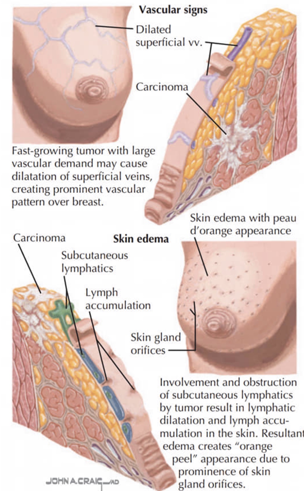
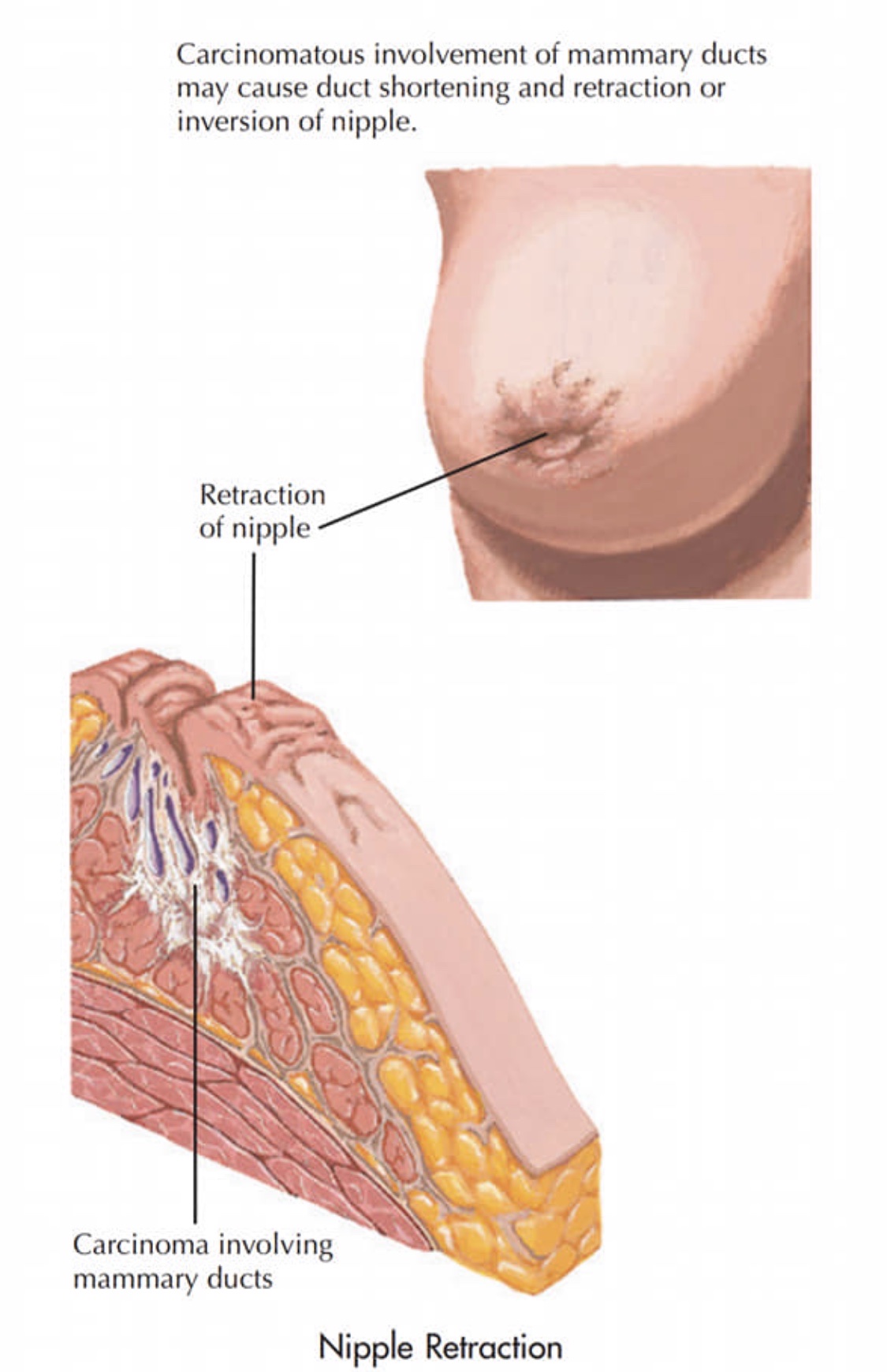
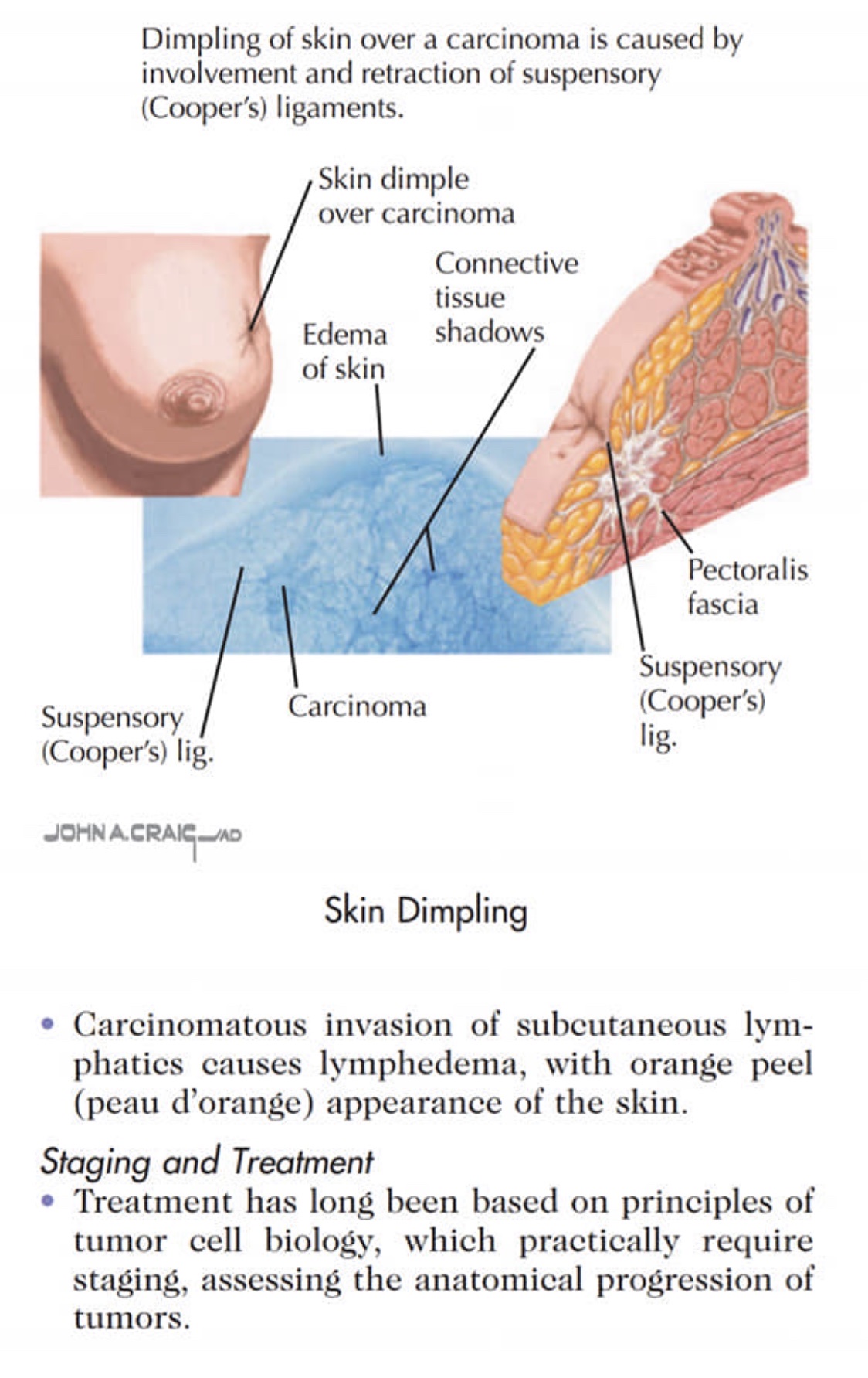
Signos de CĆ”ncer de MamĆ” / Clinical Signs of Breast Cancer

šŸ‘‰Rodrigo Arrangoiz MS, MD, FACS es cirujano oncólogo experto en mamĆ” y es miembro Center for Advanced Surgical Oncology en el Hospital Palmetto General Hospital: ā€©ā€©šŸ‘‰Es un experto en el manejo del cĆ”ncer de seno. ā€©ā€©šŸ‘‰Si tiene alguna pregunta sobre el examen de detección de cĆ”ncer de seno, no dude en comunicarse con el Dr. Arrangoiz.


šŸ‘‰

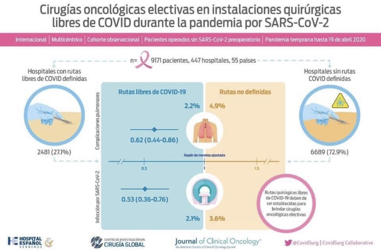
Elective Cancer Surgery in COVID-19–Free Surgical Pathways During the SARS-CoV-2 Pandemic: An International, Multicenter, Comparative Cohort Study

šŸ‘‰Rodrigo Arrangóiz MS, MD, FACS collaborated in the COVIDSurg-Cancer study published in the Journal of Clinical Oncology, which highlights the importance of having hospitals with protocols and “COVID-free routes” for the surgical care of cancer patients.

šŸ‘‰Millions of surgeries in the world have been postponed due to the pandemic.

šŸ‘‰Surgery for many conditions must continue despite the threat of a second wave.

šŸ‘‰Hospital areas with ā€œCOVID-19 free routesā€ should be established to allow surgery to be carried out safely.

šŸ‘‰Data from 9,171 patients from 55 countries were examined since the onset of the pandemic.

šŸ‘‰Pulmonary complications (2.2% vs 4.9%) and post-op mortality (0.7% vs 1.7%) were lower in patients treated in hospitals with protocols and ā€œCOVID-19 free routesā€.

šŸ‘‰Surgical care must continue during the pandemic. Hospitals must have protocols for the care of COVID and NON-COVID patients, detection programs for SARS-CoV-2 infection in asymptomatic personnel, and differentiated routes.

šŸ‘‰Hospitals without protocols represent a higher risk of complications and mortality for patients undergoing cancer surgeries. The Hospital EspaƱol de Veracruz has the COVID SAFE program to protect our patients and continue safe surgical care.

šŸ‘‰Full article: šŸ‘‰šŸ¼ https://ascopubs.org/doi/pdf/10.1200/JCO.20.01933

#Arrangoiz #CancerSurgeon #BreastSurgeon #ThyroidSurgeon #ParathyroidSurgeon #HeadandNeckSurgeon #SurgicalOncologist #CASO #CenterforAdvancedSurgicalOncology #PalmettoGeneralHospital

Breast Screening Guidelines

#Arrangoiz #CancerSurgeon #BreastSurgeon #SurgicalOncology #CASO #CenterforAdvancedSurgicalOncology #PalmettoGeneralHospital

CDH1 Mutation – Breast and Gastric Cancer

  • RecognizingĀ family history patternsĀ associated with specific gene mutations is important:
    • A family history ofĀ invasive lobular cancerĀ andĀ gastric cancer:
      • May be associated withĀ CDH1 mutations
        • These mutations confer aĀ 6.6 times greater risk of breast cancerĀ than the general population:
          • And areĀ associated with aĀ high risk of hereditary diffuse gastric cancer (55% toĀ 80%)
    • Patients withĀ CDH1 mutations:
      • Should be offeredĀ prophylactic gastrectomy
      • Consideration ofĀ bilateral prophylactic mastectomy:
        • Based onĀ family history
      • If surgery is not pursued, guidelines for screening for gastric cancer include:
        • Yearly endoscopic surveillanceĀ withĀ random mucosal biopsies;
      • Breast screening consists of:
        • Annual mammogram and breast MRI
  • References:
    • Hansford S, Kaurah P, Li-Chang H, Woo M, Senz J, Pinheiro H, et al. Hereditary diffuse gastric cancer syndrome: CDH1 mutations and beyond.Ā JAMA Oncol.Ā 2015;1(1):23-32.
    • Weiss A, Garber JE, King T. Breast cancer surgical risk reduction for patients with inherited mutations in moderate penetrance genes.Ā JAMA Surg.Ā 2018;153(12):1145-1146
    • van der Post RS, Vogelaar IP, Carneiro F, Guilford P, Huntsman D, Hoogerbrugge N, et al . Hereditary diffuse gastric cancer: updated clinical guidelines with an emphasis on germline CDH1 mutation carriers.Ā J Med Genet. 2015;52(6):361-374.
    • National Comprehensive Cancer Network Guidelines. Genetic/familial high-risk assessment: breast and ovarian, Version 1/2018.Ā https://www.nccn.org/professionals/physician_gls/pdf/genetics_screening.pdf. Accessed November 9, 2019.

#Arrangoiz #CancerSurgeon #BreastSurgeon #SurgicalOncology #BreastCancer #CASO #CenterforAdvancedSurgicalOncology #PalmettoGeneralHospital

CHEK2 Mutations and Breast Cancer

  • Truncating CHEK2 mutationsĀ (such as the c.1100delC mutation):
    • Are considered rare:
      • Moderate penetrance mutations
  • TheĀ lifetime risk of developing breast cancerĀ with these mutations:
    • IsĀ three times higherĀ than the general population:
      • This means that theĀ majority of patientsĀ with these mutations willĀ not develop breast cancer:
        • Many of the cancers that do develop may be:
          • Sporadic and notĀ related to the mutation
  • Patients with these mutations should beĀ counseled about their risk and the available risk management strategies:
    • Those with aĀ significant family history of breast cancerĀ should be counseled regarding high risk breast cancer screening:
      • Annual mammogramĀ with consideration ofĀ annual MRI starting at age 40Ā orĀ modified to an earlier age based on family history)Ā 
    • Counseled regarding high risk breast cancer prevention strategies, including:
      • Chemoprevention and prophylactic surgery:
        • It should be emphasized that there isĀ less absolute risk-reduction benefitĀ with:
          • Prophylactic surgeryĀ compared toĀ high-penetrance mutations like BRCA mutations
        • Recommendations should be based on:
          • Patient’s family history
      • National Comprehensive Cancer Network guidelines should be followed with recommendation for high-risk screening, including:
        • Colon cancer screeningĀ recommendations:
          • Among individuals with aĀ CHEK2 mutation
  • References:
  • Stratton MR, Rahman N. The emerging landscape of breast cancer susceptibility.Ā Nat Genet.Ā 2008;40(1):17-22.
  • Desmond A, Kurian AW, Gabree M, Mills MA, Anderson MJ, Kobayashi Y, et al. Clinical actionability of multigene panel testing for hereditary breast and ovarian cancer risk assessment.Ā JAMA Oncol. 2015;1(7):943-951.
  • Weiss A, Garber JE, King T. Breast cancer surgical risk reduction for patients with inherited mutations in moderate penetrance genes.Ā JAMA Surg.Ā 2018;153(12):1145-1146.
  • Tung N, Domchek SM, Stadler Z, Nathanson KL, Couch F, Garber JE, et al. Counselling framework for moderate-penetrance cancer-susceptibility mutations.Ā Nat Rev Clin Oncol. 2016;13(9):581-588.

#Arrangoiz #CancerSurgeon #BreastSurgeon #SurgicalOncology #BreastCancer #HereditaryBreastCancer #CASO #CenterforAdvancedSurgicalOncology #PalmettoGeneralHospital